Key Takeaways
- Heart valve disease often develops silently, so a new heart murmur, unexplained breathlessness, or reduced stamina deserves assessment even when you feel generally well
- The main types of valve problem are stenosis (narrowing), regurgitation (leaking), prolapse (bulging), and the rare congenital atresia
- Many heart murmurs are innocent, but only an ultrasound of the heart can reliably separate a harmless murmur from a structural valve problem
- Fainting on exertion, chest pain at rest, sudden severe breathlessness, or fever with a known valve problem are red flags that need emergency care
- Mild to moderate valve disease is usually managed with monitoring and medication; repair or replacement is advised when severe disease causes symptoms or affects heart function
- DCDC offers cardiology consultation from AED 250 with cardiovascular risk assessment included, plus on-site ECG, echo, stress test, and Holter monitoring, often within a single visit
- Dr. Shahoo Mazhari has expertise in valve disease, heart failure, and arrhythmia, and DCDC holds a 4.8/5 Google rating from 1000+ reviews with direct billing across 20+ insurers
A whooshing sound heard through a stethoscope, a flight of stairs that suddenly leaves you breathless, or fluttering in the chest at night can all point to a problem with one of the four valves that keep blood flowing the right way through your heart. Because valve disease often develops slowly and quietly, recognising heart valve disease symptoms early can make the difference between simple monitoring and a more serious problem later on. This guide explains how the valves work, the main types of valve disease, the warning signs, what a heart murmur really means, how the condition is diagnosed, and what treatment looks like. It also walks you through what to expect when you book a cardiology consultation at DCDC in Dubai Healthcare City, where testing is available on-site and results are discussed the same day.
This guide covers the four heart valves, types of valve disease, symptoms by severity, innocent versus pathological murmurs, risk factors, diagnosis, treatment options, and what to expect at DCDC in Dubai.

What Is Heart Valve Disease?
Your heart is a four-chambered pump, and its four valves act as one-way doors that keep blood moving in the right direction. The tricuspid valve sits between the right atrium and right ventricle, the pulmonary valve lets blood leave the right ventricle toward the lungs, the mitral valve sits between the left atrium and left ventricle, and the aortic valve opens to let oxygen-rich blood leave the left ventricle for the rest of the body. Each valve has thin, flexible flaps called leaflets (or cusps) that open fully with each heartbeat and then close tightly to prevent backflow. In a healthy adult this cycle repeats roughly 100,000 times a day without any noticeable effort.
Heart valve disease means that one or more of these valves is not working as it should. The problem is usually mechanical: either the valve does not open wide enough, so the heart must push harder to move blood through, or it does not close fully, so some blood leaks backward. Both situations make the heart work harder over time. The left-sided valves (mitral and aortic) carry the highest pressures and are affected far more often than the right-sided valves, which is why most of the symptoms discussed in this guide relate to them.
What makes valve disease tricky is how slowly it can develop. Many people feel completely well for years while a valve gradually narrows or leaks, and the heart compensates by thickening its walls or enlarging its chambers. By the time breathlessness or fatigue appears, the heart may already have adapted significantly. That is why recognising early heart valve disease symptoms, and getting a murmur or unexplained breathlessness checked promptly, matters so much. A cardiology consultation can identify the problem at a stage when monitoring or well-timed treatment protects the heart muscle.
According to the American Heart Association, valvular heart disease affects a substantial share of adults as they age, and degenerative aortic and mitral disease are the most common forms in developed countries. In Dubai, where the population includes many expatriates from regions where rheumatic fever remains more common, clinicians see both age-related and rheumatic valve problems, as well as congenital valve variations that are discovered for the first time in adulthood.
Types of Heart Valve Disease
Doctors classify valve problems by what is going wrong mechanically and by which valve is involved. Understanding the types of heart valve disease helps explain why symptoms, murmurs, and treatment plans differ so much from one person to another. A single valve can have more than one problem at once, for example a valve that is both narrowed and leaky.
| Valve Problem | Description | Common Causes |
|---|---|---|
| Stenosis | The valve opens too narrowly because the leaflets become thick, stiff, or fused. The heart must generate more pressure to push blood through the smaller opening. | Age-related calcium build-up (especially aortic), rheumatic fever (especially mitral), bicuspid aortic valve, radiation exposure |
| Regurgitation (insufficiency) | The valve does not close tightly, so blood leaks backward into the chamber it just left. The heart must pump extra volume with each beat. | Mitral valve prolapse, endocarditis damage, dilated heart chambers, ruptured supporting cords, connective tissue disorders, rheumatic disease |
| Prolapse | The valve leaflets bulge or flop back into the upper chamber as the heart contracts. Most often affects the mitral valve and may or may not cause leaking. | Myxomatous (floppy) degeneration, inherited connective tissue conditions such as Marfan syndrome, often no clear cause |
| Atresia | The valve fails to form at all, leaving no opening for blood to pass. It is a congenital condition present from birth and is usually found in infancy. | Developmental abnormality of the fetal heart; tricuspid and pulmonary atresia are the most commonly described forms |
Stenosis is most often seen in the aortic valve in older adults, where years of calcium deposition stiffen the leaflets. Mitral stenosis, by contrast, is most strongly linked to previous rheumatic fever. Because the valve opening is restricted, pressure builds up behind it, which can cause fluid to back up into the lungs and produce breathlessness.
Regurgitation can affect any valve. Mitral regurgitation is the most common form of significant valve leakage, while aortic regurgitation often results from a dilated aortic root or a damaged valve. Mild leakage is extremely common and often harmless, and many healthy people have trivial leakage detected incidentally on an ultrasound. Severe regurgitation, however, causes the heart chambers to enlarge and can eventually weaken the pumping muscle.
Prolapse deserves its own mention because mitral valve prolapse is one of the most commonly diagnosed valve conditions, affecting an estimated 2 to 3 percent of the population. Most people with it have no symptoms and a normal life expectancy, although a subset develops significant leakage over time. We cover mitral valve prolapse symptoms in more detail in the next section.
Atresia is rare and is almost always diagnosed in newborns or before birth, so adults will not usually encounter it as a new diagnosis. It is included here because it completes the picture of how valves can fail, and because adults who had surgery for congenital valve problems in childhood still need lifelong cardiology follow-up.
Heart Valve Disease Symptoms to Watch For
Symptoms depend on which valve is affected, whether the problem is narrowing or leaking, and how severe it has become. It is entirely possible to have a moderately abnormal valve and no symptoms at all, which is why the absence of symptoms should not be taken as proof that the heart is fine, particularly if a murmur has ever been heard. The signs of heart valve problems below are grouped by how urgently they should be assessed.
Early and Mild Signs
- Unusual tiredness: feeling worn out after activities that used to be easy, such as climbing a flight of stairs or carrying shopping
- Mild breathlessness on exertion: noticing that you get short of breath sooner than other people your age, or sooner than you used to
- Occasional palpitations: a fluttering, pounding, or skipped-beat sensation, common with mitral valve prolapse
- Reduced exercise tolerance: needing to slow down or stop during a walk, a game of padel, or a gym session
- A heart murmur found on routine examination: sometimes the only sign in the early stage
Moderate Symptoms That Need Prompt Assessment
- Breathlessness when lying flat or waking at night: a sign that fluid is backing up into the lungs
- Swollen ankles, feet, or abdomen: fluid retention caused by the heart struggling to keep up
- Dizziness or light-headedness: especially on standing or exertion, which can occur with aortic stenosis
- Chest tightness or pressure with activity: because a thickened heart muscle needs more oxygen than the narrowed valve allows it to receive
- Persistent racing or irregular heartbeat: atrial fibrillation is common in mitral valve disease and raises stroke risk
- Rapid weight gain over days: often fluid rather than fat, and a classic marker of worsening heart failure
Severe Symptoms and Red Flags
- Fainting or near-fainting, especially during exertion: a serious warning sign in aortic stenosis
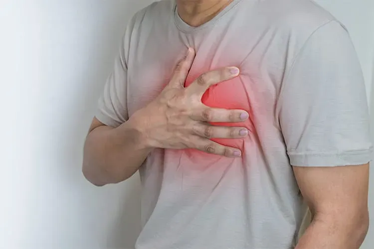
- Chest pain at rest or severe pain lasting more than a few minutes: needs emergency care
- Sudden severe breathlessness or coughing up pink, frothy sputum: may signal acute pulmonary oedema
- Sudden weakness, facial droop, or speech difficulty: a valve-related clot may have caused a stroke
- Fever with a known valve problem: could point to endocarditis, which needs urgent evaluation
If you have severe symptoms, go to the nearest emergency department or call 998 for an ambulance in Dubai. For the milder patterns above, do not wait for symptoms to become dramatic. Our guide to heart failure warning signs explains how untreated valve disease can progress, and our overview of heart disease symptoms places valve problems in the wider context of cardiac health.
Mitral Valve Prolapse Symptoms
Mitral valve prolapse symptoms vary widely. Most people have none and learn about the condition only when a doctor hears a click or murmur. When symptoms do occur, they typically include palpitations, brief sharp or stabbing chest discomfort that is unrelated to exertion, fatigue, light-headedness, and anxiety-like sensations. These symptoms can be unsettling but are often not dangerous. What matters medically is whether the prolapse causes significant leakage, since it is the leak, not the prolapse alone, that can enlarge the heart over time. If you have been told you have mitral valve prolapse, periodic review is sensible, and any change in breathlessness, palpitations, or stamina should prompt an earlier visit.
Heart Murmur: What It Means
A heart murmur is an extra or unusual sound heard through a stethoscope during the heartbeat, often described as whooshing, swishing, or blowing. It is a clinical finding, not a diagnosis, and it simply tells the doctor that blood flow in the heart is turbulent. Understanding heart murmur causes is the key to knowing when a murmur is nothing to worry about and when it needs investigation.
Innocent (Functional) Murmurs
Innocent murmurs occur in a structurally normal heart. They are very common in children and young adults and can also appear in adults when blood flow is temporarily increased. Typical triggers include fever, anaemia, pregnancy, thyroid overactivity, vigorous exercise, and dehydration. Innocent murmurs are usually soft, short, change with position or breathing, and are not accompanied by symptoms. They often disappear once the underlying trigger resolves.
Pathological (Abnormal) Murmurs
Pathological murmurs arise from a structural problem, most often a narrowed or leaking valve, but also from holes between heart chambers (septal defects), thickened heart muscle, or abnormal connections between vessels. These murmurs tend to be louder, longer, and may be heard throughout the whole of systole or diastole. Their timing and location help the doctor guess which valve is involved: for instance, a harsh murmur heard in the upper right chest that radiates to the neck suggests aortic stenosis, while a blowing murmur at the apex radiating to the armpit suggests mitral regurgitation.
| Feature | More Likely Innocent | More Likely Pathological |
|---|---|---|
| Symptoms | None | Breathlessness, chest discomfort, fainting, swelling, palpitations |
| Loudness and duration | Soft, short, mid-systolic | Loud, harsh, long, or occurs during diastole |
| Behaviour | Changes with posture or breathing; may vanish | Constant and unaffected by position |
| Associated signs | Normal pulses and heart size | Thrill (vibration), abnormal pulses, enlarged heart |
| Age at discovery | Childhood or young adulthood, or with a temporary trigger | New murmur in an older adult, or a murmur that changes |
When to Worry About a Heart Murmur
Knowing when to worry about a heart murmur comes down to context. A murmur deserves formal assessment if it is new in an adult, if it is accompanied by any symptom listed above, if you have a family history of valve disease or sudden cardiac death, if you have had rheumatic fever or endocarditis, or if you have been told your murmur has become louder. Even in the absence of symptoms, an adult who has never had a murmur evaluated should consider doing so, because it is the only way to separate a harmless flow murmur from an early valve problem. The test that settles the question is an ultrasound of the heart, which is why doctors so often recommend an echocardiogram after hearing one.
For athletes and active people, an unexplained murmur is particularly worth clarifying before continuing high-intensity training, as some conditions such as hypertrophic cardiomyopathy and severe aortic stenosis can increase risk during exertion. Our article on when to see a cardiologist in Dubai gives a broader checklist of reasons to book a heart review.
Risk Factors for Heart Valve Disease
Valve disease can develop in anyone, but certain factors raise the likelihood considerably. Some can be modified, others cannot, but knowing your own risk profile helps decide when to start screening.
- Increasing age: the leaflets gradually thicken and calcify over decades. Degenerative (age-related) disease is now the most common cause of aortic stenosis and a leading cause of mitral regurgitation
- Congenital valve abnormalities: a bicuspid aortic valve (two leaflets instead of three) affects roughly 1 to 2 percent of people and often begins to narrow or leak in midlife
- Rheumatic fever: an inflammatory reaction to untreated streptococcal throat infection that can scar the valves, particularly the mitral, years after the original illness. It remains an important cause in parts of South Asia, Africa, and the Middle East
- Infective endocarditis: infection of the valve or heart lining, more likely in people with existing valve disease, prosthetic valves, injection drug use, or poor dental health
- High blood pressure and high cholesterol: long-standing hypertension strains the aortic valve and root, and raised cholesterol is associated with faster aortic valve calcification
- Previous heart attack or cardiomyopathy: damage to the heart muscle can distort the mitral valve apparatus and cause functional leakage
- Chronic kidney disease and diabetes: both are linked with accelerated valve calcification
- Chest radiation or certain medications: prior radiotherapy to the chest and some appetite-suppressant or ergot-derived drugs have been linked with valve damage
- Connective tissue disorders and family history: conditions such as Marfan syndrome affect valve and aortic structure, and some forms of mitral valve prolapse run in families
Several of these risks overlap with general heart disease risk factors, which is why the cardiovascular risk assessment included with a DCDC cardiology consultation looks at blood pressure, lipids, blood sugar, lifestyle, and family history together rather than in isolation.
How Heart Valve Disease Is Diagnosed
The diagnosis of heart valve disease follows a stepwise path, beginning with a conversation and a physical examination and moving to imaging that shows exactly how the valves are working. Most patients do not need every test below. The cardiologist selects them according to what the earlier steps reveal.
Medical History and Physical Examination
The doctor will ask about your symptoms, exercise tolerance, past infections such as rheumatic fever, family history, medications, and any previous murmur. The key physical step is auscultation, listening to the heart with a stethoscope in several positions. The pitch, timing, loudness, and location of a murmur, together with other clues such as pulse character, blood pressure, neck veins, lung sounds, and leg swelling, give an experienced cardiologist a strong initial idea of which valve is involved and how significant the problem might be.
Echocardiogram
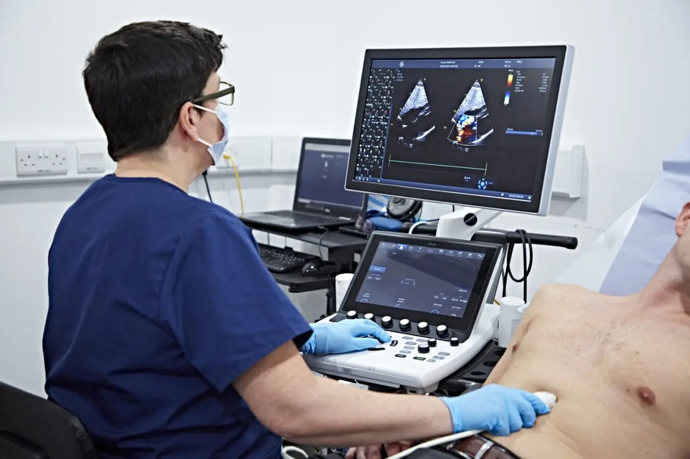
The echocardiogram is the cornerstone of heart valve disease diagnosis. It is a painless ultrasound that shows the valve leaflets moving in real time and uses Doppler technology to measure blood velocity, calculate pressure gradients across narrowed valves, and grade the severity of leaks. It also measures heart chamber size and pumping strength, which tell the doctor how the heart is coping. Our guide to the echocardiogram in Dubai explains the different types, including transthoracic and transoesophageal studies, in more detail.
Electrocardiogram (ECG)
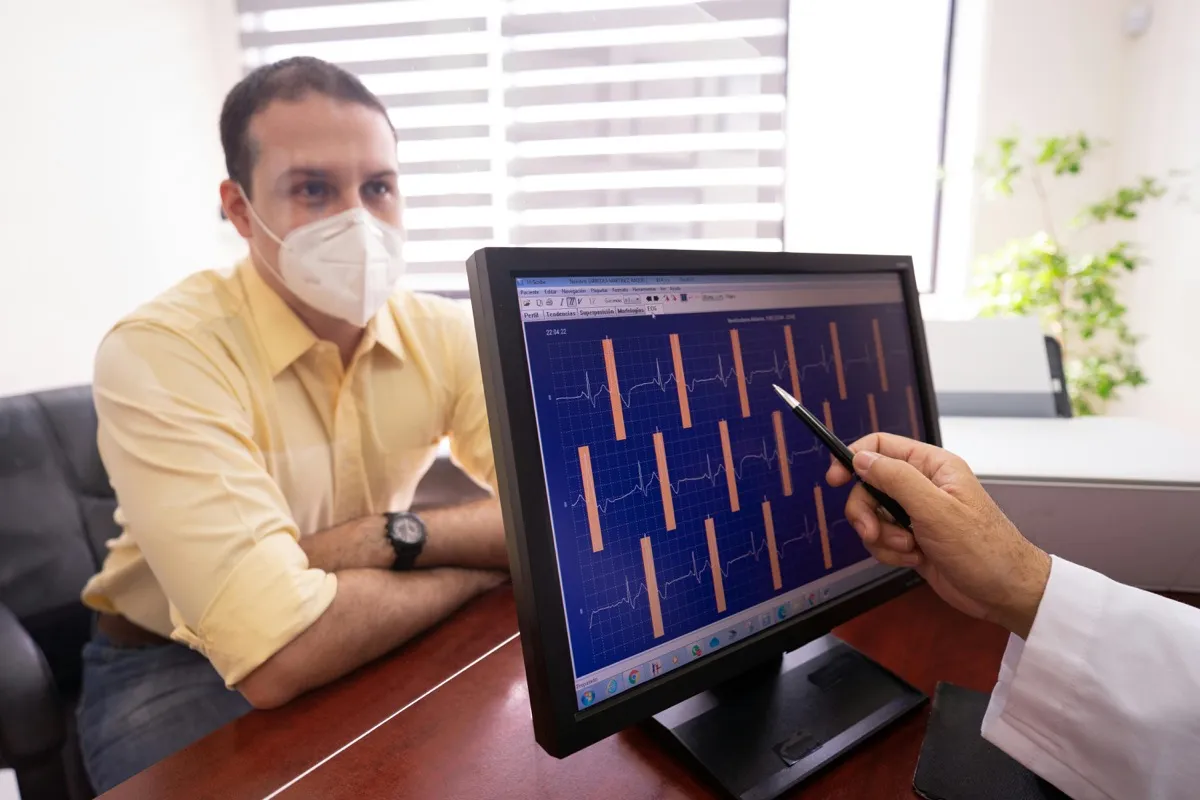
An ECG records the heart's electrical activity and takes only a few minutes. It cannot diagnose a valve problem directly, but it can reveal rhythm disturbances such as atrial fibrillation, signs of chamber enlargement or thickening, and conduction problems that often accompany valve disease. See our comparison of ECG versus echo for how the two tests complement each other.
Exercise Stress Test
A stress test is valuable when symptoms and echo findings do not seem to match, for example when someone with severe valve narrowing insists they have no symptoms. Walking on a treadmill under monitoring shows how blood pressure, heart rhythm, and symptoms respond to exertion, and can unmask limitation that the person has unconsciously adapted to. In severe aortic stenosis, however, stress testing in symptomatic patients is avoided, and the cardiologist will judge when it is safe.
24-Hour Holter Monitoring
If palpitations are a feature, as in mitral valve prolapse or mitral stenosis, a 24-hour Holter monitor records every heartbeat during normal activities and can capture intermittent arrhythmias that a short ECG misses.
Cardiac MRI and CT
When echo images are limited or more detail is required, advanced imaging helps. Cardiac MRI gives precise measurements of leak volume, chamber size, and scarring of the heart muscle. Cardiac CT is used to measure the aortic root and valve calcium, and to plan transcatheter valve procedures. These tests are reserved for selected patients and are arranged through referral when the cardiologist feels they will change management.
Treatment Options for Heart Valve Disease
Treatment depends on the type and severity of valve disease, the presence of symptoms, and the effect on the heart. Many people never need an operation. The guiding principle in modern guidelines from the American College of Cardiology and American Heart Association is to treat valve disease at the point when intervention offers a clear advantage, before the heart muscle is permanently damaged but not so early that surgical risk outweighs the benefit.
Watchful Monitoring
Mild to moderate disease without symptoms is usually managed with regular clinical review and periodic echocardiograms. The interval depends on the severity, from every few years for mild disease to every six to twelve months for severe but asymptomatic disease. Monitoring also includes advice on keeping fit, maintaining healthy blood pressure and cholesterol, dental hygiene, and reporting new symptoms promptly.
Medication
No tablet can reverse a damaged valve, but medications can control the consequences and protect the heart while you wait. Depending on the situation, a cardiologist may prescribe diuretics to reduce fluid congestion, blood pressure medicines to lower the heart's workload, beta-blockers or other rate-control drugs for atrial fibrillation, and anticoagulants (blood thinners) to prevent clots, particularly in atrial fibrillation with rheumatic mitral stenosis. Antibiotic prophylaxis before dental procedures is reserved for the highest-risk groups, such as those with prosthetic valves or previous endocarditis, rather than all people with a murmur.
Valve Repair Versus Replacement
When severe disease causes symptoms or begins to impair the heart, a procedure is recommended. The two principal options are repair and replacement.
| Option | What It Involves | Key Considerations |
|---|---|---|
| Valve repair | The surgeon or interventional cardiologist reshapes or supports the patient's own valve, for example with an annuloplasty ring or a catheter-based clip. | Preferred for mitral regurgitation when feasible because it preserves the natural valve, usually avoids lifelong blood thinners, and maintains better heart function |
| Mechanical valve replacement | The damaged valve is replaced with a durable artificial valve. | Lasts decades but requires lifelong warfarin therapy; often chosen for younger patients |
| Bioprosthetic (tissue) valve replacement | A valve made from animal tissue replaces the diseased valve, either surgically or through a catheter (TAVI/TAVR). | Does not usually need long-term warfarin but may wear out over 10 to 20 years; transcatheter options suit many older or higher-risk patients |
| Balloon valvuloplasty | A balloon is inflated across a narrowed valve to open it, performed through a catheter. | Most useful for suitable rheumatic mitral stenosis; less durable for calcified aortic stenosis |
The choice depends on the valve involved, the cause of the disease, your age and other health conditions, your preferences about blood thinners, and the expertise available. These decisions are made by a heart team that typically includes a cardiologist, a cardiac surgeon, and an imaging specialist. When a patient at DCDC needs a procedure, Dr. Shahoo Mazhari helps coordinate the referral and then resumes follow-up after the operation.
Recovery after valve surgery typically involves a hospital stay of about a week, several weeks to a few months of gradual return to normal activity, and cardiac rehabilitation. Transcatheter procedures generally have shorter hospital stays and quicker recovery. Whatever the approach, lifelong follow-up with periodic echocardiograms is part of the plan.
What to Expect at DCDC
Doctors Clinic Diagnostic Center in Dubai Healthcare City (Building 64, Block A, Al Razi Medical Complex) brings consultation and cardiac testing under one roof, so the process from first concern to a clear answer can often be completed in a single visit. Dr. Shahoo Mazhari, our cardiologist, has particular expertise in heart failure, valve disease, and arrhythmia.
- Step 1, book your appointment: call +971 54 219 5008 or message us on WhatsApp at +971 50 970 3884. We are open Saturday to Thursday 8AM to 10PM and Friday 9AM to 9PM, with free dedicated parking
- Step 2, cardiology consultation (from AED 250): Dr. Mazhari reviews your symptoms, history, and medications, and performs a careful cardiac examination including auscultation
- Step 3, cardiovascular risk assessment (included): blood pressure, cholesterol, diabetes, lifestyle, and family history are reviewed as part of your consultation at no additional charge
- Step 4, on-site ECG (from AED 200): a quick tracing of your heart rhythm and electrical activity, available immediately
- Step 5, echocardiogram same visit: if a murmur or valve concern is suspected, the heart ultrasound is performed on-site, so you do not need to book a separate imaging appointment (pricing varies by study)
- Step 6, stress test if needed (from AED 800): a treadmill test is arranged when exertion-related symptoms or borderline findings need clarification. 24-hour Holter monitoring is available for palpitations
- Step 7, same-day results discussion: Dr. Mazhari explains what the images show in plain language, including which valve is involved and how severe the problem is
- Step 8, treatment plan and follow-up: you leave with a clear plan, whether that is reassurance, medication, scheduled monitoring, or referral for a valve procedure, and a follow-up date
For patients who are worried about a cardiac event, DCDC also offers troponin testing at AED 149 for an individual test or AED 299 for a panel. DCDC holds a 4.8 out of 5 Google rating from over 1,000 reviews, and we work with more than 20 insurance partners with direct billing, so many patients pay little or nothing out of pocket. Check your policy or ask our team to verify coverage when you book.
Worried About a Heart Murmur or Valve Symptoms?
Book a cardiology consultation with Dr. Shahoo Mazhari at DCDC Dubai Healthcare City. Consultation from AED 250 with cardiovascular risk assessment included, on-site ECG, echocardiogram, and stress testing, plus direct billing with 20+ insurers.
Call +971 54 219 5008 or WhatsApp +971 50 970 3884 to book today.
Related Services at DCDC
Expert care and advanced diagnostics at Dubai Healthcare City
Frequently Asked Questions
Final Thoughts
Heart valve disease ranges from a harmless leaflet variation that never causes trouble to a severe narrowing or leak that needs a procedure. Because the early stages are frequently silent, the most useful step you can take is to have a murmur, breathlessness, or reduced stamina properly evaluated rather than assuming it is age, stress, or fitness. Modern imaging allows doctors to measure the problem precisely and plan treatment at the right moment, protecting the heart muscle from long-term strain.
If you have a known murmur, a family history of valve disease, a past of rheumatic fever, or any of the warning signs described here, book a cardiology consultation at DCDC. Dr. Shahoo Mazhari and our team can examine you, perform an ECG and echocardiogram on-site, and explain the findings the same day. Call +971 54 219 5008 or message us on WhatsApp at +971 50 970 3884 to arrange your visit. For chest pain at rest, fainting, or sudden severe breathlessness, call 998 or go to the nearest emergency department immediately.
Sources & References
This article references the following sources:
- American Heart Association — Problem: Heart Valve Disease
- 2020 ACC/AHA Guideline for the Management of Patients With Valvular Heart Disease (PubMed)
- 2021 ESC/EACTS Guidelines for the Management of Valvular Heart Disease (PubMed)
- Burden of Valvular Heart Diseases: A Population-Based Study (PubMed)
- National Heart, Lung, and Blood Institute — Heart Valve Diseases
- Mayo Clinic — Heart Murmurs: Symptoms and Causes
This information is educational and does not replace individual medical advice. See our editorial policy for more information.

Written by
Dr. Shahoo Mazhari
Cardiology
MD, Cardiology
Dr. Shahoo Mazhari is a Cardiologist at Doctors Clinic Diagnostic Center (DCDC) in Dubai Healthcare City.
Related Articles
Echocardiogram Dubai: Types & What It Shows (2026)
Heart Disease Symptoms Dubai: 8 Signs to Watch (2026)
Heart Failure Warning Signs Dubai (2026)
ECG vs Echocardiogram: Differences Explained (2026)
When to See a Cardiologist Dubai (2026)
More in Cardiology

Heart-Healthy Foods: Diet Tips Dubai (2026)
Read MorePeripheral Artery Disease Dubai: Symptoms (2026)
Read More
Smartwatch ECG vs Medical ECG Dubai (2026)
Read MoreHeart Disease in Women Dubai: Warning Signs (2026)
Read MoreABPM Test Dubai: 24-Hour BP Monitoring (2026)
Read More
Heart Tests Dubai: ECG, Echo & Stress Test Cost (2026)
Read More© 2026 Doctors Clinic Diagnostic Center (DCDC), Dubai Healthcare City. Originally published at https://doctorsclinicdubai.ae/blog/heart-valve-disease-symptoms-dubai. All rights reserved. Unauthorized reproduction is prohibited.