Introduction
Skin cancer is the most common form of cancer worldwide, and among its types, melanoma is particularly deadly if not caught early. The rising incidence of melanoma has led to an increased focus on early detection methods, with mole mapping emerging as a key tool. This article delves into the science behind melanoma and mole mapping, exploring how early detection can save lives and the role advanced technologies play in this process.
What is Melanoma?
Melanoma is a type of skin cancer that develops from melanocytes, the cells responsible for producing melanin, the pigment that gives skin its color. Unlike other skin cancers, melanoma is more likely to spread to other parts of the body, making it more dangerous if not treated promptly.
Types of Melanoma
1. Superficial Spreading Melanoma: The most common type, it typically grows horizontally across the skin before penetrating deeper layers.
2. Nodular Melanoma: This type grows vertically from the outset and can spread rapidly, making early detection crucial.
3. Lentigo Maligna Melanoma: Often occurring in older adults, this type appears as a flat or slightly raised brownish patch.
4. Acral Lentiginous Melanoma: This type occurs on the palms, soles, or under the nails and is more common in people with darker skin.
Risk Factors for Melanoma
Several factors can increase the risk of developing melanoma:
– UV Exposure: Prolonged exposure to ultraviolet (UV) radiation from the sun or tanning beds is a major risk factor.
– Skin Type: People with fair skin, light hair, and light-coloured eyes are at higher risk.
– Moles: Having many moles or unusual moles (dysplastic nevi) increases the risk.
– Family History: A family history of melanoma can indicate a genetic predisposition.
– Weakened Immune System: Individuals with weakened immune systems are more susceptible to melanoma.
The Importance of Early Detection
Early detection of melanoma dramatically improves survival rates. When caught in its earliest stages, melanoma is often curable with surgical removal. However, if the cancer spreads (metastasizes) to other parts of the body, treatment becomes more challenging, and the prognosis worsens.
Melanoma Survival Rates
Survival rates for melanoma vary depending on the stage at which it is diagnosed:
– Stage 0 (In Situ): The melanoma is confined to the epidermis and has not spread. The 5-year survival rate is nearly 100%.
– Stage I and II: The melanoma is still localized, but it has penetrated deeper into the skin. The 5-year survival rate ranges from 70% to 95%.
– Stage III: The cancer has spread to nearby lymph nodes or tissues. The 5-year survival rate drops to 40% to 70%.
– Stage IV: The melanoma has metastasized to distant organs. The 5-year survival rate is around 15% to 20%.
These statistics underscore the critical importance of early detection and the role it plays in improving patient outcomes.
Traditional Methods of Skin Cancer Detection
Traditionally, skin cancer detection has relied on visual examinations by dermatologists. The ABCDE rule is commonly used to assess moles:
– A – Asymmetry: One half of the mole does not match the other.
– B – Border: The edges are irregular, scalloped, or poorly defined.
– C – Color: The mole has varying colors, such as shades of brown, black, or even red, white, or blue.
– D – Diameter: Melanomas are usually larger than 6mm in diameter (about the size of a pencil eraser).
– E – Evolving: The mole is changing in size, shape, or color.
While these guidelines are useful, they are not foolproof. Early-stage melanomas may not fit these criteria, and benign moles can sometimes resemble melanoma. This is where mole mapping and advanced technologies come into play.
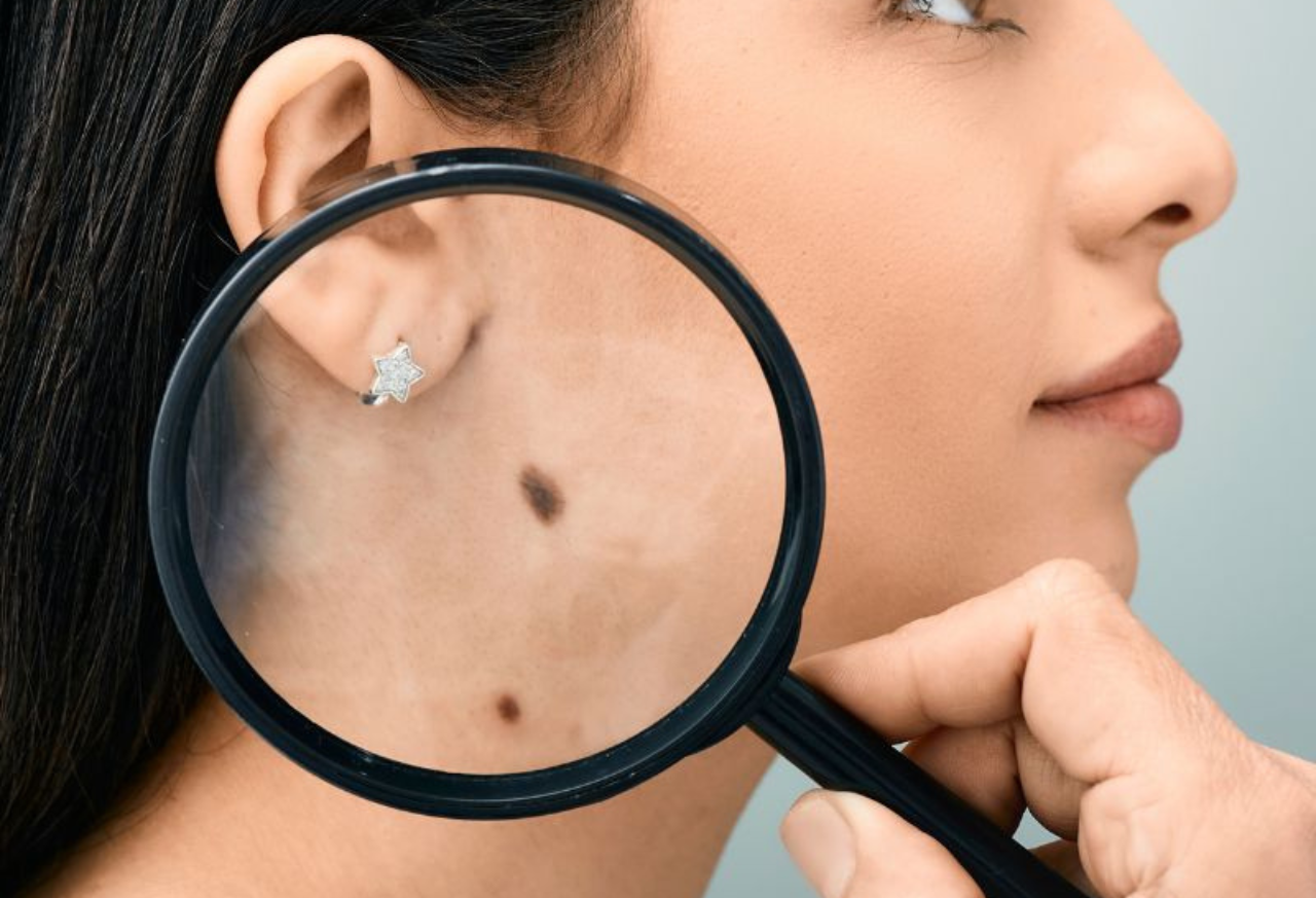
What is Mole Mapping?
Mole mapping is a technique used to monitor moles and detect changes that could indicate melanoma. It involves taking high-resolution photographs of the entire body to create a visual record of a patient’s moles. These images can be compared over time to identify any changes in existing moles or the appearance of new ones.
How Mole Mapping Works
1. Baseline Photography: During the initial visit, high-resolution images are taken of the patient’s entire body. These images serve as a baseline for future comparisons.
2. Regular Monitoring: At subsequent visits, new images are taken and compared to the baseline. Any changes in the moles’ size, shape, color, or number can be detected early.
3. Digital Dermoscopy: In addition to whole-body photography, digital dermoscopy (also known as dermatoscopy) may be used. This involves using a special device to magnify and photograph individual moles, allowing for detailed analysis.
4. Computer-Assisted Analysis: Advanced software can analyze the images, detecting subtle changes that may not be visible to the naked eye. This assists dermatologists in making more accurate assessments.
The Science Behind Mole Mapping
The science behind mole mapping is rooted in the understanding that melanoma often develops from pre-existing moles. By closely monitoring these moles for changes, it is possible to catch melanoma in its earliest stages when it is most treatable.
Technology Used in Mole Mapping
1. High-Resolution Photography: Modern mole mapping systems use high-resolution cameras to capture detailed images of the skin. These images can reveal subtle changes in moles that might indicate the development of melanoma.
2. Dermatoscopes: These handheld devices magnify the skin’s surface, allowing for detailed examination of moles. Some dermatoscopes are equipped with polarized light to reduce reflection and enhance visibility of skin structures.
3. Computer-Assisted Diagnosis (CAD): CAD systems use algorithms to analyze images of moles and compare them to databases of known benign and malignant lesions. These systems can help dermatologists identify suspicious moles that warrant further investigation.
4. Artificial Intelligence (AI): AI is increasingly integrating into mole mapping systems. Machine learning algorithms train on vast datasets of mole images, learning to recognize patterns associated with melanoma.AI can assist in identifying changes in moles over time and flagging those that are likely to be malignant.
Benefits of Mole Mapping
Mole mapping offers several advantages over traditional skin cancer detection methods:
1. Accuracy and Reliability: Mole mapping allows for precise monitoring of moles over time, increasing the likelihood of detecting melanoma early. By comparing images side by side, even subtle changes can be identified.
2. Non-Invasive: The procedure is non-invasive and painless, involving nothing more than photography. This makes it an ideal screening tool for patients at high risk of melanoma.
3. Convenience: Once the baseline images are taken, follow-up appointments are quick and easy. This convenience encourages regular monitoring, which is essential for early detection.
Peace of Mind: Mole mapping gives patients with many moles or a family history of melanoma peace of mind by promptly detecting any changes.
Who Should Consider Mole Mapping?
While mole mapping can be beneficial for anyone, certain individuals may be at higher risk for melanoma and should consider regular monitoring:
1. Individuals with Multiple Moles: People with numerous moles are at a higher risk of developing melanoma, particularly if they have atypical or dysplastic nevi.
2. Family History of Melanoma: A family history of melanoma increases the risk of developing the disease. These individuals should consider mole mapping as a precautionary measure.
3. Fair Skin and High UV Exposure: Those with fair skin, light hair, and a history of sunburns or excessive UV exposure are at increased risk.
4. Previous History of Skin Cancer: Individuals who have had skin cancer in the past are more likely to develop it again, making regular monitoring essential.
5. Immunosuppressed Individuals: Those with weakened immune systems, such as organ transplant recipients, are at higher risk of melanoma and should be vigilant about skin monitoring.
The Procedure: What to Expect During Mole Mapping
Understanding what to expect during a mole mapping session can help alleviate any concerns and ensure a smooth process.
Step-by-Step Process of Mole Mapping
1. Initial Consultation: The process begins with an initial consultation, where the patient’s medical history and risk factors are reviewed. The dermatologist explains the mole mapping procedure and answers any questions.
2. Baseline Photography: The patient undresses to allow for full-body photography. We take high-resolution images of the entire body, including close-ups of any notable moles. We may ask the patient to change positions to ensure that we cover all areas.
Digital Dermoscopy: If necessary, a dermatoscope examines individual moles, and these images are recorded for future comparison.
Image Analysis: Computer-assisted tools analyze the images, flagging any suspicious moles for further examination or biopsy.
Follow-Up Appointments: Schedule regular follow-up appointments, typically every 6 to 12 months. During these visits, the dermatologist takes new images and compares them to the baseline to detect any changes.
Preparing for a Mole Mapping Session
To ensure accurate results, patients should follow these guidelines before a mole mapping session:
– Avoid Tanning: Refrain from tanning or excessive sun exposure for several weeks before the session. A consistent skin tone is important for accurate comparisons.
– Remove Makeup and Jewelry: Remove all makeup, jewelry, and any other items that may obstruct the view of the skin.
– Wear Comfortable Clothing: Wear loose, comfortable clothing that is easy to remove, as full-body photography requires undressing.
Inform the Dermatologist of Any Changes: If you notice any new or changing moles, inform the dermatologist so they can closely examine them.
Advances in Mole Mapping Technology
As technology continues to evolve, mole mapping is becoming more advanced and accessible.
Artificial Intelligence and Machine Learning
AI and machine learning are revolutionizing mole mapping. These technologies can analyze images with incredible precision, identifying patterns and changes that may not be visible to the human eye. AI-driven tools can provide dermatologists with a second opinion, enhancing the accuracy of diagnoses.
Integration with Wearable Devices
The future of mole mapping may include integration with wearable devices that continuously monitor the skin. Smartwatches and other wearable tech could equip sensors that detect changes in moles, alerting users to potential issues in real-time.
Telemedicine and Remote Monitoring
Telemedicine is expanding the reach of mole mapping by allowing patients to submit images of their moles remotely. Dermatologists can review these images and provide consultations without the need for in-person visits. This is particularly beneficial for patients in remote or underserved areas.
The Role of Patients in Early Detection
While mole mapping is a powerful tool, patients play a crucial role in early detection. Self-examination and awareness of one’s skin are essential components of a comprehensive skin cancer prevention strategy.
How to Perform a Self-Examination
1. Examine the Entire Body: Use a full-length mirror to examine the entire body, including hard-to-see areas like the back, scalp, and soles of the feet.
2. Check for the ABCDEs: Use the ABCDE rule to assess moles and look for any that are asymmetric, have irregular borders, multiple colors, a large diameter, or are evolving.
3. Take Note of New Moles: Keep track of any new moles that appear and monitor existing ones for changes.
4. Photograph Suspicious Moles: Take photographs of any moles that seem unusual and compare them over time.
When to Seek Medical Advice
If you notice any moles that are changing in size, shape, color, or texture, or if you have a mole that looks different from your other moles (the “ugly duckling” sign), seek medical advice promptly. Early consultation with a dermatologist can lead to early detection and better outcomes.
Conclusion
Melanoma is a serious and potentially deadly form of skin cancer, but with early detection, it is often treatable. Mole mapping offers a cutting-edge approach to monitoring skin changes and catching melanoma in its earliest stages. By combining advanced technology with regular self-examinations and professional monitoring, individuals can take control of their skin health and reduce their risk of melanoma.
As technology continues to advance, the future of mole mapping looks promising, with AI, wearable devices, and telemedicine poised to enhance the accuracy and accessibility of this life-saving tool. Whether you are at high risk for melanoma or simply want to take a proactive approach to your skin health, mole mapping is a valuable resource that empowers you to stay ahead of skin cancer and ensure early detection.
Visit for More Details: https://doctorsclinicdubai.ae/