Key Takeaways
- A spine MRI in Dubai costs between AED 1,000 and AED 1,600 per region (cervical, thoracic, or lumbar), with whole-spine MRI ranging from AED 2,500 to AED 4,000
- Spine MRI is the gold standard for diagnosing herniated discs, spinal stenosis, nerve compression, sciatica, and degenerative spine conditions
- Lumbar spine MRI is the most commonly ordered spine study, accounting for the majority of back-related MRI referrals in Dubai
- Your doctor may order a spine MRI for persistent back or neck pain, radiating leg or arm pain, numbness, tingling, or weakness in the extremities
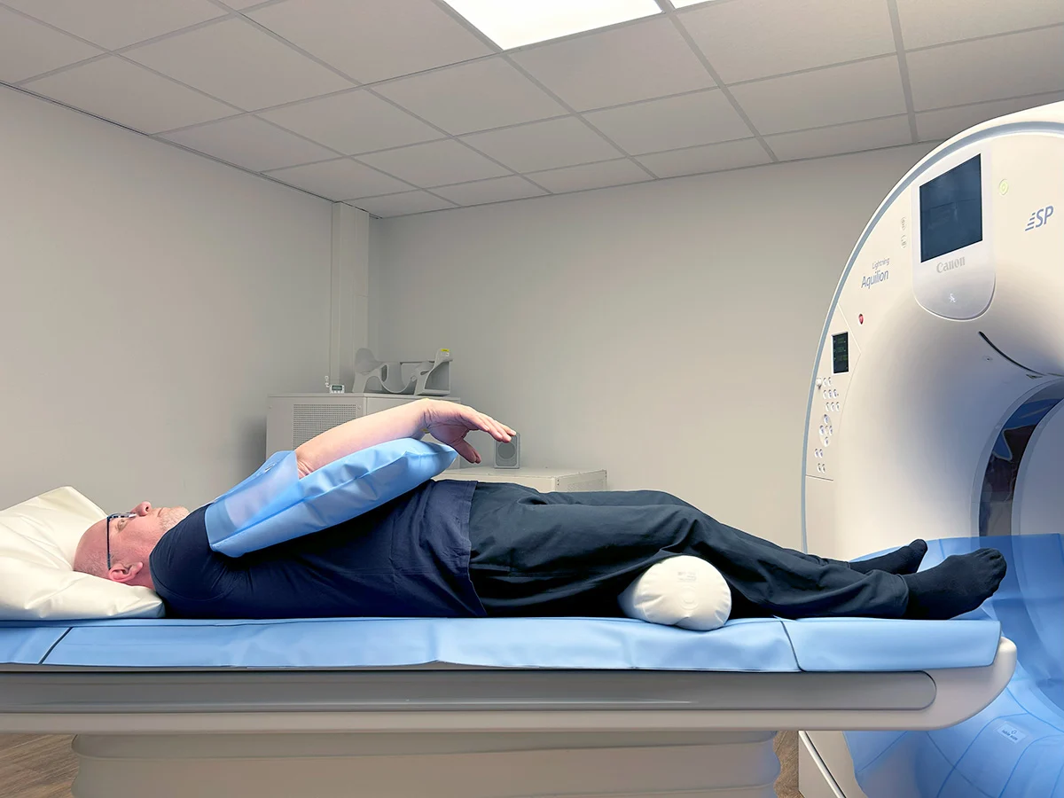
- No special preparation is needed for most spine MRI scans, and the procedure takes 30 to 45 minutes per spinal region
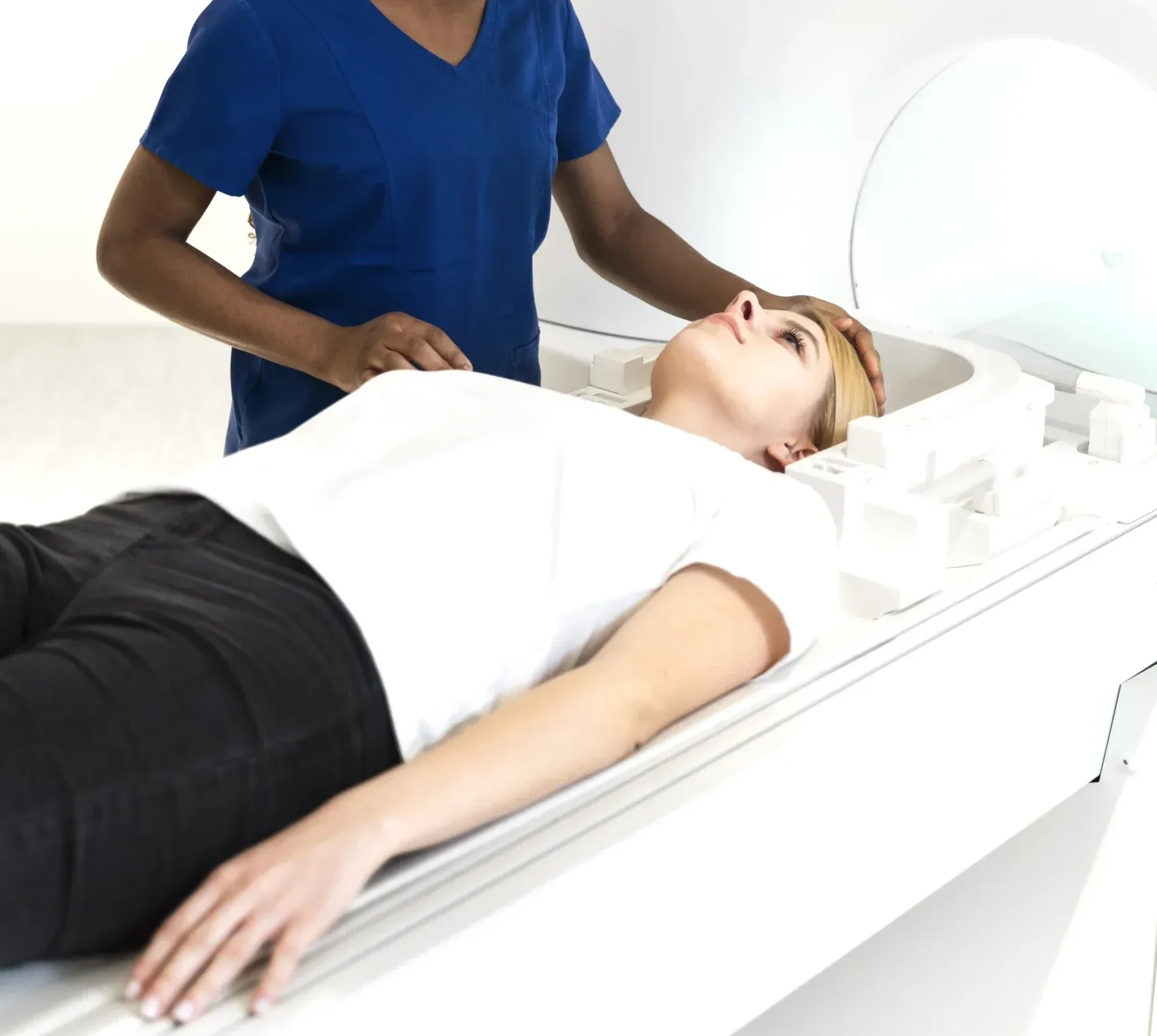
A spine MRI scan provides the most detailed view of the spinal cord, intervertebral discs, nerve roots, and surrounding structures available in modern medicine. Whether you are suffering from chronic back pain, sciatica, neck stiffness with radiating arm pain, or have been told you may have a herniated disc, spine MRI delivers the precise diagnostic information your physician needs to recommend the right treatment. In Dubai, where sedentary office work and an active sporting culture both contribute to a high prevalence of spinal conditions, spine MRI is one of the most frequently requested imaging studies.


What Does a Spine MRI Show?
A spine MRI creates highly detailed images of the vertebrae, intervertebral discs, spinal cord, nerve roots, facet joints, ligaments, and paraspinal muscles. Unlike X-rays, which show only the bones, MRI reveals the soft tissue structures that are responsible for the majority of spinal symptoms. The following conditions are commonly diagnosed with spine MRI:
- Herniated disc (slipped disc): A herniated disc occurs when the soft inner core of an intervertebral disc pushes through the outer ring and compresses a nearby nerve root. MRI clearly shows the size, location, and direction of the herniation, and whether it is compressing the spinal cord or nerve roots. This information is critical for determining whether conservative treatment or surgery is appropriate.
- Spinal stenosis: Narrowing of the spinal canal or neural foramina that compresses the spinal cord or nerve roots. MRI measures the degree of stenosis and identifies the cause — whether it is due to disc bulging, ligament thickening, bone spurs, or a combination of factors.
- Sciatica and nerve compression: Sciatica — pain radiating from the lower back down the leg — is most commonly caused by a herniated lumbar disc or stenosis compressing the sciatic nerve roots. MRI pinpoints the exact level and cause of nerve compression, guiding targeted treatment.
- Degenerative disc disease and spondylosis: Age-related changes including disc dehydration, disc height loss, osteophyte formation, and facet joint arthropathy are clearly demonstrated on MRI. The scan shows which levels are most affected and whether the degeneration is causing nerve compression.
- Spinal cord abnormalities: MRI is the only imaging modality that directly visualizes the spinal cord. It detects myelopathy (spinal cord compression), syringomyelia (fluid-filled cavities within the cord), tumors, demyelination, and vascular malformations.
- Spinal infections: Discitis (disc infection) and vertebral osteomyelitis are serious conditions that MRI can detect early, often before significant bone destruction occurs. Contrast-enhanced MRI is particularly useful for identifying the extent of infection and any associated abscess formation.
- Spinal fractures: While X-ray and CT are excellent for detecting fractures, MRI adds crucial information about whether a fracture is acute or chronic (by detecting bone marrow edema), whether there is spinal cord compression, and whether there are associated ligamentous injuries.
- Spinal tumors: Primary spinal tumors and metastatic disease are clearly visualized on MRI. Contrast-enhanced studies help characterize tumors and differentiate them from other conditions.
"The spine is one of the areas where MRI truly excels," explains Dr. Osama Elzamzami, Consultant Radiologist at DCDC. "I can evaluate every disc, every nerve root, and the spinal cord itself in a single study. For a patient with sciatica, for example, I can identify precisely which disc is herniated, which nerve root is compressed, and how severe the compression is — information that directly determines treatment."
How Much Does a Spine MRI Cost in Dubai?
Spine MRI pricing in Dubai depends primarily on how many spinal regions are scanned. The spine is divided into three regions — cervical (neck), thoracic (mid-back), and lumbar (lower back) — and each is typically ordered and priced separately. At Doctors Clinic Diagnostic Center, all spine MRI pricing includes the scan, a comprehensive consultant radiologist report, and digital image copies.
| Spine MRI Type | Approximate Cost (AED) |
|---|---|
| Cervical spine MRI (neck) | 1,000 – 1,400 |
| Thoracic spine MRI (mid-back) | 1,000 – 1,400 |
| Lumbar spine MRI (lower back) | 1,000 – 1,400 |
| Two regions (e.g., cervical + lumbar) | 1,800 – 2,500 |
| Whole spine MRI (all three regions) | 2,500 – 4,000 |
| Spine MRI with contrast (any region) | Add AED 300 – 500 |
Prices are approximate and include the radiologist report at DCDC. Contact us for exact pricing.
The lumbar spine is by far the most commonly scanned region, as lower back pain and sciatica are among the most frequent reasons patients seek spine MRI. The cervical spine is the second most common, typically ordered for neck pain with radiating arm symptoms. Thoracic spine MRI is less frequently needed but is essential when mid-back symptoms, suspected cord compression, or thoracic disc herniations are involved.
For patients who need multiple regions scanned, combined packages offer significant cost savings compared to booking each region individually. Whole spine MRI — covering cervical, thoracic, and lumbar regions in a single session — is particularly cost-effective and is often ordered for patients with widespread symptoms, suspected inflammatory conditions like ankylosing spondylitis, or as part of a full body MRI screening.
Cervical vs Thoracic vs Lumbar MRI: Which Do You Need?
The region of the spine your doctor orders for MRI depends entirely on your symptoms and clinical examination findings. Understanding which spinal region corresponds to which symptoms helps patients understand their doctor's recommendation. For related information, see our guide on Back Pain Dubai: Causes & When You Need an MRI.
| Spinal Region | Common Symptoms | Typical Conditions Found |
|---|---|---|
| Cervical (C1-C7) | Neck pain, arm pain/numbness/tingling, hand weakness, headaches originating from neck | Cervical disc herniation, cervical stenosis, cervical spondylosis, myelopathy |
| Thoracic (T1-T12) | Mid-back pain, band-like pain around the chest or abdomen, weakness in the legs | Thoracic disc herniation, compression fractures, tumors, cord lesions |
| Lumbar (L1-L5/S1) | Lower back pain, sciatica (leg pain), leg numbness/tingling, difficulty walking | Lumbar disc herniation, lumbar stenosis, spondylolisthesis, cauda equina compression |
Your doctor selects the appropriate region based on a neurological examination that includes testing your reflexes, muscle strength, and sensation. The distribution of symptoms provides reliable clues — for example, pain or numbness radiating into the hand points to the cervical spine, while pain shooting down the back of the leg points to the lumbar spine. If there is diagnostic uncertainty, your physician may order two regions or a whole spine study.
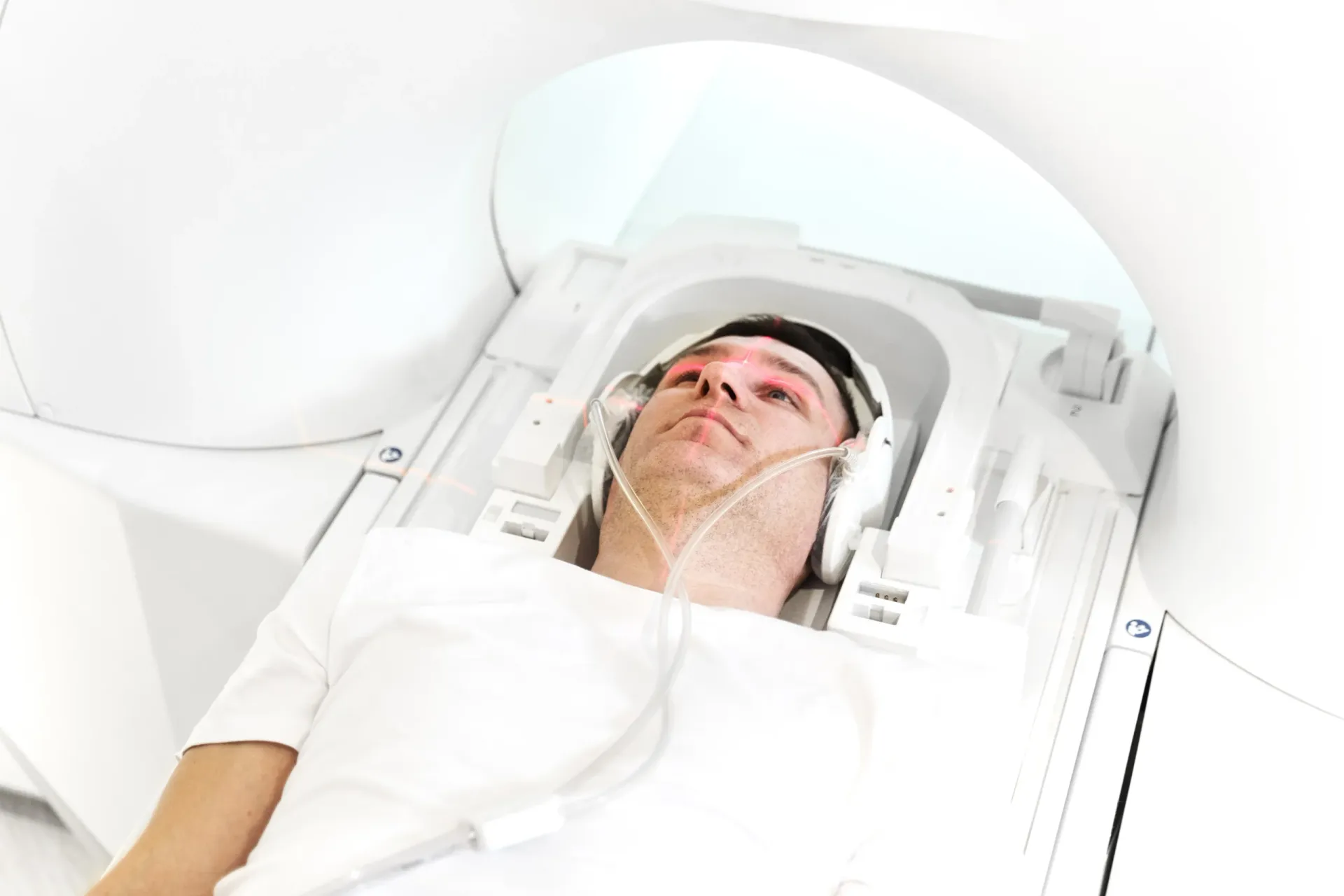
Preparing for Your Spine MRI
One of the advantages of spine MRI is that it requires minimal preparation. The following guidelines will help ensure your scan goes smoothly:
- No fasting required: For a standard non-contrast spine MRI, you can eat and drink normally before the appointment. If contrast is being used, you may be asked to avoid eating for 2 to 4 hours beforehand.
- Remove all metal: Before entering the MRI room, remove all jewellery, watches, belts, hairpins, and any clothing with metal components. You will be provided with a gown if needed.
- Complete the safety questionnaire: You will be asked about metal implants, pacemakers, surgical clips, and other potential contraindications. Bring a list of any implants or medical devices you have.
- Inform the team about claustrophobia: If you experience claustrophobia, let the radiology team know when booking your appointment. Options to improve comfort include mild sedation (prescribed by your doctor), relaxation techniques, and music through headphones.
- Bring your referral and previous imaging: If you have a physician referral letter, bring it along with any previous X-rays, CT scans, or MRI images of the same region. These allow the radiologist to compare current findings with prior studies.
- Plan for the duration: A single-region spine MRI takes approximately 30 to 45 minutes. Whole spine MRI may take 60 to 90 minutes. Allow extra time for check-in and preparation.
Book Your Spine MRI at DCDC
At Doctors Clinic Diagnostic Center in Dubai Healthcare City, we offer comprehensive spine MRI with detailed consultant radiologist reports. Same-week appointments available.
When Does a Doctor Order a Spine MRI?
Not all back or neck pain requires an MRI. In fact, most episodes of acute back pain resolve within a few weeks with conservative management. However, there are specific clinical situations where a spine MRI is clearly indicated and provides essential diagnostic information:
- Back or neck pain with radiating limb symptoms: Pain that travels down the arm (from the cervical spine) or down the leg (from the lumbar spine) suggests nerve root compression, which MRI can confirm and localize.
- Progressive neurological symptoms: Worsening weakness, numbness, or loss of coordination in the arms or legs may indicate spinal cord or nerve root compression that requires urgent evaluation.
- Cauda equina syndrome symptoms: Loss of bladder or bowel control, saddle-area numbness, or sudden bilateral leg weakness is a medical emergency that requires immediate spine MRI to assess for cauda equina compression.
- Back pain lasting more than 6 weeks: When back pain persists despite conservative treatment (rest, physiotherapy, medication), MRI helps identify the structural cause and guide further management.
- Suspected spinal infection or tumor: Fever with back pain, unexplained weight loss with new back symptoms, or a history of cancer with new spinal complaints all warrant urgent MRI.
- Pre-surgical planning: Before spinal surgery — whether for disc herniation, stenosis, or fusion — surgeons require detailed MRI to plan the surgical approach and confirm the target level.
- Post-surgical evaluation: After spinal surgery, MRI (usually with contrast) assesses healing, graft integrity, and rules out complications such as recurrent disc herniation, infection, or scar tissue formation.
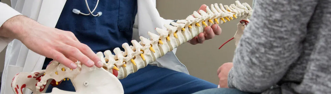
Understanding Your Spine MRI Report
Spine MRI reports contain specialized terminology that can be confusing for patients. The following are common terms you may encounter in your report and what they mean: You may also find our Lateral Recess Stenosis: Symptoms & CT Scan helpful.
- Disc bulge: A broad-based outward extension of the disc beyond the normal vertebral margin. Small disc bulges are extremely common and often asymptomatic, particularly in patients over 30.
- Disc protrusion/herniation: A focal extension of disc material beyond the vertebral margin. Protrusions are more localized than bulges and more likely to compress a nerve root, though not all herniations cause symptoms.
- Disc extrusion: A more severe form of herniation where disc material extends significantly beyond the disc space. Extrusions are more likely to cause nerve compression and symptoms.
- Spinal stenosis (mild/moderate/severe): Narrowing of the spinal canal. The grading indicates how much the canal has narrowed and helps determine treatment urgency.
- Neural foraminal narrowing: Narrowing of the openings through which nerve roots exit the spine. Moderate to severe narrowing may compress the exiting nerve root, causing radiculopathy.
- Degenerative changes: Age-related wear and tear including disc dehydration, disc height loss, osteophytes, and facet joint arthropathy. These are nearly universal in adults over 40 and do not always correlate with symptoms.
It is important to discuss your MRI results with your referring physician, who will correlate the imaging findings with your symptoms and clinical examination. Not all MRI findings are clinically significant — studies consistently show that many adults without any back pain have disc bulges and degenerative changes on MRI. The clinical context is essential for determining which findings are relevant to your symptoms.
Get Expert Spine MRI Interpretation
At DCDC, every spine MRI is interpreted by a consultant radiologist who provides a detailed, structured report with clear descriptions of findings at each spinal level. Our reports are designed to give your treating physician the precise information they need. Learn more about our MRI services.
Related Services at DCDC
Expert care and advanced diagnostics at Dubai Healthcare City
Frequently Asked Questions
Final Thoughts
Spine MRI is an indispensable diagnostic tool for evaluating back pain, neck pain, sciatica, and neurological symptoms originating from the spinal column. By providing detailed images of discs, nerves, the spinal cord, and surrounding structures, it gives physicians the information needed to make accurate diagnoses and recommend targeted treatment — whether that is physiotherapy, injections, or surgical intervention.
At Doctors Clinic Diagnostic Center in Dubai Healthcare City, we perform spine MRI on advanced equipment with interpretation by experienced consultant radiologists. Our detailed, level-by-level reports ensure your treating physician has the precise information they need. Contact us to book your spine MRI or to discuss which spinal region should be scanned based on your symptoms.
Sources & References
This article was reviewed by our medical team and references the following sources:
- American College of Radiology - Appropriateness Criteria for Low Back Pain
- Radiological Society of North America - Spine MRI
- North American Spine Society - Imaging Guidelines
- The New England Journal of Medicine - MRI of the Lumbar Spine in Asymptomatic Adults
- Dubai Health Authority - Diagnostic Imaging Regulations
Medical content on this site is reviewed by DHA-licensed physicians. See our editorial policy for more information.

Written by
Dr. Osama Elzamzami
Diagnostic Radiology
MD, FRCR
Dr. Osama Elzamzami is a Consultant Radiologist specializing in diagnostic imaging including MRI, CT, and ultrasound at DCDC Dubai Healthcare City.
Related Articles
MRI Cost in Dubai: Complete Guide to Types & Pricing (2026)
Brain MRI in Dubai: What It Detects, Cost & When You Need One

Back Pain: Causes, Treatment & When to Get an MRI
Herniated Disc Rehabilitation: Complete Recovery Timeline
MRI with Contrast: What to Expect, Safety & Cost
More in Diagnostic Imaging
CT Scan Cost Dubai: Prices & Types (2026)
Read More
DEXA Scan Dubai: Cost, T-Score & Who Needs It (2026)
Read More![Full Body MRI Cost Dubai: AED 5,000-15,000 [2026]](/wp-media/blog/full-body-mri-scan.webp)
Full Body MRI Cost Dubai: AED 5,000-15,000 [2026]
Read MoreMRI Preparation Dubai: Complete Guide (2026)
Read More
CBCT Scan: 3D Dental Imaging Guide Dubai (2026)
Read More
Ultrasound Preparation Dubai: What to Expect (2026)
Read More© 2026 Doctors Clinic Diagnostic Center (DCDC), Dubai Healthcare City. Originally published at https://doctorsclinicdubai.ae/blog/spine-mri-cost-dubai. All rights reserved. Unauthorized reproduction is prohibited.