Key Takeaways
- A thyroid ultrasound is a painless, radiation-free imaging test that uses high-frequency sound waves to produce detailed images of the thyroid gland, detecting nodules as small as 2-3 millimeters
- Thyroid nodules are found in up to 68% of the general population on high-resolution ultrasound, but fewer than 5% of all thyroid nodules are malignant, making accurate classification essential to avoid unnecessary procedures
- The TI-RADS (Thyroid Imaging Reporting and Data System) classification standardizes how radiologists assess thyroid nodules, scoring features such as composition, echogenicity, shape, margin, and echogenic foci to guide whether a biopsy is needed
- A thyroid ultrasound requires no preparation, takes 15 to 20 minutes, involves no needles or contrast, and produces results that are typically available on the same day at DCDC
- Thyroid ultrasound is the first-line imaging tool for evaluating thyroid abnormalities, but it complements rather than replaces blood tests (TSH, T3, T4) and nuclear medicine thyroid scans, each of which provides different clinical information
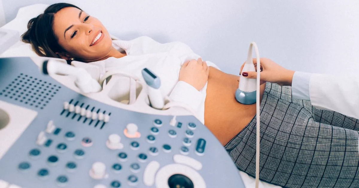
A thyroid ultrasound is the gold-standard imaging test for evaluating the thyroid gland, a butterfly-shaped organ at the front of the neck that regulates metabolism, energy, heart rate, and body temperature. Using high-frequency sound waves, a thyroid ultrasound in Dubai produces real-time, high-resolution images of the thyroid's size, shape, texture, and blood flow without exposing the patient to any ionizing radiation. It is the most reliable method for detecting thyroid nodules, determining whether those nodules require further investigation, and monitoring known thyroid conditions over time.
This guide explains what a thyroid ultrasound is, the clinical situations that warrant one, what it can and cannot detect, how the TI-RADS classification system works, how to prepare, how it compares to other thyroid tests, and what a thyroid scan in Dubai costs at Doctors Clinic Diagnostic Center (DCDC) in Dubai Healthcare City.


What Is a Thyroid Ultrasound?
A thyroid ultrasound, also called thyroid sonography, is a non-invasive diagnostic imaging examination that uses a handheld transducer emitting high-frequency sound waves (typically 7.5 to 15 MHz) to create detailed cross-sectional and longitudinal images of the thyroid gland and surrounding cervical structures. The transducer is placed directly on the skin of the neck over a thin layer of gel, and the reflected sound waves are converted by a computer into real-time images displayed on a monitor.
The thyroid gland sits just below the Adam's apple and consists of two lobes connected by a thin bridge of tissue called the isthmus. A normal adult thyroid measures approximately 4 to 6 centimeters in length per lobe and weighs between 15 and 25 grams. Because the thyroid is a superficial structure located just beneath the skin and the thin strap muscles of the neck, it is ideally suited for ultrasound evaluation. High-frequency transducers produce images with sub-millimeter resolution, allowing radiologists to identify nodules as small as 2 to 3 millimeters and to characterize internal features that help distinguish benign lesions from those that warrant further investigation.
"Thyroid ultrasound is the single most important imaging tool we have for evaluating the thyroid gland," says Dr. Osama Elzamzami, Head of Radiology at DCDC. "It gives us a level of anatomical detail that no blood test or physical examination can provide, and it does so without any radiation exposure to the patient."
Unlike nuclear medicine thyroid scans, which use radioactive tracers to assess gland function, ultrasound provides structural and morphological information. It shows the size of each thyroid lobe, the overall gland volume, the echotexture of the parenchyma (the internal tissue pattern), the presence and characteristics of any nodules, the vascularity of the gland and its nodules, and the condition of nearby cervical lymph nodes. This structural information is essential for clinical decision-making and is used by endocrinologists, surgeons, and primary care physicians worldwide. Patients with thyroid conditions often benefit from coordinated care with our endocrine and metabolic disorders specialists alongside imaging follow-up.
The prevalence of thyroid nodules makes thyroid ultrasound one of the most commonly performed neck imaging studies. Research published in the journal Thyroid indicates that high-resolution ultrasound detects thyroid nodules in up to 68% of randomly selected adults, a figure far higher than the 4 to 7% detection rate achieved by physical palpation alone. The vast majority of these nodules are benign, but the ability to identify the small percentage that harbor malignancy, and to do so before the cancer has grown or spread, is what gives thyroid ultrasound its critical clinical value. In the UAE and the wider Gulf region, thyroid disorders are particularly common due to a combination of iodine status variations, genetic factors, and environmental influences, making thyroid ultrasound in Dubai one of the most frequently requested radiology examinations at centers like DCDC.
When Do You Need a Thyroid Ultrasound?
A thyroid ultrasound is recommended in a range of clinical scenarios, from evaluating an incidental finding to monitoring a previously diagnosed condition. The American Thyroid Association (ATA), the European Thyroid Association (ETA), and the American College of Radiology (ACR) all endorse ultrasound as the first-line imaging modality for thyroid evaluation. The following are the most common reasons a physician orders a thyroid ultrasound.
Palpable Neck Lump or Thyroid Nodule
If you or your doctor feel a lump in your neck, a thyroid ultrasound is the immediate next step. A palpable nodule needs to be evaluated for size, internal characteristics, and any features that suggest malignancy. Not every lump in the neck originates from the thyroid; enlarged lymph nodes, parathyroid adenomas, thyroglossal duct cysts, branchial cleft cysts, and lipomas can all present as neck masses, and ultrasound differentiates between these possibilities with a high degree of accuracy. For nodules that are confirmed to arise from the thyroid, the ultrasound appearance guides all subsequent management, from simple observation to biopsy to surgical referral.
Abnormal Thyroid Blood Tests
When thyroid blood tests reveal abnormal thyroid-stimulating hormone (TSH), free T4, or free T3 levels, ultrasound helps identify the structural cause. Hyperthyroidism (overactive thyroid) may be caused by Graves' disease, a toxic multinodular goiter, or a solitary toxic adenoma, each of which has distinct ultrasound features. Hypothyroidism (underactive thyroid) may be associated with Hashimoto's thyroiditis, which produces characteristic changes in the gland's echotexture that are clearly visible on ultrasound. Even when thyroid function tests are normal (euthyroid state), a thyroid ultrasound may still be warranted if there is a clinical suspicion of structural disease, because many patients with thyroid nodules, including some with thyroid cancer, have completely normal thyroid hormone levels.
Incidental Thyroid Finding (Thyroid Incidentaloma)
Thyroid nodules are increasingly discovered incidentally during imaging performed for other reasons, such as CT scans of the neck, carotid Doppler ultrasound, MRI of the cervical spine, or PET scans. These incidental findings, known as thyroid incidentalomas, require dedicated thyroid ultrasound to characterize the nodule properly and determine whether biopsy is needed.
Family or Personal History of Thyroid Cancer
Patients with a first-degree relative who has had thyroid cancer, or those with a personal history of head and neck radiation exposure during childhood, are at increased risk and benefit from periodic thyroid ultrasound screening. Similarly, patients with certain genetic syndromes such as Multiple Endocrine Neoplasia (MEN) type 2 or familial medullary thyroid carcinoma undergo regular surveillance ultrasound.
Monitoring Known Thyroid Nodules or Post-Surgical Follow-Up
Patients with known thyroid nodules that do not meet criteria for biopsy are typically monitored with repeat ultrasound at intervals of 6 to 24 months, depending on the nodule's TI-RADS category. After thyroid surgery for cancer, ultrasound of the thyroid bed and cervical lymph nodes is an essential part of long-term surveillance to detect local recurrence or lymph node metastasis.
Guidance for Fine-Needle Aspiration Biopsy
When a thyroid nodule requires tissue sampling, ultrasound provides real-time guidance for fine-needle aspiration (FNA) biopsy. The radiologist uses the ultrasound image to direct the needle precisely into the target nodule, ensuring an adequate sample while avoiding blood vessels and other critical structures. Ultrasound-guided FNA has a significantly higher diagnostic accuracy than palpation-guided biopsy, with studies showing that image-guided sampling produces adequate specimens in more than 90% of cases compared to approximately 60 to 70% for blind palpation-guided techniques. At DCDC, ultrasound-guided FNA can be performed immediately following the diagnostic ultrasound if the nodule meets criteria for biopsy, saving the patient a second visit.
What Does a Thyroid Ultrasound Show?
A thyroid ultrasound provides detailed structural information about the thyroid gland and the surrounding neck anatomy that cannot be obtained from any other non-invasive test. The radiologist evaluates multiple parameters in a systematic, standardized approach to produce a comprehensive report that serves as the foundation for all clinical decisions regarding thyroid management.
It is important to note what thyroid ultrasound does not show. Ultrasound cannot measure thyroid hormone levels, cannot determine whether a nodule is producing excess thyroid hormone (functional autonomy), and cannot provide a tissue diagnosis of cancer. These questions are answered by blood tests, nuclear medicine scans, and fine-needle aspiration biopsy, respectively. However, as a structural imaging tool, thyroid ultrasound is unsurpassed in its ability to detect, characterize, and monitor thyroid lesions.
- Thyroid gland size and volume: Each lobe is measured in three dimensions (length, width, depth), and the total gland volume is calculated. An enlarged thyroid (goiter) is identified when the volume exceeds approximately 18 mL in women or 25 mL in men.
- Parenchymal echotexture: The normal thyroid has a homogeneous, mildly hyperechoic (brighter than muscle) appearance. Diffuse heterogeneity, hypoechogenicity, and increased vascularity suggest inflammatory or autoimmune conditions such as Hashimoto's thyroiditis or Graves' disease.
- Thyroid nodules: Each nodule is measured and characterized by its composition (solid, cystic, or mixed), echogenicity (hyperechoic, isoechoic, hypoechoic, or very hypoechoic relative to normal thyroid), shape (wider-than-tall or taller-than-wide), margins (smooth, ill-defined, lobulated, or irregular with extra-thyroidal extension), and the presence of echogenic foci (microcalcifications, macrocalcifications, or comet-tail artifacts).
- Vascularity: Color Doppler and power Doppler ultrasound assess blood flow within the gland and within individual nodules. Increased central vascularity in a nodule may raise concern, although this finding alone is not diagnostic of malignancy.
- Cervical lymph nodes: The ultrasound examination includes evaluation of the lymph nodes in the central and lateral neck compartments. Features such as a rounded shape, loss of the normal fatty hilum, cystic change, microcalcifications, or abnormal vascularity raise suspicion for metastatic lymphadenopathy.
- Surrounding structures: The examination assesses the relationship of the thyroid to the trachea, carotid arteries, jugular veins, strap muscles, and esophagus. Any displacement or compression of these structures by an enlarged thyroid or nodule is documented.
"A thorough thyroid ultrasound is more than just finding nodules," says Dr. Osama Elzamzami, Head of Radiology at DCDC. "We evaluate the entire gland, the lymph nodes, and the surrounding anatomy. This comprehensive approach ensures nothing is missed and gives the referring physician a complete picture to guide treatment decisions."
Patient Story: From Routine Check-Up to Early Detection
A 38-year-old woman living in Dubai visited her internal medicine physician for a routine annual health check. Her blood tests showed a mildly elevated TSH level but no other abnormalities, and she had no symptoms. Her doctor ordered a thyroid ultrasound at DCDC as part of the workup. The ultrasound revealed a 12-millimeter solid hypoechoic nodule in the right thyroid lobe with irregular margins and several punctate echogenic foci consistent with microcalcifications. The nodule was classified as ACR TI-RADS 5 (highly suspicious), and an ultrasound-guided fine-needle aspiration biopsy was performed on the same day.
The cytology results confirmed papillary thyroid carcinoma, the most common type of thyroid cancer and one that carries an excellent prognosis when detected early. Because the cancer was identified at stage I, confined to the thyroid with no evidence of lymph node involvement or distant spread, the patient underwent a straightforward total thyroidectomy followed by radioactive iodine therapy. She is now disease-free on follow-up surveillance, with regular thyroid ultrasound scans of the neck showing no evidence of recurrence. Without that initial thyroid ultrasound, this cancer would likely have gone undetected for months or years, potentially growing larger, invading surrounding tissues, and spreading to cervical lymph nodes before diagnosis. Papillary thyroid carcinoma has a 10-year survival rate exceeding 98% when caught early, but outcomes are less favorable when the disease has spread beyond the thyroid at the time of diagnosis. This case illustrates the life-saving value of thyroid ultrasound in detecting malignancy at its earliest, most treatable stage.
Book a Thyroid Ultrasound at DCDC
At Doctors Clinic Diagnostic Center in Dubai Healthcare City, our radiology team provides expert thyroid ultrasound with high-resolution imaging, same-day results, and comprehensive reporting including TI-RADS classification.
Thyroid Nodule Classification: TI-RADS Explained
The Thyroid Imaging Reporting and Data System (TI-RADS) is a standardized classification system developed by the American College of Radiology (ACR) that provides a consistent framework for radiologists to assess thyroid nodules and recommend appropriate management. TI-RADS assigns points based on five ultrasound feature categories: composition, echogenicity, shape, margin, and echogenic foci. The total point score determines the TI-RADS category, which in turn dictates whether a nodule should be biopsied, monitored, or left alone.
The ACR TI-RADS system was introduced in 2017 and has since been widely adopted by radiology departments worldwide, including at DCDC. The system reduces unnecessary biopsies of benign nodules while ensuring that nodules with suspicious features are appropriately investigated. Each ultrasound feature is assigned a point value, and the sum of points determines the final TI-RADS category. Features associated with malignancy, such as a solid composition, marked hypoechogenicity, a taller-than-wide shape, irregular margins, and punctate echogenic foci (microcalcifications), receive higher point scores.
The five feature categories evaluated in TI-RADS are: Composition (cystic or almost completely cystic = 0 points; spongiform = 0; mixed cystic and solid = 1; solid or almost completely solid = 2), Echogenicity (anechoic = 0; hyperechoic or isoechoic = 1; hypoechoic = 2; very hypoechoic = 3), Shape (wider-than-tall = 0; taller-than-wide = 3), Margin (smooth = 0; ill-defined = 0; lobulated or irregular = 2; extra-thyroidal extension = 3), and Echogenic Foci (none or large comet-tail artifacts = 0; macrocalcifications = 1; peripheral rim calcifications = 2; punctate echogenic foci = 3). The radiologist assigns points from each category, and the total determines the TI-RADS level. This systematic approach ensures that nodule assessment is reproducible and minimizes subjective variation between different radiologists.
| TI-RADS Category | Points | Suspicion Level | Management Recommendation |
|---|---|---|---|
| TR1 - Benign | 0 points | Benign | No FNA or follow-up needed |
| TR2 - Not Suspicious | 2 points | Not suspicious | No FNA or follow-up needed |
| TR3 - Mildly Suspicious | 3 points | Mildly suspicious | FNA if nodule is 2.5 cm or larger; follow-up if 1.5 cm or larger |
| TR4 - Moderately Suspicious | 4-6 points | Moderately suspicious | FNA if nodule is 1.5 cm or larger; follow-up if 1.0 cm or larger |
| TR5 - Highly Suspicious | 7 or more points | Highly suspicious | FNA if nodule is 1.0 cm or larger; follow-up if 0.5 cm or larger |
ACR TI-RADS categories with corresponding suspicion levels and recommended management. FNA = Fine-Needle Aspiration biopsy.
TI-RADS Risk Assessment: Malignancy Risk and Biopsy Thresholds
To help patients understand what their TI-RADS category means in practical terms, the following table summarizes the estimated malignancy risk at each level and the corresponding biopsy thresholds. These figures are based on published data from the American College of Radiology and large international studies.
| TI-RADS Level | Suspicion | Estimated Malignancy Risk | FNA Biopsy Threshold | Typical Action |
|---|---|---|---|---|
| TI-RADS 1 | Benign | ~0% | No FNA needed | No follow-up required |
| TI-RADS 2 | Not Suspicious | <2% | No FNA needed | No follow-up required |
| TI-RADS 3 | Mildly Suspicious | 2-5% | FNA if nodule >= 2.5 cm | Follow-up ultrasound if >= 1.5 cm |
| TI-RADS 4 | Moderately Suspicious | 5-20% | FNA if nodule >= 1.5 cm | Follow-up ultrasound if >= 1.0 cm |
| TI-RADS 5 | Highly Suspicious | >20% | FNA if nodule >= 1.0 cm | Follow-up ultrasound if >= 0.5 cm |
TI-RADS risk stratification with estimated malignancy rates and biopsy size thresholds. FNA = Fine-Needle Aspiration. Percentages are approximations based on published literature.
It is important to understand that a higher TI-RADS score does not automatically mean cancer. It indicates a higher statistical probability that the nodule is malignant and therefore a lower size threshold at which biopsy is recommended. Research shows that the malignancy rate for TR2 nodules is less than 2%, while the rate for TR5 nodules is approximately 35 to 50%, meaning that even among the most suspicious category, the majority of nodules are still benign. Many TR4 and even some TR5 nodules turn out to be benign on biopsy. Conversely, TI-RADS is not infallible; a small percentage of cancers, particularly the follicular variant of papillary thyroid carcinoma, can appear deceptively benign on ultrasound. This is why the system is used as a risk-stratification tool to guide clinical decision-making, not as a standalone definitive diagnosis.
"TI-RADS brings objectivity and consistency to thyroid nodule assessment," says Dr. Osama Elzamzami, Head of Radiology at DCDC. "Every radiologist evaluating the same nodule should arrive at the same score and the same recommendation. At DCDC, we include the TI-RADS category in every thyroid ultrasound report so that the referring physician has a clear, evidence-based recommendation for the next step."
Patient Story: Finding a Thyroid Nodule and Learning Not to Panic
Mariam, a 35-year-old school teacher living in Jumeirah, went for her annual women's health screening at DCDC. She felt perfectly healthy and had no neck symptoms. During a routine physical examination, her doctor noticed a small, painless lump on the right side of her neck. A thyroid ultrasound was ordered the same day.
The ultrasound revealed an 18-millimeter isoechoic nodule in the right thyroid lobe with smooth margins, wider-than-tall shape, and no microcalcifications. It was classified as TI-RADS 3 (mildly suspicious), meaning the estimated malignancy risk was only 2-5%. Because it was under 2.5 cm, a fine-needle aspiration biopsy was not required. Her doctor explained that the vast majority of nodules like hers are completely benign, and the recommended plan was simply to monitor it with a follow-up ultrasound in 6 months.
"When I first heard the word 'nodule', I was terrified," Mariam recalls. "I immediately thought cancer. But my radiologist at DCDC took the time to explain the TI-RADS scoring system and show me on the screen exactly why my nodule looked reassuring. That conversation made all the difference." Her 6-month follow-up showed the nodule was completely stable in size and appearance. She has now been monitored for two years with annual ultrasounds, and the nodule has not changed. Her thyroid blood work has remained normal throughout.
Mariam's experience is extremely common. Thyroid nodules are found in up to 68% of adults on high-resolution ultrasound, yet fewer than 5% are cancerous. The key is proper characterization using TI-RADS, so that genuinely suspicious nodules are investigated promptly while the vast majority of benign nodules are simply monitored without unnecessary anxiety or invasive procedures. For a complete understanding of what your ultrasound findings mean, see our guide on how to understand your ultrasound results.
Worried About a Thyroid Nodule?
At DCDC in Dubai Healthcare City, our radiology team uses the ACR TI-RADS system to classify every thyroid nodule objectively. Most nodules are benign and require only monitoring. If a biopsy is needed, we perform ultrasound-guided FNA on the same day. Get clarity and peace of mind with an expert evaluation.
No special preparation needed. Same-day results. Most insurance accepted.
How to Prepare for a Thyroid Ultrasound
A thyroid ultrasound is one of the simplest diagnostic imaging procedures to prepare for because, in most cases, no special preparation is required. Unlike abdominal ultrasound, which may require fasting, or pelvic ultrasound, which may require a full bladder, a thyroid ultrasound has no preparatory requirements. The examination is completely external, non-invasive, and uses no radiation, contrast agents, or needles (unless an FNA biopsy is planned as a combined procedure). This makes it suitable for patients of all ages, including pregnant women and individuals with allergies to contrast agents.
- No fasting required: You can eat and drink normally before and after the examination. There are no dietary restrictions.
- Take your medications as usual: Continue all regular medications, including thyroid medications such as levothyroxine. Do not stop any medication for the ultrasound.
- Wear appropriate clothing: Wear a top with an open or accessible neckline. You may be asked to remove necklaces, scarves, or high-collared garments to give the sonographer clear access to your neck.
- Bring previous imaging: If you have had previous thyroid ultrasound or other neck imaging, bring the reports and images (or the CD/USB containing them) so the radiologist can compare findings over time.
- Bring your referral and blood work: Bring your doctor's referral letter and any recent thyroid blood test results (TSH, free T4, free T3, thyroid antibodies). This information helps the radiologist provide a more clinically relevant interpretation.
- Allow 30 minutes: While the scan itself takes 15 to 20 minutes, allow 30 minutes for registration, the examination, and any questions you may have afterward.
What Happens During the Examination
You will lie on your back on a padded examination table with a small pillow or rolled towel placed behind your shoulders to gently extend your neck, bringing the thyroid gland closer to the skin surface. The sonographer or radiologist applies a warm, water-based gel to the front of your neck and places the ultrasound transducer on the skin. The transducer is moved methodically across the neck, capturing images of both thyroid lobes, the isthmus, and the cervical lymph node stations.
You may be asked to swallow during certain parts of the examination; swallowing causes the thyroid to move upward briefly, which can help the radiologist evaluate the lower poles of the gland and assess whether a nodule is fixed or mobile. The examination is painless, though gentle pressure from the transducer may feel slightly uncomfortable if you have a tender thyroid. The gel is wiped off at the end, and you can resume all normal activities immediately.
During the ultrasound, the radiologist captures both grayscale (B-mode) images and color Doppler images. The grayscale images show the anatomy and structural characteristics of the gland and any nodules. The color Doppler assessment maps blood flow patterns, showing whether the gland has normal, increased, or decreased vascularity, and whether any nodules have predominantly peripheral, central, or mixed blood flow. This vascular information adds another layer of diagnostic detail that helps the radiologist refine the assessment of each nodule.
If an FNA biopsy is indicated based on the ultrasound findings and TI-RADS classification, it may be performed immediately following the diagnostic scan or scheduled for a separate appointment. The biopsy uses a thin needle (25 to 27 gauge), similar to one used for drawing blood. Local anesthetic is applied to the skin, and the needle is guided into the nodule under continuous ultrasound visualization. The entire biopsy procedure takes approximately 10 to 15 minutes, and patients can return to their normal routine the same day with only mild tenderness at the puncture site.
Thyroid Ultrasound vs Other Thyroid Tests
A thyroid ultrasound answers structural questions: what does the gland look like, are there nodules, how large are they, and do they have suspicious features? However, it does not directly measure thyroid function. A complete thyroid evaluation often requires a combination of ultrasound, blood tests, and sometimes nuclear medicine studies, each providing different and complementary information. Understanding the role and limitations of each test helps patients make informed decisions about their thyroid health.
- Thyroid blood tests (TSH, free T4, free T3): These blood tests measure thyroid hormone levels and pituitary feedback. They tell your doctor whether the thyroid is overactive (hyperthyroidism), underactive (hypothyroidism), or functioning normally (euthyroid). Blood tests are essential but cannot detect structural abnormalities, nodules, or cancer.
- Thyroid antibody tests (anti-TPO, anti-thyroglobulin): These detect autoimmune thyroid conditions such as Hashimoto's thyroiditis and Graves' disease. They complement ultrasound findings by confirming an autoimmune etiology when the gland shows characteristic ultrasound changes.
- Nuclear medicine thyroid scan (scintigraphy): This uses a small amount of radioactive iodine or technetium to assess thyroid function at the tissue level. It identifies whether a nodule is "hot" (actively producing thyroid hormone), "warm" (functioning normally), or "cold" (not producing hormone). Hot nodules are almost always benign, while cold nodules have a higher probability of malignancy. A thyroid scan answers the functional question that ultrasound cannot.
- Fine-needle aspiration (FNA) biopsy: This is a tissue sampling procedure, not an imaging test, but it is performed under ultrasound guidance. FNA provides cytological (cellular) information that allows a pathologist to determine whether a nodule is benign, malignant, or indeterminate.
- CT or MRI of the neck: These cross-sectional imaging modalities are not first-line tools for thyroid evaluation but may be ordered when a large goiter extends behind the breastbone (substernal goiter), when thyroid cancer has spread to surrounding structures, or when surgical planning requires detailed anatomical mapping beyond what ultrasound provides.
In clinical practice, the standard initial workup for a suspected thyroid problem includes a TSH blood test and a thyroid ultrasound. The results of these two tests guide all subsequent decisions, including whether additional blood work, a nuclear medicine scan, or an FNA biopsy is needed. For example, if a patient presents with a thyroid nodule and the TSH is low (suggesting hyperthyroidism), a nuclear medicine scan is typically ordered next to determine whether the nodule is autonomously functioning ("hot"). If the TSH is normal or elevated, the ultrasound appearance alone, classified using TI-RADS, determines whether the nodule should be biopsied or simply monitored.
Understanding the complementary nature of these tests helps patients appreciate why their doctor may order multiple investigations. A thyroid ultrasound showing a suspicious nodule does not mean you have cancer; it means the nodule has features that warrant further evaluation. Equally, normal blood test results do not rule out structural disease. The combination of structural imaging (ultrasound), functional assessment (blood tests and nuclear medicine), and tissue diagnosis (FNA biopsy when indicated) provides the most complete and accurate picture of thyroid health.
Thyroid Ultrasound Cost in Dubai
The cost of a thyroid ultrasound in Dubai varies depending on the facility, the complexity of the examination, and whether additional procedures such as Doppler assessment or fine-needle aspiration biopsy are performed at the same visit. At most diagnostic imaging centers in Dubai, a standard thyroid ultrasound ranges from AED 300 to AED 800. Factors that influence the price include whether the examination is performed by a sonographer with a separate radiologist report or directly by a consultant radiologist, whether color Doppler assessment is included as standard or billed separately, and whether the facility is located in a medical free zone such as Dubai Healthcare City or in a general commercial area.
At DCDC (Doctors Clinic Diagnostic Center) in Dubai Healthcare City, thyroid ultrasound pricing includes the full examination of both thyroid lobes, the isthmus, and the cervical lymph nodes, along with a detailed radiology report with TI-RADS classification of any nodules detected. Results are typically available on the same day and are shared digitally with your referring physician.
- Standard thyroid ultrasound: Includes bilateral thyroid evaluation, isthmus assessment, cervical lymph node survey, and comprehensive radiologist report with TI-RADS scoring
- Thyroid ultrasound with Doppler: Adds color and power Doppler flow assessment for detailed vascularity evaluation of the gland and any nodules
- Ultrasound-guided FNA biopsy: Performed when a nodule meets TI-RADS criteria for tissue sampling; includes the biopsy procedure, cytology specimen preparation, and pathology report (billed separately from the ultrasound)
Most major insurance plans in Dubai cover thyroid ultrasound when ordered by a referring physician with a valid clinical indication. DCDC accepts a wide range of insurance providers. For patients without insurance or those paying out of pocket, the center offers competitive self-pay rates. To confirm current pricing and insurance coverage, contact DCDC directly by phone, WhatsApp, or through the online booking form.
DCDC is located in Dubai Healthcare City, one of the region's premier medical free zones, easily accessible from Oud Metha, Karama, Bur Dubai, Downtown Dubai, and the wider UAE via major road networks and the Dubai Metro. The center is open six days a week, and thyroid ultrasound appointments can be booked by phone, WhatsApp, or through the online booking system. Walk-in patients are also accommodated subject to availability. With over 13 years of operation and more than 1,000 diagnostic imaging examinations performed every month, DCDC has established a reputation for accurate, timely, and patient-centered radiology services.
Thyroid Imaging and Test Costs at DCDC (2026)
The following table provides estimated costs for the most commonly requested thyroid tests and imaging studies in Dubai. Prices reflect typical ranges at diagnostic centers in Dubai Healthcare City and may vary based on insurance coverage and clinical requirements.
| Test / Procedure | Estimated Cost (AED) | What It Includes |
|---|---|---|
| Thyroid Ultrasound | 500 - 800 | Bilateral thyroid evaluation, isthmus, lymph nodes, TI-RADS classification report |
| Thyroid Ultrasound with Doppler | 700 - 1,000 | Standard thyroid ultrasound plus color and power Doppler flow assessment |
| Ultrasound-Guided FNA Biopsy | 1,500 - 2,500 | Biopsy procedure, cytology specimen preparation, and pathology report |
| Thyroid Blood Panel (TSH, T3, T4) | 200 - 400 | Basic thyroid function assessment; essential complement to ultrasound |
| Complete Thyroid Panel with Antibodies | 400 - 600 | TSH, free T3, free T4, anti-TPO, anti-thyroglobulin antibodies |
| Nuclear Thyroid Scan (Scintigraphy) | External Referral | Radioactive iodine or technetium scan; not performed at DCDC, requires nuclear medicine facility |
Estimated thyroid test and imaging costs in Dubai (2026). Prices are approximate and may vary. Insurance coverage applies for most tests when ordered by a referring physician.
For a broader overview of ultrasound pricing across different examination types, see our comprehensive guide on ultrasound cost in Dubai. If you are considering a comprehensive screening that includes thyroid assessment alongside other essential tests, explore our executive health checkup guide.
Schedule Your Thyroid Ultrasound Today
DCDC in Dubai Healthcare City offers expert thyroid ultrasound with high-resolution imaging, TI-RADS nodule classification, and same-day results. No special preparation is needed. Walk-ins are welcome, or book your appointment in advance.
Conveniently located in Dubai Healthcare City. Most major insurance plans accepted. Contact us by phone, WhatsApp, or online booking.
Related Services at DCDC
Expert care and advanced diagnostics at Dubai Healthcare City
Frequently Asked Questions
Final Thoughts
A thyroid ultrasound is the cornerstone of modern thyroid evaluation, providing detailed structural information that no blood test or physical examination can match. Whether you have a palpable neck lump, abnormal thyroid blood work, a thyroid incidentaloma found on another scan, or a family history of thyroid disease, ultrasound delivers the diagnostic clarity needed to guide the next step in your care. The TI-RADS classification system ensures that nodule assessment is objective, consistent, and evidence-based, helping to avoid unnecessary biopsies while ensuring that genuinely suspicious lesions are investigated promptly.
At Doctors Clinic Diagnostic Center in Dubai Healthcare City, thyroid ultrasound is performed by experienced radiology specialists using high-resolution equipment, with same-day reporting that includes comprehensive TI-RADS scoring. If your doctor has recommended a thyroid ultrasound in Dubai, or if you are experiencing neck swelling, difficulty swallowing, or have concerns about your thyroid health, booking a scan at DCDC is a straightforward and reliable step toward answers. For more information about ultrasound imaging, explore our guide on what is an ultrasound scan or learn about how to understand your ultrasound results.
Sources & References
This article was reviewed by our medical team and references the following sources:
- American College of Radiology - ACR TI-RADS: Thyroid Imaging Reporting and Data System
- American Thyroid Association - Management Guidelines for Adult Patients with Thyroid Nodules and Differentiated Thyroid Cancer (2015)
- Tessler FN, et al. ACR Thyroid Imaging, Reporting and Data System (TI-RADS): White Paper of the ACR TI-RADS Committee. Journal of the American College of Radiology, 2017
- RadiologyInfo.org - Ultrasound of the Thyroid
Medical content on this site is reviewed by DHA-licensed physicians. See our editorial policy for more information.

Written by
Dr. Osama Elzamzami
Diagnostic Radiology
MD, FRCR
Dr. Osama Elzamzami is Head of Radiology at DCDC Dubai Healthcare City, specializing in diagnostic imaging including ultrasound, CT, MRI, and interventional procedures such as ultrasound-guided biopsies.
Related Articles
What Is an Ultrasound Scan? Complete Guide to Diagnostic Ultrasound
Ultrasound Results Explained: How to Understand Your Report
What Is Doppler Ultrasound?
More in Diagnostic Imaging

3D vs 2D Mammogram Dubai: Key Differences (2026)
Read More
X-Ray at Home Dubai: Cost & Guide (2026)
Read More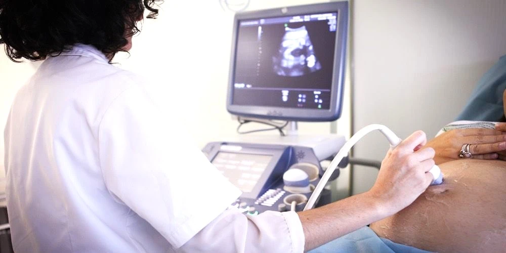
Ultrasound at Home Dubai: From AED 599 (2026)
Read More
Neck MRI Dubai: What It Shows & Cost (2026)
Read More
X-Ray Scan Dubai: Complete Guide (2026)
Read More
MRI vs Ultrasound Dubai: Which Scan? (2026)
Read More© 2026 Doctors Clinic Diagnostic Center (DCDC), Dubai Healthcare City. Originally published at https://doctorsclinicdubai.ae/blog/thyroid-ultrasound-guide. All rights reserved. Unauthorized reproduction is prohibited.