
Key Takeaways
- Doppler ultrasound uses the Doppler effect to measure blood flow speed and direction — it is painless, radiation-free, and takes 15–45 minutes
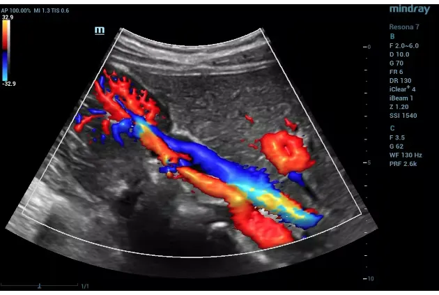
- Three types of Doppler exist: colour Doppler (maps flow direction), power Doppler (detects slow flow in small vessels), and spectral Doppler (measures exact velocities)
- Most Doppler scans require no special preparation; abdominal and renal Doppler require 6–8 hours fasting to reduce bowel gas interference
- Doppler ultrasound costs in Dubai range from AED 400 to AED 1,500 depending on type — most are covered by major insurance when medically indicated
- Unlike CT angiography or MR angiography, Doppler uses no radiation, no contrast injection, and is fully repeatable for serial monitoring
- DCDC in Dubai Healthcare City offers all Doppler types with same-day appointments, competitive pricing, and rapid reporting
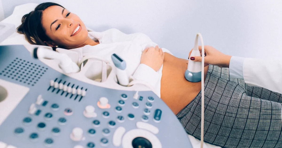
A Doppler ultrasound is a specialised diagnostic test that uses sound waves to evaluate how blood flows through your arteries and veins. While a standard ultrasound scan shows the structure of organs and tissues, a Doppler goes further — it detects blood flow speed, direction, and any blockages or abnormalities in the circulatory system. This makes it an essential tool for diagnosing blood clots, narrowed arteries, vein problems, and organ blood supply issues.
This guide covers everything you need to know about Doppler ultrasound in Dubai: how it works (in plain English), the different types available, how Doppler differs from regular ultrasound, how to prepare for each scan type, what happens during the procedure, what each scan costs, and insurance coverage details.


How Does a Doppler Ultrasound Work?
Doppler ultrasound relies on a physics principle called the Doppler effect. You have experienced this without realising it: when an ambulance approaches, its siren sounds higher in pitch; as it drives away, the pitch drops. This change in sound frequency occurs because the sound waves are compressed as the source approaches and stretched as it recedes.
In medical Doppler, the ultrasound probe sends sound waves into your body. When those sound waves bounce off moving red blood cells, they return at a slightly different frequency. The machine calculates this frequency shift to determine how fast the blood is moving, which direction it is flowing, and whether any areas of abnormal flow (turbulence, blockage, or reversal) are present.
Think of it like watching a river: a standard ultrasound shows you the shape of the riverbank (organ structure), while a Doppler ultrasound shows you the speed and direction of the water current (blood flow). Both types of information are diagnostically valuable, and modern ultrasound machines can perform both simultaneously.
Three Types of Doppler Ultrasound
| Type | How It Works | What It Shows | Best For |
|---|---|---|---|
| Colour Doppler | Assigns colours to blood flow direction — typically red for flow toward the probe, blue for flow away | Real-time colour map overlaid on the anatomical image showing flow patterns | Identifying areas of blockage, turbulence, or abnormal flow direction in arteries and veins |
| Power Doppler | Detects the amplitude (strength) of blood flow signals rather than velocity | More sensitive to slow flow in small vessels; does not show flow direction | Evaluating blood supply to organs, detecting inflammation, assessing small-vessel flow in thyroid, testes, and kidneys |
| Spectral (Pulsed-Wave) Doppler | Measures exact flow velocities at a specific point in a vessel, displayed as a waveform graph over time | Peak systolic velocity (PSV), end-diastolic velocity, pulsatility index (PI), resistance index (RI) | Grading stenosis severity, assessing fetal blood flow indices, calculating vessel-specific measurements |
The three types of Doppler ultrasound. Most clinical exams use a combination of all three for a complete assessment.
In practice, most Doppler examinations use all three types together. The radiologist starts with colour Doppler to get a visual overview of flow, then uses spectral Doppler to obtain precise velocity measurements at specific locations, and power Doppler to evaluate areas where slow flow might be present.
Doppler vs Regular Ultrasound: What Is the Difference?
This is one of the most common questions we receive. Both use the same basic technology (sound waves), but they provide fundamentally different information.
| Feature | Standard (B-Mode) Ultrasound | Doppler Ultrasound |
|---|---|---|
| What It Shows | Organ structure, tissue density, fluid collections | Blood flow speed, direction, and patterns |
| Primary Purpose | Structural/anatomical assessment | Functional/vascular assessment |
| Image Type | Greyscale image of organs and tissues | Colour-coded flow maps + velocity waveforms overlaid on greyscale |
| Detects | Masses, cysts, stones, organ enlargement, fluid | Blood clots, artery narrowing, vein reflux, abnormal flow |
| Radiation | None | None |
| Pain | None | None |
| Duration | 15–30 minutes | 15–45 minutes (slightly longer due to flow analysis) |
| Requires Contrast | No | No (unlike CT or MR angiography) |
| Cost (AED) | 300–800 | 400–1,500 |
| Example | Checking kidney size and structure | Checking blood flow to the kidney (renal artery stenosis) |
Standard ultrasound vs Doppler ultrasound comparison. Both are safe, painless, and radiation-free. Many modern exams combine both in a single session.
Can One Scan Do Both?
Yes. Modern ultrasound machines are capable of performing both standard B-mode imaging and Doppler analysis in a single session. This is called a duplex ultrasound. For example, a "renal Doppler" actually includes B-mode images of kidney structure plus Doppler analysis of blood flow to the kidneys. You do not need two separate appointments.
Doppler vs CT Angiography vs MR Angiography
When vascular problems are suspected, your doctor may choose between Doppler ultrasound, CT angiography (CTA), or MR angiography (MRA). Each has advantages.
| Feature | Doppler Ultrasound | CT Angiography | MR Angiography |
|---|---|---|---|
| Radiation | None | Yes (moderate) | None |
| Contrast Agent | None required | Iodinated IV contrast | Gadolinium IV contrast |
| Duration | 15–45 min | 5–15 min | 30–60 min |
| Cost (AED) | 400–1,500 | 1,500–4,000 | 3,000–6,000 |
| Real-Time Flow | Yes (live assessment) | No (snapshot in time) | Limited |
| Repeatability | Unlimited (no radiation) | Limited by cumulative radiation | Unlimited but expensive |
| Operator Dependent | Yes (skill matters) | Less so | Less so |
| Kidney Safety | No concerns | Contrast can affect kidneys | Gadolinium risk in kidney disease |
| Best For | Initial screening, serial monitoring, pregnancy, bedside assessment | Comprehensive vessel mapping, surgical planning, pulmonary embolism | Soft-tissue vascular detail, gadolinium-free sequences available |
Doppler ultrasound vs CT angiography vs MR angiography. Doppler is typically the first-line test due to its safety, accessibility, and cost-effectiveness.
In most clinical scenarios, Doppler ultrasound is the first-line diagnostic tool for vascular assessment. If the Doppler findings are inconclusive or if more detailed vessel mapping is needed (for example, before surgery), CTA or MRA may be requested as a follow-up.
Types of Doppler Scans Available in Dubai
Doppler ultrasound is used across many medical specialties. Here is what each type evaluates and who typically needs it.
Carotid Doppler
Evaluates blood flow through the carotid arteries in the neck, which supply blood to the brain. Critical for stroke prevention screening in patients with hypertension, diabetes, high cholesterol, smoking history, or prior stroke/TIA. Measures stenosis (narrowing) percentage and plaque characteristics.
Lower Extremity Venous Doppler
The primary test for deep vein thrombosis (DVT) and varicose vein assessment. Uses the compression technique (non-compressible vein = clot) and maps valve function in the saphenous veins. Essential for patients with sudden leg swelling, calf pain, or post-surgical clot risk.
Lower Extremity Arterial Doppler
Assesses arterial blood flow from the aortic bifurcation to the foot. Includes the ankle-brachial index (ABI) measurement — an ABI below 0.90 confirms peripheral arterial disease (PAD). Analyses waveform patterns (triphasic, biphasic, monophasic) and velocity ratios to grade stenosis severity.
Leg Doppler: Arterial vs Venous
| Feature | Venous Leg Doppler | Arterial Leg Doppler |
|---|---|---|
| Target Vessels | Deep and superficial veins (femoral, popliteal, saphenous, calf veins) | Arteries (iliac, femoral, popliteal, tibial, dorsalis pedis) |
| Conditions Detected | DVT, varicose veins, chronic venous insufficiency, valve reflux | PAD, arterial stenosis, aneurysm, post-bypass graft surveillance |
| Key Measurement | Compressibility, reflux duration, flow augmentation | ABI, peak systolic velocity (PSV), waveform pattern |
| Common Symptoms | Leg swelling, calf pain, visible varicose veins, skin discolouration | Walking pain (claudication), cold feet, weak pulses, non-healing wounds |
| Duration | 20–30 min per leg | 30–40 min per leg |
Arterial vs venous leg Doppler comparison. Your doctor will specify which type based on your symptoms.
Upper Extremity Venous Doppler
Evaluates veins in the arms. Used for arm DVT (which can occur with central venous catheters, thoracic outlet syndrome, or pacemaker leads) and for dialysis access assessment in haemodialysis patients.
Renal Doppler
Assesses blood flow to the kidneys. Detects renal artery stenosis (a cause of resistant hypertension), monitors transplanted kidneys for vascular complications, and evaluates kidney damage using the resistance index (RI). Requires 6–8 hours fasting.
Abdominal / Hepatic Doppler
Examines blood flow through the liver, portal vein, hepatic veins, and splenic vessels. Essential for patients with liver cirrhosis, portal hypertension, Budd-Chiari syndrome, or liver transplant monitoring. Requires 6–8 hours fasting.
Pregnancy / Obstetric Doppler
Measures blood flow in the umbilical artery, middle cerebral artery (MCA), ductus venosus, and uterine arteries. Monitors fetal wellbeing in high-risk pregnancies including pre-eclampsia, intrauterine growth restriction (IUGR), and gestational diabetes. Completely safe for mother and baby. For detailed obstetric Doppler information, see our pregnancy Doppler guide.
Penile & Testicular Doppler
Penile Doppler evaluates arterial and venous blood flow during erection (often pharmacologically induced) for erectile dysfunction workup. Testicular Doppler assesses blood flow to the testes for varicocele evaluation, testicular torsion (emergency), and undescended testes.
How to Prepare for a Doppler Ultrasound
Most Doppler scans require little or no preparation. The exceptions are abdominal and renal Doppler, which require fasting. Here is a complete preparation guide by scan type.
| Doppler Type | Fasting Required? | Special Preparation | Duration |
|---|---|---|---|
| Carotid | No | None. Wear a top with an open neckline or V-neck. | 20–30 min |
| Leg Venous | No | Wear loose trousers or shorts. Remove compression stockings before the exam. | 20–30 min per leg |
| Leg Arterial | No | No smoking for 2 hours before (nicotine constricts arteries). Wear loose trousers. | 30–40 min per leg |
| Renal | Yes (6–8 hours) | Avoid carbonated drinks and gas-producing foods the day before. Morning appointment preferred. | 20–30 min |
| Abdominal / Hepatic | Yes (6–8 hours) | Same as renal. Water permitted in small sips. | 20–30 min |
| Pregnancy / Obstetric | No | None. A moderately full bladder may help in early pregnancy. | 20–30 min |
| Testicular | No | None. Wear comfortable underwear. | 15–20 min |
| Upper Extremity | No | Wear short sleeves or loose-fitting top. | 20–30 min |
Doppler ultrasound preparation by type. Continue all prescribed medications unless specifically instructed otherwise by your doctor.
General Preparation Checklist
- Continue all medications unless your doctor specifically tells you to stop. If you take anticoagulants (blood thinners), mention this to the sonographer as it affects interpretation
- Wear loose, comfortable clothing that allows easy access to the area being scanned. Two-piece outfits are best
- Bring your referral letter and insurance card. For insurance coverage, pre-authorisation may be required
- Bring prior imaging results if available — comparison with previous scans is valuable
- For fasting scans (renal, abdominal): no food for 6–8 hours. Small sips of water are permitted. Schedule morning appointments to minimise fasting discomfort
- Avoid smoking for 2 hours before arterial Doppler scans — nicotine causes arterial constriction that can affect velocity measurements
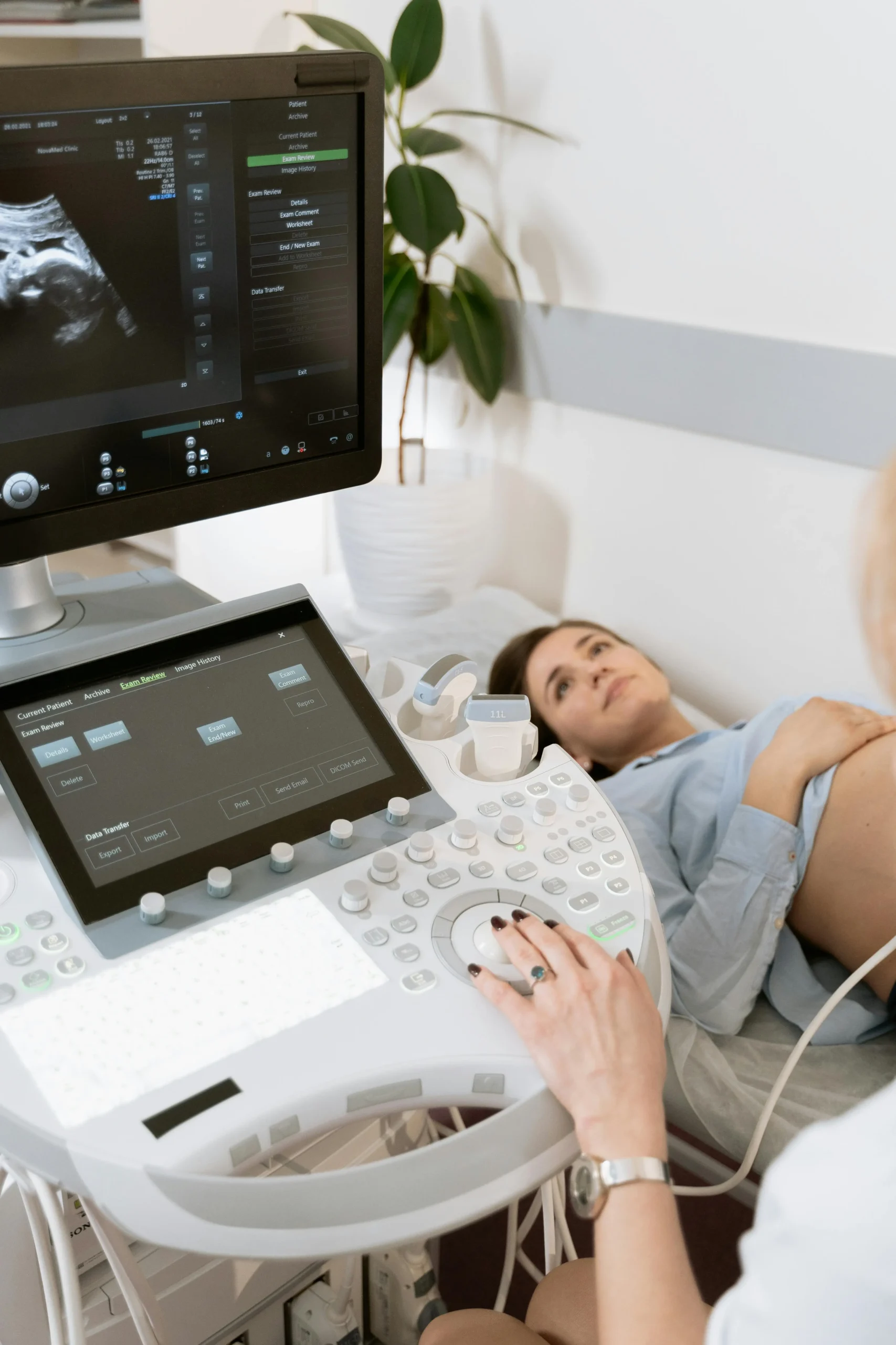
What Happens During a Doppler Ultrasound: Step by Step
- Step 1 — Check-in: You change into a gown if needed (depends on scan area) and the sonographer confirms your details and the reason for the exam
- Step 2 — Positioning: You lie on the examination table. Position varies by scan: on your back for carotid/abdominal, on your back or side for leg scans, semi-reclined for testicular
- Step 3 — Gel application: Warm water-based gel is applied to the scan area. This gel conducts sound waves and prevents air gaps between the probe and your skin
- Step 4 — Scanning: The sonographer moves the probe over the area, adjusting pressure and angle. You may hear a "whooshing" sound — this is the audio representation of your blood flow, which helps the examiner identify vessels
- Step 5 — Measurements: The sonographer takes specific velocity and flow measurements at key points. You may be asked to take deep breaths, perform a Valsalva manoeuvre (bearing down), or have blood pressure cuffs inflated on your limbs
- Step 6 — Completion: The gel is wiped off, you dress, and the images are sent to the radiologist for interpretation. There are no post-scan restrictions — you can eat, drive, and resume normal activities immediately
The entire process is painless. You may feel mild pressure from the probe, and for venous leg scans, the sonographer will compress each vein segment with the probe (the compression technique used to detect blood clots), which feels like brief firm pressure.
Doppler Ultrasound Cost in Dubai by Type (2026)
Doppler ultrasound pricing in Dubai varies based on the type of scan, the body region examined, and the facility. Below is a comprehensive breakdown of typical costs for 2026.
| Doppler Ultrasound Type | Cost (AED) | Common Indications |
|---|---|---|
| Carotid Doppler | 500–900 | Stroke risk screening, neck artery narrowing, plaque assessment |
| Lower Extremity Venous Doppler | 500–1,000 | DVT, varicose veins, leg swelling, chronic venous insufficiency |
| Lower Extremity Arterial Doppler | 600–1,100 | PAD, claudication, cold feet, non-healing wounds, ABI |
| Upper Extremity Venous Doppler | 450–800 | Arm DVT, swelling, dialysis access assessment |
| Renal Doppler | 600–1,200 | Renal artery stenosis, transplant monitoring, resistant hypertension |
| Abdominal / Hepatic Doppler | 500–1,000 | Portal hypertension, liver cirrhosis, Budd-Chiari, transplant monitoring |
| Pregnancy / Obstetric Doppler | 400–900 | Fetal blood flow, placental health, IUGR, pre-eclampsia |
| Penile Doppler | 1,500–2,500 | Erectile dysfunction vascular evaluation |
| Testicular Doppler | 400–800 | Varicocele, testicular torsion, scrotal pain |
Doppler ultrasound pricing in Dubai (2026). Prices are approximate and may vary by facility, reporting urgency, and package inclusions.
"Many patients delay vascular testing because they assume it will be expensive," says Dr. Hadi Komshi, Specialist in Internal Medicine at DCDC. "But a Doppler scan costing AED 500–900 can detect a life-threatening blood clot or a critically narrowed artery — conditions where early detection dramatically changes outcomes. The cost of not testing is far higher than the cost of the test."
Insurance Coverage for Doppler Ultrasound in Dubai
Most major health insurance providers in the UAE cover Doppler ultrasound when it is deemed medically necessary and referred by a physician. Key providers include:
- Daman: Covers medically indicated Doppler with physician referral and pre-authorisation
- Oman Insurance: Covers diagnostic Doppler for specified conditions
- AXA: Covers with referral; some plans require pre-approval for certain scan types
- MetLife: Covers as part of diagnostic imaging benefits with co-pay varying by plan
- Cigna: Covers medically necessary Doppler with pre-authorisation
Out-of-pocket costs depend on your plan, deductible, and co-pay structure. Patients without insurance can benefit from competitive self-pay pricing at DCDC, where transparent pricing and no hidden fees are standard.
Safety of Doppler Ultrasound
Doppler ultrasound is one of the safest diagnostic tests available:
- No radiation: Uses only sound waves, making it completely safe for serial monitoring, pregnancy, and paediatric patients
- No contrast injection: Unlike CT or MR angiography, Doppler requires no intravenous contrast agents, eliminating the risk of contrast reactions or kidney damage
- No sedation: Fully awake, no recovery time needed
- Infinitely repeatable: Because there is no radiation or contrast, Doppler can be repeated as often as clinically needed without cumulative risk
- Safe in pregnancy: Doppler ultrasound has been used in obstetric care for decades with no evidence of harm to mother or baby. Major organisations (ACOG, RCOG, ISUOG) endorse its use in pregnancy monitoring
Note on consumer handheld Doppler devices: Over-the-counter fetal Doppler monitors sold online are not the same as clinical Doppler ultrasound. They lack the precision, imaging capability, and diagnostic value of clinical systems and can provide false reassurance or unnecessary anxiety. Clinical Doppler should always be performed by qualified sonographers and interpreted by radiologists.
Doppler Ultrasound at DCDC Dubai Healthcare City
Doctors Clinic Diagnostic Center (DCDC) offers comprehensive Doppler ultrasound services in Dubai Healthcare City:
- All Doppler types available: Carotid, venous, arterial, renal, abdominal, obstetric, penile, and testicular
- Experienced radiologists specialising in vascular imaging with advanced ultrasound equipment
- Same-day appointments — walk-ins welcome for most scan types
- Rapid reporting: Results typically available same day or within 24 hours
- All major insurance accepted with transparent self-pay pricing for uninsured patients
- Convenient location: Dubai Healthcare City, easily accessible from Oud Metha, Karama, and Business Bay
Book Your Doppler Ultrasound
Whether you need a carotid Doppler for stroke screening, a venous Doppler for DVT evaluation, or a renal Doppler for kidney assessment, our team at Doctors Clinic Diagnostic Center in Dubai Healthcare City provides all types of Doppler ultrasound with competitive pricing and same-day appointments.
Related Services at DCDC
Expert care and advanced diagnostics at Dubai Healthcare City
Frequently Asked Questions
Final Thoughts
Doppler ultrasound is one of the most versatile, safe, and accessible diagnostic tools in modern medicine. Whether you need stroke risk screening, blood clot detection, kidney vascular assessment, fetal monitoring, or peripheral artery disease evaluation, there is a Doppler scan designed for your specific clinical question — and all are available in Dubai at reasonable cost with minimal preparation.
At Doctors Clinic Diagnostic Center, we provide all types of Doppler ultrasound with competitive pricing, experienced radiologists, and same-day reporting. For condition-specific information, see our detailed guides on carotid Doppler for stroke prevention, DVT detection, renal Doppler, peripheral vascular disease, and pregnancy Doppler.
Sources & References
This article was reviewed by our medical team and references the following sources:
- Dubai Health Authority — Diagnostic Imaging Standards
- American Institute of Ultrasound in Medicine (AIUM) — Doppler Guidelines
- Society for Vascular Ultrasound (SVU) — Practice Standards
- Radiological Society of North America (RSNA) — Doppler Ultrasound
- American Heart Association (AHA) — Peripheral Vascular Disease
- European Society of Cardiology (ESC) — Vascular Guidelines
Medical content on this site is reviewed by DHA-licensed physicians. See our editorial policy for more information.

Written by
Dr. Osama Elzamzami
Consultant Radiologist
MD, Radiology
Dr. Osama Elzamzami is a Consultant Radiologist specialising in diagnostic imaging including MRI, CT, ultrasound, and Doppler studies at DCDC Dubai Healthcare City. He performs all types of vascular Doppler examinations with a focus on accurate diagnosis and patient-centred care.
Related Articles

Carotid Doppler Ultrasound and Stroke Prevention
Doppler Ultrasound for DVT Detection
Ultrasound Cost in Dubai: Complete Guide
Renal Doppler Ultrasound: Kidney Blood Flow Guide
More in Diagnostic Imaging

X-Ray at Home Dubai: Cost & Guide (2026)
Read More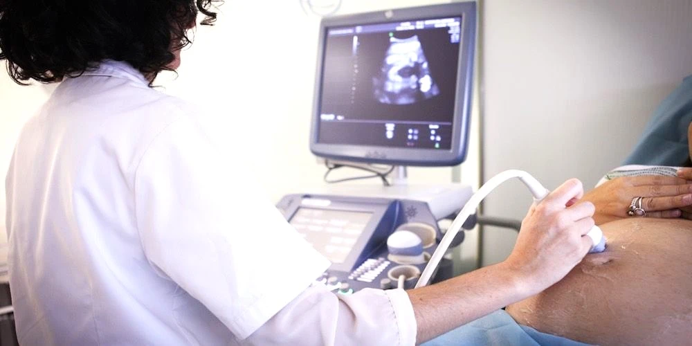
Ultrasound at Home Dubai: From AED 599 (2026)
Read More
Neck MRI Dubai: What It Shows & Cost (2026)
Read More
X-Ray Scan Dubai: Complete Guide (2026)
Read More
MRI vs Ultrasound Dubai: Which Scan? (2026)
Read More
MRI vs X-Ray Dubai: Which Scan Do You Need? (2026)
Read More© 2026 Doctors Clinic Diagnostic Center (DCDC), Dubai Healthcare City. Originally published at https://doctorsclinicdubai.ae/blog/doppler-ultrasound-cost-dubai. All rights reserved. Unauthorized reproduction is prohibited.