Key Takeaways
- A brain MRI in Dubai costs between AED 1,200 and AED 2,000, depending on whether contrast dye is used and the imaging center
- Brain MRI is the gold standard for detecting tumors, stroke, multiple sclerosis, aneurysms, and the structural causes of seizures and chronic headaches
- MRI is preferred over CT for most brain conditions because it provides superior soft tissue contrast with zero radiation exposure
- A neurologist, internist, or emergency physician may order a brain MRI for persistent headaches, vision changes, seizures, memory problems, or neurological symptoms
- The scan takes 30 to 60 minutes, is completely painless, and most patients can resume normal activities immediately afterward
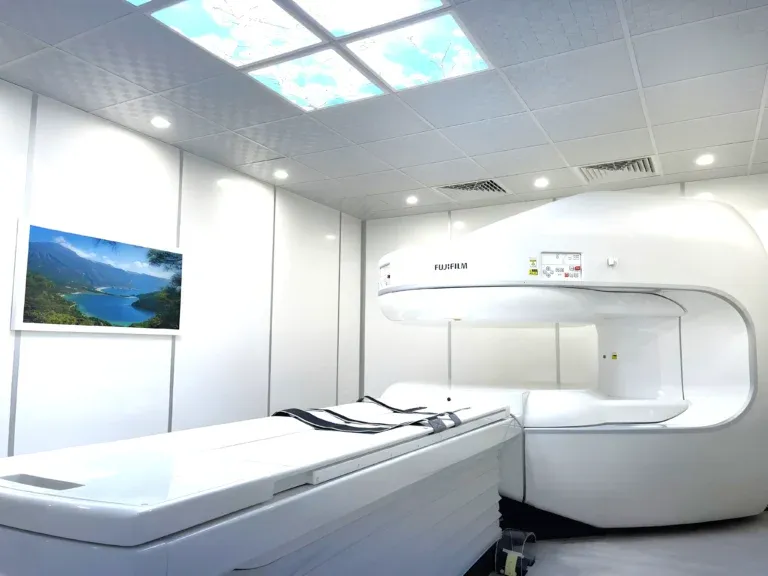
A brain MRI scan is the most detailed and sensitive imaging tool available for evaluating the brain and surrounding structures. From detecting tumors and stroke to investigating the causes of chronic headaches and seizures, brain MRI provides neurologists and physicians with the precise structural information they need to make accurate diagnoses. In Dubai, brain MRI is widely accessible and represents a critical component of neurological evaluation for patients of all ages.


What Does a Brain MRI Detect?
A brain MRI generates detailed cross-sectional images of the brain, brain stem, cerebellum, and surrounding structures including the pituitary gland, sinuses, and inner ear. The exceptional soft tissue contrast of MRI makes it far superior to CT for evaluating most brain conditions. The following are among the most important conditions that brain MRI can detect:
- Brain tumors: MRI is the primary imaging modality for detecting, characterizing, and monitoring brain tumors. It can identify tumor location, size, margins, and relationship to surrounding brain structures. Contrast-enhanced MRI further helps differentiate tumor types and grade their aggressiveness.
- Stroke and cerebrovascular disease: MRI with diffusion-weighted imaging (DWI) can detect ischemic stroke within minutes of onset — far earlier than CT. It also identifies chronic stroke changes, transient ischemic attack (TIA) sequelae, and cerebral small vessel disease.
- Multiple sclerosis (MS): Brain MRI is essential for diagnosing and monitoring MS. It reveals demyelinating plaques in the white matter, which are the hallmark of the disease. Serial MRI scans track disease activity and response to treatment over time.
- Brain aneurysms: MRI angiography (MRA) can detect intracranial aneurysms — weakened, balloon-like areas in blood vessel walls that carry a risk of rupture. Early detection through screening MRI can be life-saving, particularly for patients with a family history of aneurysms.
- Seizures and epilepsy: MRI identifies structural abnormalities that may cause seizures, including cortical malformations, hippocampal sclerosis, tumors, vascular malformations, and post-traumatic changes. Specialized epilepsy protocols enhance detection of subtle findings.
- Headaches and migraines: While most headaches have no structural cause, MRI is ordered when headaches are severe, progressive, associated with neurological symptoms, or have changed in character. MRI can rule out tumors, aneurysms, hydrocephalus, and Chiari malformations as potential causes.
- Concussion and traumatic brain injury: MRI is more sensitive than CT for detecting the subtle brain changes associated with concussion, including microhemorrhages, diffuse axonal injury, and contusions that may not be visible on CT.
- Neurodegenerative diseases: MRI can reveal patterns of brain atrophy associated with Alzheimer's disease, Parkinson's disease, and other neurodegenerative conditions. Volumetric MRI measurements help track disease progression.
"Brain MRI is one of the most powerful diagnostic tools in modern medicine," explains Dr. Osama Elzamzami, Consultant Radiologist at DCDC. "The level of anatomical detail it provides allows us to detect lesions as small as a few millimeters and differentiate between conditions that might look identical on other imaging modalities. For neurological symptoms, there is simply no substitute for a high-quality brain MRI."
How Much Does a Brain MRI Cost in Dubai?
The cost of a brain MRI in Dubai ranges from approximately AED 1,200 to AED 2,000. The variation depends on whether contrast dye (gadolinium) is needed, the specific protocols ordered, and the imaging center. At Doctors Clinic Diagnostic Center, all brain MRI pricing includes the scan, a comprehensive consultant radiologist report, and digital image copies.
| Brain MRI Type | Approximate Cost (AED) |
|---|---|
| Brain MRI without contrast | 1,200 – 1,500 |
| Brain MRI with contrast (gadolinium) | 1,500 – 2,000 |
| Brain MRI with MRA (angiography) | 1,800 – 2,200 |
| Brain + cervical spine MRI (combined) | 2,200 – 2,800 |
Prices are approximate and include the radiologist report at DCDC. Contact us for exact pricing.
Contrast-enhanced brain MRI is typically ordered when a tumor, infection, or inflammatory condition is suspected. The gadolinium contrast agent highlights areas of abnormal blood-brain barrier breakdown, making certain lesions far more visible. For most headache evaluations, screening for aneurysms, or follow-up studies, a non-contrast brain MRI is sufficient.
Brain MRI is covered by most health insurance plans in the UAE when ordered by a licensed physician with appropriate clinical justification. Common indications that insurers accept include persistent headaches, seizures, neurological deficits, suspected stroke, and follow-up of known brain pathology.
When Does a Doctor Order a Brain MRI?
Brain MRI is not a routine screening test for the general population — it is ordered when a physician identifies symptoms or clinical findings that warrant detailed brain imaging. A neurologist, internist, emergency physician, or other specialist may order a brain MRI in the following clinical scenarios: For related information, see our guide on MRI Cost Dubai: AED 900–15,000 by Type.
- New-onset seizures: Any adult experiencing seizures for the first time requires a brain MRI to identify or exclude structural causes such as tumors, vascular malformations, or cortical abnormalities.
- Severe or progressive headaches: Headaches that are sudden in onset (thunderclap headache), progressively worsening, associated with vomiting or visual changes, or that wake the patient from sleep may indicate serious pathology requiring MRI evaluation.
- Neurological deficits: Weakness, numbness, difficulty speaking, vision changes, balance problems, or coordination difficulties may suggest stroke, MS, or a space-occupying lesion. Brain MRI is the primary investigation in these cases.
- Memory loss or cognitive decline: Progressive memory problems, confusion, or personality changes may prompt MRI to assess for neurodegenerative disease, normal-pressure hydrocephalus, or chronic subdural hematoma.
- Head trauma with persistent symptoms: After a concussion or traumatic brain injury, MRI is ordered if symptoms persist beyond the expected recovery period or if there are signs of complications.
- Suspected brain tumor: When clinical features suggest a brain tumor — such as new headaches with neurological signs, unexplained seizures, or progressive neurological deterioration — MRI with contrast is the definitive investigation.
- Screening for aneurysms: Patients with a strong family history of intracranial aneurysms, polycystic kidney disease, or certain connective tissue disorders may be offered screening MRA to detect aneurysms before they rupture.
Book Your Brain MRI at DCDC
At Doctors Clinic Diagnostic Center in Dubai Healthcare City, we offer comprehensive brain MRI with expert consultant radiologist interpretation. Reports are delivered within 24 to 48 hours.
Brain MRI vs CT Scan: Which Is Better for the Brain?
Both MRI and CT scan can image the brain, but they serve different clinical purposes. Understanding their respective strengths helps patients and physicians choose the right modality for each situation.
| Factor | Brain MRI | Brain CT Scan |
|---|---|---|
| Cost | AED 1,200 – 2,000 | AED 500 – 900 |
| Radiation | None (zero radiation) | Yes (2-4 mSv) |
| Scan time | 30 – 60 minutes | 5 – 10 minutes |
| Soft tissue detail | Excellent (gold standard) | Limited |
| Bone and skull detail | Moderate | Excellent |
| Acute hemorrhage detection | Good (with specialized sequences) | Excellent (first-line) |
| Tumor detection | Superior | Limited sensitivity |
| Best for | Tumors, MS, seizures, chronic headaches, follow-up | Acute trauma, acute hemorrhage, skull fractures, emergencies |
CT is preferred for acute emergencies. MRI is preferred for most other brain conditions.
In emergency settings — such as suspected acute stroke with hemorrhage, severe head trauma, or sudden neurological deterioration — CT is typically performed first because it is faster and excels at detecting acute bleeding and skull fractures. However, for the detailed evaluation of most neurological conditions, MRI is vastly superior. Its ability to differentiate between gray and white matter, detect subtle lesions, and provide multiple tissue contrasts makes it indispensable for diagnosing tumors, MS, epilepsy-related abnormalities, and chronic conditions.
Another significant advantage of brain MRI is the absence of ionizing radiation. This makes it the preferred choice for follow-up imaging, particularly in patients requiring serial scans to monitor disease progression or treatment response. A patient with a known brain tumor, for example, may undergo MRI scans every 3 to 6 months — exposing them to cumulative CT radiation in this scenario would be inadvisable.
What to Expect During a Brain MRI
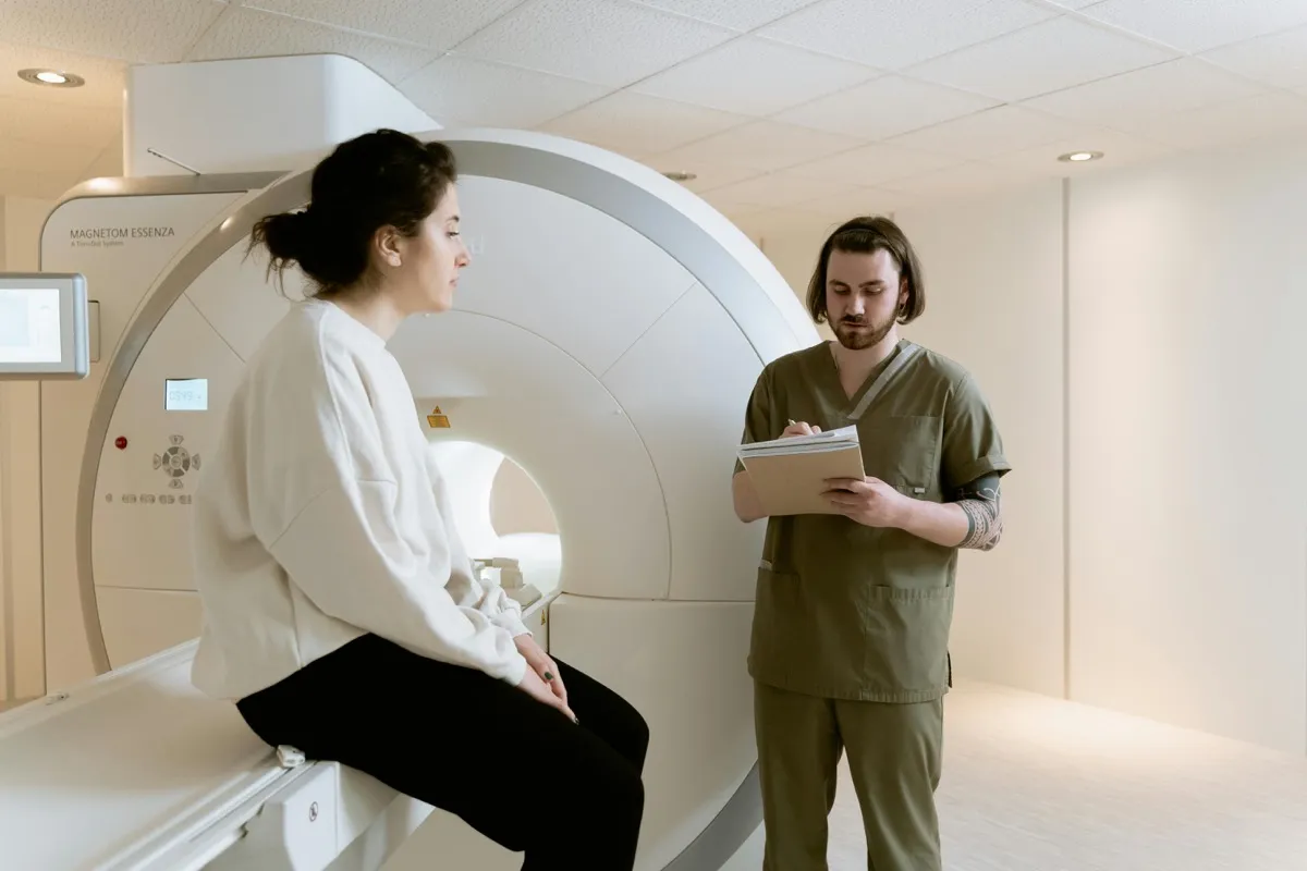
A brain MRI is a non-invasive, painless procedure that requires no special preparation in most cases. Understanding the process helps reduce any anxiety about the examination.
- Before the scan: You will complete a detailed safety screening questionnaire to identify any metal implants, pacemakers, or other contraindications. All metal objects must be removed, including jewellery, hairpins, hearing aids, and dentures with metal components. If contrast is being used, a small IV line will be placed in your arm.
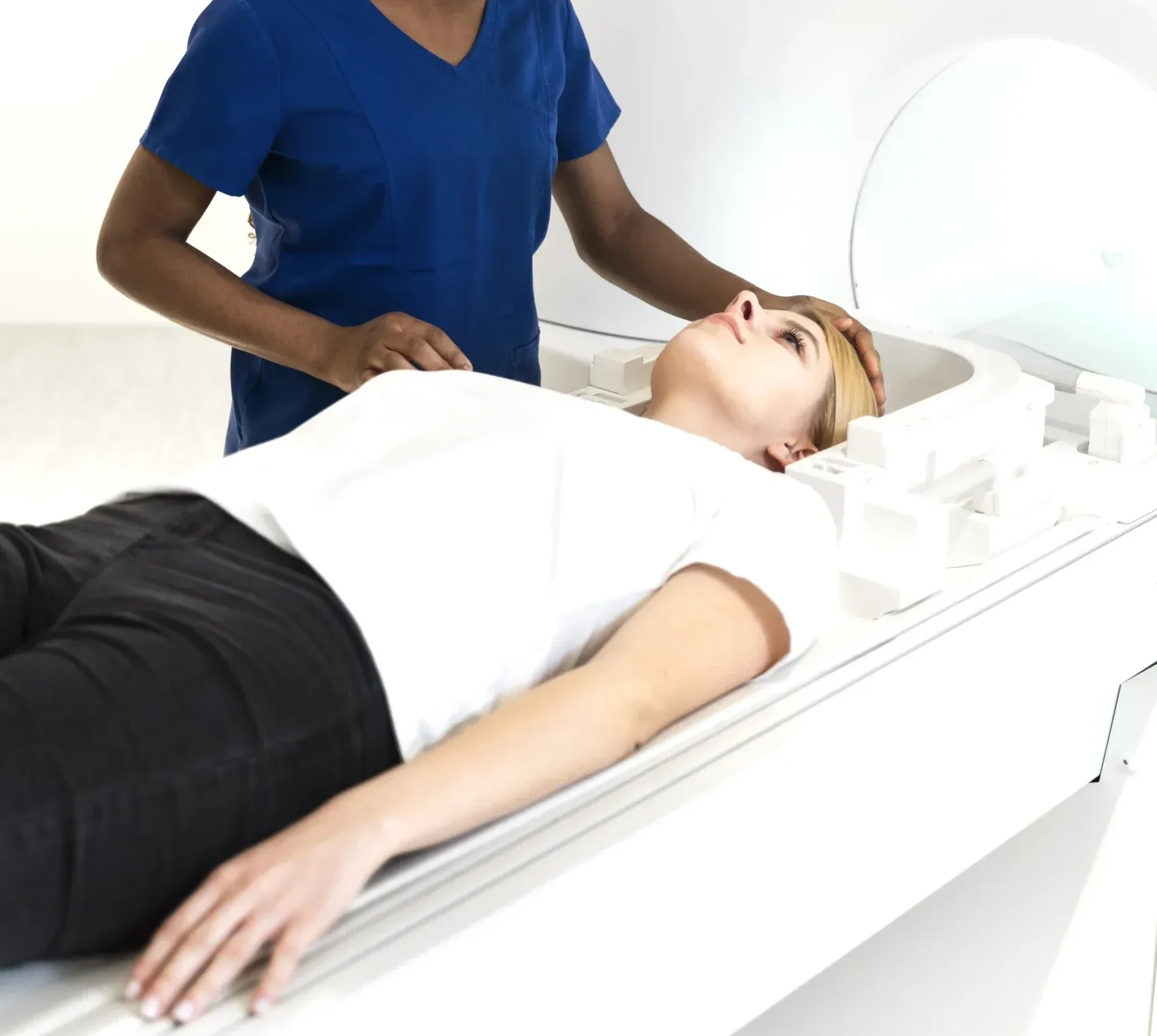
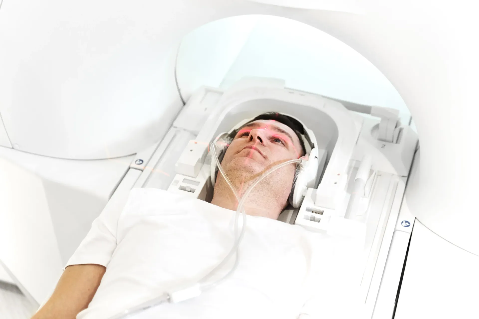
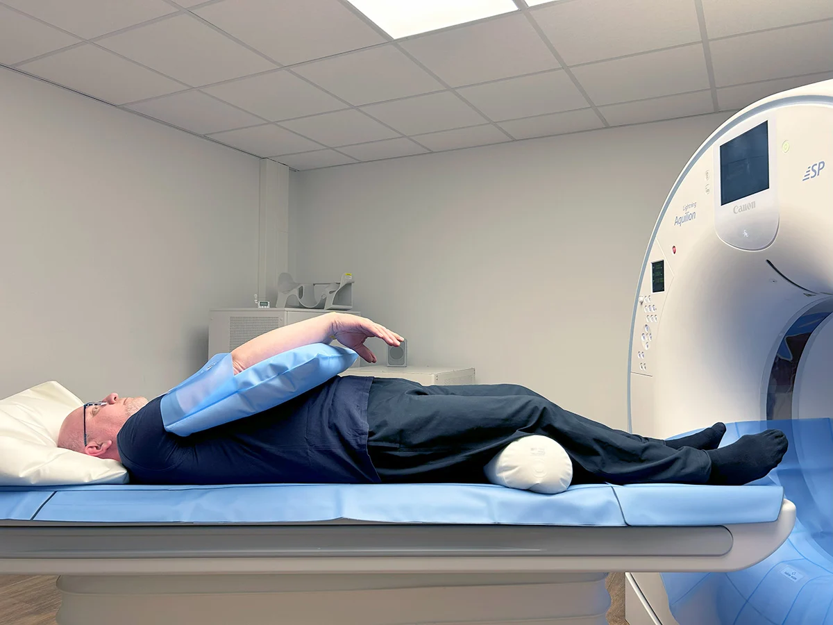
- Positioning: You will lie on your back on the MRI table with your head positioned in a padded head coil. The coil sits around your head like a cage and is essential for producing high-quality brain images. Foam pads may be placed beside your head to minimize movement.
- During the scan: The table slides into the MRI machine. The machine produces a series of loud knocking, tapping, and buzzing sounds as it captures images from different angles and with different tissue contrasts. You will be given earplugs or headphones. A technologist monitors you throughout the scan and communicates via intercom.
- Duration: A standard brain MRI without contrast takes approximately 30 minutes. With contrast, the total scan time is typically 45 to 60 minutes. Specialized protocols (epilepsy protocol, MRA) may take slightly longer.
- After the scan: You can get up and leave immediately. There is no recovery period, and you can drive, eat, and return to normal activities right away. If contrast was used, you are encouraged to drink extra water to help flush the agent from your system.
Patients who experience claustrophobia should inform the radiology team in advance. Several options are available to improve comfort, including guided relaxation techniques, music through the headphones, and in some cases a mild sedative prescribed by your physician. At DCDC, our technologists are experienced in managing claustrophobic patients and will work with you to ensure the scan is completed successfully.
Who Should Consider a Brain MRI in Dubai?
Brain MRI is recommended based on clinical symptoms and risk factors rather than age alone. However, certain groups of patients in Dubai are more likely to benefit from brain imaging: You may also find our CT Scan Cost Dubai: Prices & Types helpful.
- Patients with chronic or worsening headaches: If you have headaches that are increasing in frequency or severity, associated with nausea or visual disturbances, or are fundamentally different from your typical headache pattern, a brain MRI can rule out serious causes.
- Patients with neurological symptoms: Any new neurological symptom — weakness, numbness, tingling, visual changes, speech difficulty, balance problems, or confusion — warrants medical evaluation that may include brain MRI.
- Patients with a family history of brain aneurysms: If a first-degree relative (parent, sibling) has had an intracranial aneurysm, screening MRA is recommended, as the risk of aneurysm is significantly elevated in these families.
- Cancer patients: Brain MRI is performed to screen for metastases or to monitor known brain tumors during and after treatment. It is the most sensitive modality for detecting even small brain metastases.
- Patients seeking comprehensive health screening: As part of an executive health checkup or full body MRI screening, brain MRI provides baseline imaging that can detect incidental findings such as silent infarcts, small meningiomas, or aneurysms.
Get Expert Brain MRI Interpretation at DCDC
Every brain MRI at Doctors Clinic Diagnostic Center is interpreted by a consultant radiologist with extensive neuroradiology experience. Our structured reports provide your referring physician with detailed findings and clinical correlation. Learn more about our MRI services.
Related Services at DCDC
Expert care and advanced diagnostics at Dubai Healthcare City
Frequently Asked Questions
Final Thoughts
Brain MRI is an invaluable diagnostic tool that provides unmatched detail of brain anatomy and pathology. Whether your physician is investigating headaches, seizures, neurological symptoms, or screening for aneurysms, a high-quality brain MRI with expert interpretation is essential for accurate diagnosis and appropriate treatment planning.
At Doctors Clinic Diagnostic Center in Dubai Healthcare City, our consultant radiologists bring extensive experience in neuroimaging to every brain MRI interpretation. Contact us to schedule your brain MRI or to discuss whether this examination is appropriate for your symptoms.
Sources & References
This article was reviewed by our medical team and references the following sources:
- American College of Radiology - Appropriateness Criteria for Headache
- Radiological Society of North America - Brain MRI
- American Academy of Neurology - Neuroimaging Guidelines
- The Lancet Neurology - MRI in Neurological Diagnosis
- Dubai Health Authority - Diagnostic Imaging Regulations
Medical content on this site is reviewed by DHA-licensed physicians. See our editorial policy for more information.

Written by
Dr. Osama Elzamzami
Diagnostic Radiology
MD, FRCR
Dr. Osama Elzamzami is a Consultant Radiologist specializing in diagnostic imaging including MRI, CT, and ultrasound at DCDC Dubai Healthcare City.
Related Articles
MRI Cost in Dubai: Complete Guide to Types & Pricing (2026)
Full Body MRI Cost Dubai: Complete Price Guide (2026)
Spine MRI in Dubai: Cost, What It Shows & When You Need One
MRI for Headaches: When You Need a Brain Scan for Migraines
MRI for Vertigo: When Dizziness Needs Brain & Spine Imaging
More in Diagnostic Imaging
CT Scan Cost Dubai: Prices & Types (2026)
Read More
DEXA Scan Dubai: Cost, T-Score & Who Needs It (2026)
Read MoreFull Body MRI Cost Dubai: AED 5,000-15,000 [2026]
Read More
MRI Preparation Dubai: Complete Guide (2026)
Read More
CBCT Scan: 3D Dental Imaging Guide Dubai (2026)
Read More
Ultrasound Preparation Dubai: What to Expect (2026)
Read More© 2026 Doctors Clinic Diagnostic Center (DCDC), Dubai Healthcare City. Originally published at https://doctorsclinicdubai.ae/blog/brain-mri-scan-dubai. All rights reserved. Unauthorized reproduction is prohibited.