Key Takeaways
- An MRI report has four main sections: clinical indication, technique, findings and impression. The impression at the end is the most important part, summarising what the radiologist considers clinically significant.
- Many MRI findings that sound alarming are common and often asymptomatic. Disc bulges appear in over 50 percent of adults over 40 who have no back pain at all, and small pineal cysts are found in up to 10 percent of brain MRIs.
- Key terms to know: hyperintense means brighter than surrounding tissue, hypointense means darker, enhancement means the area lights up after contrast dye, and unremarkable means normal.
- Red flags that need urgent follow-up include signs of cord compression, acute stroke on diffusion-weighted imaging, a mass with enhancement, and language such as urgent clinical review recommended.
- Subspecialty radiologist reads improve diagnostic accuracy. At DCDC in Dubai Healthcare City, brain MRIs are read by neuroradiologists and joint MRIs by musculoskeletal radiologists, not generalists.
- MRI results at DCDC are available within 18 to 24 hours via the patient portal, with same-day reporting for urgent cases. MRI scans start from AED 900 per region.
You have had an MRI scan and now you are staring at a report filled with words like hyperintense, disc protrusion, degenerative changes and clinical correlation recommended. It can be unsettling, especially when you do not know whether these terms describe something serious or something entirely routine. This guide breaks down every section of an MRI report in plain language, explains the most common findings by body area and tells you when results need urgent attention and when they are part of normal ageing.
MRI (magnetic resonance imaging) produces extraordinarily detailed pictures of soft tissues, joints, the brain and the spine without using any radiation. The report that follows is written by a radiologist, a doctor who specialises in interpreting medical images. Understanding the report's structure and vocabulary puts you in a much better position to discuss results with your treating physician and ask the right questions about next steps.


How to Read Your MRI Report: The Four Key Sections
Every MRI report follows a standardised structure regardless of which body part was scanned. Knowing how each section works makes the entire document less intimidating. If you are new to MRI and want background on the scan itself, our complete guide to MRI scans in Dubai covers preparation, the procedure and what happens during the scan.
Clinical Indication and History
This opening section states why the scan was ordered. It usually contains a brief clinical summary such as chronic low back pain, rule out disc herniation or right knee pain after sports injury. The indication matters because it tells the radiologist what to focus on and helps them prioritise their findings. If this section is vague or inaccurate, it can affect the quality of the interpretation, so always make sure your referring doctor provides a clear clinical question.
Technique
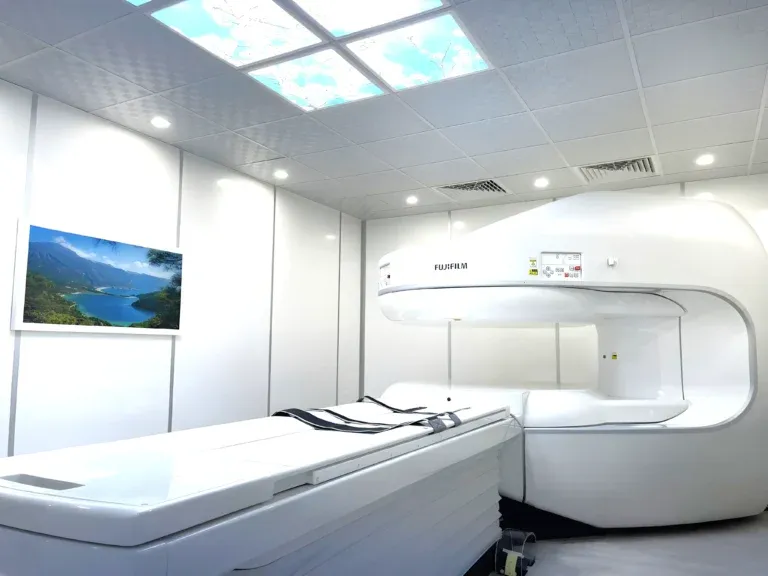
The technique section describes the scanner used, the magnetic field strength (for example, 1.5 Tesla), the imaging sequences performed (T1-weighted, T2-weighted, FLAIR, diffusion-weighted) and whether contrast dye (gadolinium) was administered. This section is primarily for other doctors and radiologists. At DCDC, scans are performed on a Siemens 1.5T wide-bore scanner with a 70 cm opening, and protocols are tailored to each clinical question. If you want to understand the different scanner types, see our comparison of 1.5T, 3T and open MRI.
Findings
The findings section is the longest part of the report and describes what the radiologist sees on each imaging sequence, organised by anatomical structure. For a spine MRI this typically moves vertebra by vertebra; for a brain MRI it covers the parenchyma, ventricles, midline structures and so on. Every observation is documented, including normal anatomy. This is where most of the technical vocabulary appears, and where patients tend to become anxious. Remember: a finding is not the same as a diagnosis. Many documented findings are incidental or age-related and have no clinical significance.
Impression (Conclusion)
The impression is the most important section for patients and treating doctors. It distils the findings into a concise summary of what is clinically significant, often numbered by priority. If the report is long and overwhelming, read the impression first. It may say something like mild degenerative disc disease at L4-L5 without significant neural foraminal narrowing, no acute abnormality, or new enhancing lesion, recommend oncology review. Any phrase like clinical correlation recommended means the radiologist is asking your treating physician to interpret the finding alongside your symptoms and history.
MRI Terminology Explained: A Patient-Friendly Glossary
The single biggest barrier to understanding MRI reports is the specialised vocabulary. Below is a glossary of the terms patients encounter most often, translated into everyday language. Knowing these words removes most of the anxiety that comes from reading a report for the first time.
| MRI Term | What It Means in Plain English |
|---|---|
| Hyperintense (high signal) | The area appears brighter than the tissue around it on the image |
| Hypointense (low signal) | The area appears darker than the tissue around it |
| Isointense | The area has the same brightness as the reference tissue — looks normal |
| Enhancement | The area lights up after contrast dye injection, suggesting increased blood flow from inflammation, infection or a tumour |
| Effusion | A collection of fluid inside a joint, often indicating inflammation or injury |
| Edema | Swelling or excess fluid within tissue, a sign of injury or inflammation |
| Herniation / Protrusion | Disc material pushing beyond its normal boundary in the spine |
| Stenosis | Narrowing of a passage such as the spinal canal or a blood vessel |
| Degenerative changes | Wear-and-tear changes associated with ageing — extremely common and often painless |
| Unremarkable | Normal; nothing abnormal was found in that structure |
| Lesion | Any area of abnormal tissue — this does NOT automatically mean cancer |
| Atrophy | A reduction in the size of tissue or an organ, sometimes normal with ageing |
| Incidental finding | Something found by chance that was not the reason for the scan |
| Clinical correlation recommended | The radiologist is asking your doctor to interpret this finding in the context of your symptoms |
| No acute abnormality | Nothing urgently wrong; no new or dangerous finding |
Common MRI report terms and their plain-language meanings
T1-Weighted vs T2-Weighted Images
You will often see references to T1 and T2 sequences in the technique or findings. T1-weighted images show anatomy well and make fat appear bright, which is useful for seeing the overall structure. T2-weighted images make fluid and areas of pathology appear bright, which is excellent for detecting inflammation, oedema and many types of lesions. FLAIR is a variation of T2 that suppresses the signal from cerebrospinal fluid, making it easier to spot brain lesions. Diffusion-weighted imaging (DWI) is particularly sensitive to acute stroke. Each sequence highlights different tissue properties, and the radiologist combines them to reach a diagnosis.
Common MRI Findings and What They Mean
One of the most important things to understand about MRI is that it is extremely sensitive. It detects structural changes that X-rays and CT scans miss, but many of those changes are part of normal ageing and are present in people with no symptoms at all. A landmark systematic review published in the American Journal of Neuroradiology found that disc degeneration appears in 68 percent of asymptomatic 40-year-olds and 88 percent of 50-year-olds. Disc bulges were present in over 50 percent of pain-free adults over 40. These are not false findings; the MRI is accurately showing what is there. But their presence does not mean they are causing your symptoms.
Understanding Brain MRI Results
Brain MRI reports are among the most anxiety-inducing because any abnormality in the brain sounds serious. In reality, a large study of over 16,000 brain MRIs in the general population found that 83 percent were entirely normal, 13.3 percent had abnormalities that needed no follow-up, and only 3.7 percent had findings that required further investigation. Common incidental findings include small pineal cysts (found in up to 10 percent of brain MRIs), arachnoid cysts and mild age-related brain volume loss (atrophy). Small scattered white matter changes after age 50 are often associated with hypertension and are not evidence of dementia. For a deeper look at brain-specific imaging, see our guide on brain MRI in Dubai.
Understanding Spine MRI Results
Spine MRI reports frequently mention disc bulges, disc protrusions, annular tears, facet joint hypertrophy and Modic changes. These terms describe structural changes in and around the vertebral discs and joints. While they sound alarming, the research is clear: disc bulges, protrusions and degenerative changes are present in the majority of adults over 40 regardless of whether they have pain. The critical question is not whether a disc bulge exists, but whether it is compressing a nerve root or the spinal cord and whether that compression matches your symptoms.
A disc herniation with nerve compression at L5-S1 that matches your left-leg sciatica is clinically significant. A mild disc bulge at L3-L4 with no nerve contact in a patient with neck pain is almost certainly unrelated. Your treating physician, often an orthopedic specialist or neurologist, correlates the MRI findings with your physical examination and symptoms to determine what matters. For cost and preparation details, see our spine MRI guide.
Understanding Joint and Knee MRI Results
Joint MRI reports, particularly for the knee, commonly mention meniscal tears, ligament signal changes, cartilage thinning, bone marrow oedema and joint effusion. Studies have shown that approximately 37 percent of people with no knee pain at all have meniscal tears visible on MRI. Similarly, chondral (cartilage) changes and small joint effusions are very common incidental findings. An ACL or PCL tear that matches clinical instability is significant; grade 1 signal change in a ligament (representing minor strain) in an asymptomatic patient usually is not. For detailed information on knee imaging, read our knee MRI guide.
Understanding Abdominal and Pelvic MRI Results
Abdominal MRI reports may mention liver haemangiomas (benign blood vessel clusters present in up to 7 percent of people), simple renal cysts (extremely common after age 50), adrenal adenomas and ovarian cysts. These are among the most frequent incidental findings in abdominal imaging. Contrast enhancement patterns help the radiologist distinguish benign from potentially concerning lesions. A finding described as a simple cyst with no enhancement is almost always benign. A complex enhancing mass requires further work-up. If your MRI was ordered with contrast, our guide on MRI with contrast explains what the dye does and why it matters.
Normal vs Abnormal MRI Results: When Not to Worry
Perhaps the most important message in this guide is that MRI detects structure, not pain. A finding on the report does not automatically mean it is the cause of your symptoms. Many structural changes coexist with perfect health, and many episodes of pain occur with a completely normal MRI. The relationship between imaging findings and symptoms requires clinical judgement.
- Degenerative disc disease is a normal part of ageing, not a disease in the traditional sense. It is present in most adults over 40 and rarely requires treatment on its own.
- Disc bulges without nerve compression are extremely common and are usually not the source of pain.
- Small pineal cysts and arachnoid cysts are almost always benign incidental findings that need no treatment.
- Mild white matter changes in the brain after age 50 are often related to hypertension and normal ageing, not dementia.
- Small joint effusions can appear after normal activity and do not necessarily indicate injury.
- Incidental findings are common. A study found that roughly 13 percent of brain MRIs in the general population showed incidental abnormalities, the vast majority requiring no intervention.
The takeaway: do not panic when you read your MRI report. Wait for your doctor to put the findings into clinical context. A finding that looks frightening in isolation may be entirely irrelevant to your symptoms.
When MRI Results Need Urgent Follow-Up
While most MRI findings are reassuring, some require immediate medical attention. If your report contains any of the following, contact your treating doctor promptly rather than waiting for a scheduled appointment.
- Spinal cord compression: if the report describes cord compression or cord signal change with symptoms like progressive weakness, numbness or difficulty walking, urgent surgical evaluation is needed.
- Cauda equina syndrome signs: MRI findings of severe nerve root compression combined with bladder or bowel dysfunction and saddle-area numbness is a surgical emergency.
- Acute stroke on diffusion-weighted imaging (DWI): restricted diffusion in the brain indicates acute stroke and is time-critical.
- Enhancing mass or tumour: a new mass that enhances after contrast needs oncology referral for further characterisation.
- Signs of infection (discitis, abscess): infection in the spine or brain requires immediate antibiotic treatment.
- Report language that signals urgency: phrases like urgent clinical review recommended, urgent referral advised or findings suggest acute process are explicit calls for prompt action.
At DCDC, the radiologist contacts the referring physician directly when critical or unexpected findings are identified, rather than waiting for the standard reporting pathway. This safety-net protocol ensures that time-sensitive conditions are acted on without delay.
Why Subspecialty Radiologist Reads Matter
Not all MRI interpretations are equal. A radiologist who subspecialises in neuroradiology reads brain and spine MRIs daily and is trained to detect subtle findings such as early multiple sclerosis plaques, small pituitary tumours and complex vascular malformations that a general radiologist might overlook. Similarly, a musculoskeletal radiologist evaluating a knee MRI is better equipped to grade the severity of a meniscal tear, detect subtle cartilage defects and identify bone marrow oedema patterns that indicate hidden fractures.
Research consistently shows that subspecialty second reads change the diagnosis or clinical recommendation in a meaningful percentage of cases. At Doctors Clinic Diagnostic Center in Dubai Healthcare City, every MRI is reviewed by a subspecialty radiologist who focuses on that specific body area. Brain MRIs are read by neuroradiologists. Joint and spine MRIs are read by musculoskeletal radiologists. Abdominal MRIs are read by body imaging specialists. This is a genuine clinical advantage that directly affects the accuracy of your report and, by extension, your treatment plan.
Questions About Your MRI Results?
Book a consultation at DCDC to discuss your MRI findings with a specialist. Every scan is read by a subspecialty radiologist, and results are available via the patient portal within 18 to 24 hours.
Call +971 54 219 5008 or WhatsApp +971 50 970 3884
What to Expect at DCDC After Your MRI
Understanding the results timeline and the process at DCDC helps set realistic expectations and reduces the anxiety of waiting.
During and After the Scan
Your MRI at DCDC is performed on a Siemens 1.5T wide-bore scanner with a 70 cm opening, which is 10 cm wider than most standard machines and significantly reduces feelings of confinement. If you find enclosed spaces difficult, an open MRI option is also available. Most single-region scans take 20 to 45 minutes. Once the scan is complete, you can leave immediately with no recovery time needed.
When Your Report Is Ready
Reports are typically available within 18 to 24 hours. For urgent or clinically critical cases, same-day reporting is available and the radiologist contacts the referring physician directly. Your report and images are accessible through the DCDC patient portal, so you can review them digitally from home without returning to the clinic. Printed reports and digital copies are also available from reception.
Discussing Results with Your Doctor
While reading your own MRI report is useful for understanding, the clinical interpretation belongs with your treating physician. They combine the imaging findings with your symptoms, physical examination and medical history to make treatment decisions. At DCDC, if your results require specialist follow-up, the multi-specialty team under one roof means you can often see a cardiologist, neurologist, orthopedic surgeon or other specialist without leaving the building. DCDC is located in Building 64, Block A at Dubai Healthcare City, with free on-site parking and extended hours until 10 PM, Saturday through Thursday.
MRI Scan Cost in Dubai (2026)
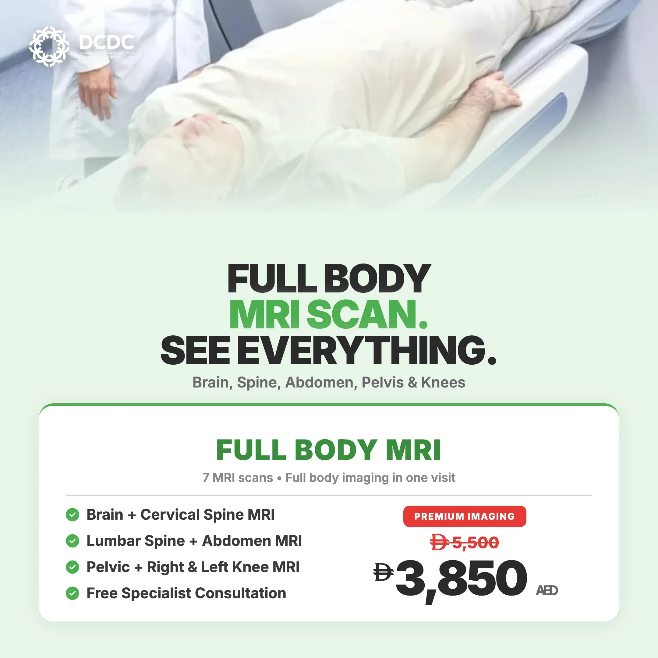
MRI pricing in Dubai varies by body region, whether contrast is needed and the facility. At DCDC, a single-region MRI starts from AED 900, which includes the subspecialty radiologist interpretation. The market range for MRI in Dubai is broadly AED 1,200 to AED 5,000 per region, depending on the hospital or imaging centre. Full-body MRI at DCDC is AED 3,850 (regularly AED 5,500). Most major insurance plans cover MRI when it is medically indicated with a physician referral, and DCDC offers direct billing with 20 or more insurance partners including Daman, AXA, Bupa and MetLife.
| MRI Type | DCDC Price | Dubai Market Range |
|---|---|---|
| Single-region MRI (brain, spine, knee, etc.) | From AED 900 | AED 1,200–5,000 |
| Full-body MRI | AED 3,850 | AED 5,000–15,000 |
| MRI with contrast (additional) | Additional fee for gadolinium | Varies by provider |
MRI pricing comparison — DCDC vs Dubai market (2026)
For a detailed breakdown of MRI costs by body region, see our dedicated pricing guides for brain MRI, spine MRI and knee MRI.
Related Services at DCDC
Expert care and advanced diagnostics at Dubai Healthcare City
Frequently Asked Questions
Final Thoughts
An MRI report is a clinical document written for doctors, and it is completely normal to feel confused or anxious when reading one for the first time. The most important things to remember: the impression section summarises what matters, many findings that sound alarming are normal age-related changes, and the clinical significance of any finding depends on whether it correlates with your symptoms. Never make treatment decisions based on an MRI report alone.
If you have had an MRI and need help understanding the results, or if you need an MRI with a subspecialty radiologist read, Doctors Clinic Diagnostic Center in Dubai Healthcare City is open Saturday to Thursday until 10 PM and Friday until 9 PM, with free parking and direct insurance billing. Call +971 54 219 5008 or WhatsApp +971 50 970 3884 to book.
Sources & References
This article references the following sources:
- Brinjikji W, et al. — Systematic Literature Review of Imaging Features of Spinal Degeneration in Asymptomatic Populations, AJNR 2015
- Morris Z, et al. — Incidental Findings on Brain Magnetic Resonance Imaging, N Engl J Med 2009
- RadiologyInfo.org — How to Read Your Radiology Report (RSNA/ACR)
- American College of Radiology — Practice Parameters for Communication of Diagnostic Imaging Findings
- Cleveland Clinic — MRI: What It Is, What It Diagnoses, Uses & Safety
This information is educational and does not replace individual medical advice. See our editorial policy for more information.

Written by
DCDC Editorial Team
Healthcare Content
Medical Editorial Board
The DCDC Editorial Team produces evidence-based health content reviewed for clinical accuracy. Our team works with specialist physicians at Doctors Clinic Diagnostic Center in Dubai Healthcare City.
Related Articles
Complete Guide to MRI Scan in Dubai
Brain MRI in Dubai: What It Detects & Cost
Spine MRI in Dubai: Cost & What It Shows
Knee MRI in Dubai: What It Shows & Cost
MRI with Contrast: What to Expect & Safety
More in Diagnostic Imaging

Chest X-Ray Cost Dubai: From AED 150 (2026)
Read More
CT Scan Cost Dubai: Prices & Types (2026)
Read More
Ultrasound Cost Dubai: AED 300-800 by Type [2026 Prices]
Read More
CT Scan vs Ultrasound Dubai: Which Test? (2026)
Read More
Open vs Closed MRI Dubai: Key Differences (2026)
Read More
X-Ray Cost Dubai: Prices by Body Part (2026)
Read More© 2026 Doctors Clinic Diagnostic Center (DCDC), Dubai Healthcare City. Originally published at https://doctorsclinicdubai.ae/blog/mri-report-results-explained-dubai. All rights reserved. Unauthorized reproduction is prohibited.