Key Takeaways
- CT uses X-rays for fast, detailed cross-sectional imaging (10-30 minutes); ultrasound uses sound waves for real-time radiation-free imaging (15-45 minutes)
- CT excels for lungs, bones, emergency trauma, kidney stones, and internal bleeding; ultrasound is preferred for pregnancy, thyroid, abdominal organs, and vascular flow
- Ultrasound is completely radiation-free and safe for repeated use, pregnant women, and children; DCDC uses low-dose 3D CT technology that reduces radiation by up to 50%
- DCDC pricing: CT scans from AED 449 and ultrasound from AED 399, with direct billing through 20+ insurance partners
- Some conditions benefit from both tests used together, such as appendicitis evaluation or liver mass characterization
- Every scan at DCDC is interpreted by a subspecialty radiologist, with results delivered within 18-24 hours and same-day for urgent cases
When your doctor suspects an internal problem, the choice between a CT scan and an ultrasound depends on what body part needs to be examined, whether radiation exposure is a concern, and how quickly your doctor needs a diagnosis. Both are essential diagnostic tools used daily across Dubai, but they rely on completely different technology and each has distinct clinical strengths. Understanding why your doctor chose one over the other helps you know what to expect and why one test may be more appropriate for your symptoms than the other.
This comprehensive comparison explains the technology behind CT and ultrasound, compares their performance across every major body region, covers cost and radiation differences, and provides a practical decision framework so you understand which test is right for your situation. Whether you have been referred for imaging or simply want to learn about your options before a consultation, this guide covers everything patients in Dubai need to know about CT versus ultrasound in 2026.


CT Scan vs Ultrasound: Quick Comparison Table
Before exploring each technology in detail, here is a side-by-side overview of the most important differences between CT and ultrasound imaging. This table covers the factors that matter most to patients: speed, radiation, comfort, cost, and diagnostic strengths.
| Feature | CT Scan | Ultrasound |
|---|---|---|
| Technology | X-ray beams rotating around the body | High-frequency sound waves via handheld probe |
| Radiation | Yes (ionizing radiation; DCDC uses low-dose 3D with up to 50% reduction) | None (completely radiation-free) |
| Scan duration | 10-30 minutes | 15-45 minutes |
| Image type | Detailed cross-sectional slices and 3D reconstructions | Real-time moving images |
| Scanner design | Large ring-shaped gantry; patient lies on a motorised table | Handheld probe moved over skin with gel |
| Patient comfort | Quick and painless; must lie still briefly | Comfortable; no enclosed space; some pressure from probe |
| Contrast agent | Iodine-based IV or oral contrast (when needed) | None required for most studies |
| Bone and lung imaging | Excellent | Limited (sound waves cannot penetrate bone or air) |
| Soft tissue / organ detail | Good, especially with contrast | Good to excellent for superficial and abdominal organs |
| Blood flow assessment | CT angiography (with contrast) | Doppler ultrasound (no contrast needed) |
| Pregnancy safety | Avoided unless clinically essential | Standard imaging throughout pregnancy |
| Pediatric use | Used selectively with dose reduction protocols | Preferred first-line test for children |
| DCDC pricing | From AED 449 | From AED 399 |
| Results at DCDC | 18-24 hours (same-day for urgent) | 18-24 hours (same-day for urgent) |
CT and ultrasound are complementary tools. The best test depends on the clinical question, the body part, and patient-specific factors such as pregnancy or age.
How a CT Scan Works
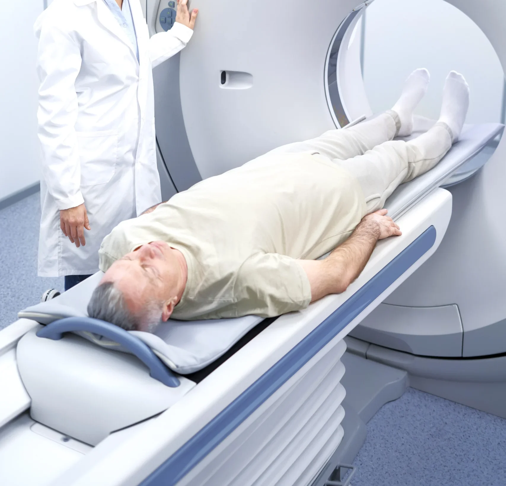
CT (Computed Tomography) creates detailed images by rotating an X-ray tube and detector array around your body. As the X-ray beam passes through different tissues, dense structures such as bone absorb more radiation and appear bright white on the image, while air-filled structures like the lungs absorb very little and appear black. A powerful computer processes thousands of these measurements to produce thin cross-sectional slices that can be stacked into 3D reconstructions.
Modern multi-slice CT scanners, including the low-dose 3D system used at DCDC, acquire images in seconds while you lie on a motorised table that slides through the scanner ring. The entire process typically takes 10 to 30 minutes depending on whether contrast dye is required. If you are preparing for a CT scan, our CT scan preparation guide explains exactly what to do before your appointment.
CT excels at imaging bones, lungs, and dense internal structures. It is the gold standard for emergency trauma evaluation, kidney stone detection, and lung disease assessment. With intravenous contrast, CT provides excellent detail of blood vessels (CT angiography), abdominal organs, and tumours.
How an Ultrasound Works
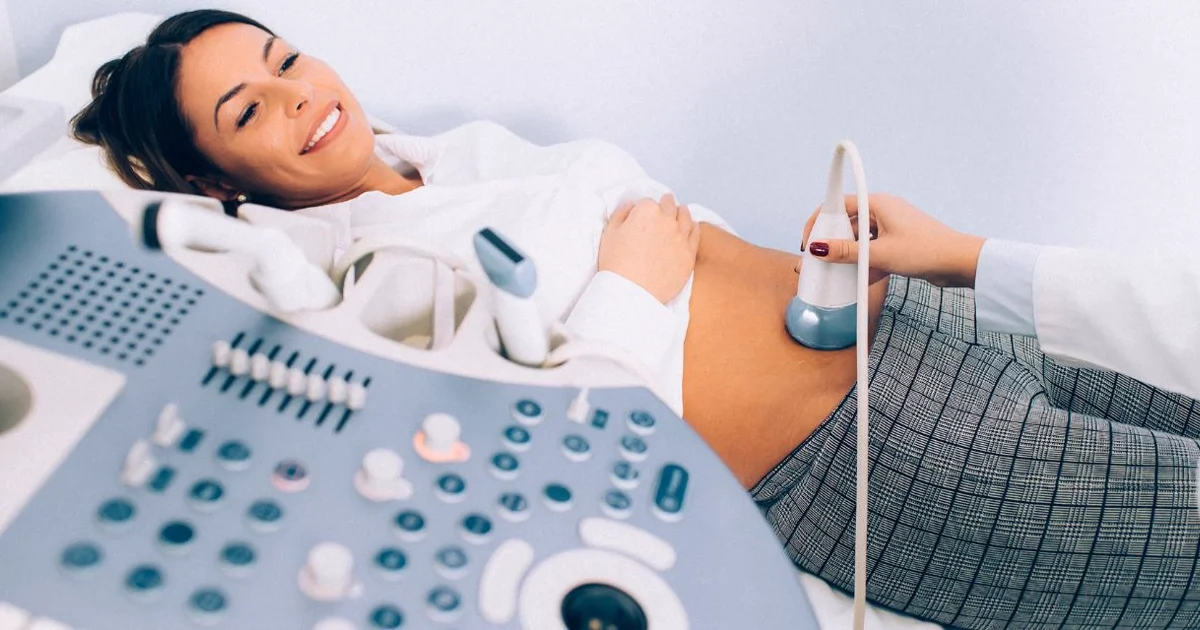
Ultrasound imaging (also called sonography) works by sending high-frequency sound waves into the body through a handheld probe (transducer) placed on the skin. When these sound waves encounter boundaries between different tissues, such as the interface between a solid organ and a fluid-filled structure, they bounce back as echoes. The ultrasound machine converts these returning echoes into a real-time moving image displayed on a screen.
A water-based gel is applied to the skin to eliminate air between the probe and the body, ensuring clear transmission of sound waves. The sonographer or radiologist moves the probe across the area of interest, adjusting angle and pressure to visualise different structures. Unlike CT, ultrasound produces images in real time, allowing the examiner to assess organ movement, blood flow, and fetal activity as it happens.
Ultrasound uses no ionizing radiation whatsoever, making it the safest imaging modality for repeated use, for pregnant women at every stage of pregnancy, and for children. It is the first-choice test for abdominal organs, thyroid, pregnancy monitoring, breast evaluation, and vascular assessment. For guidance on preparing for your scan, see our ultrasound preparation guide.
When Your Doctor Orders a CT Scan
CT is typically ordered when your doctor needs fast, highly detailed images of internal structures that ultrasound cannot adequately visualise. The following clinical scenarios are among the most common reasons for a CT scan referral in Dubai.
- Emergency trauma: After a car accident, fall, or significant impact, CT rapidly detects fractures, internal bleeding, and organ injury across the entire body
- Acute abdominal pain: CT is the standard for diagnosing appendicitis, diverticulitis, bowel obstruction, and abdominal abscesses when the cause is unclear. Our abdominal CT scan guide explains what these scans reveal
- Kidney stones: Non-contrast CT is the gold standard for detecting and measuring urinary tract stones, with near-100% sensitivity
- Lung conditions: CT is definitively superior for evaluating pneumonia complications, lung nodules, pulmonary embolism (blood clot in the lungs), and interstitial lung disease
- Cancer staging: CT of the chest, abdomen, and pelvis is routinely used to stage cancers and monitor treatment response
- Head injuries and stroke: CT is the first-line imaging for acute head trauma and suspected stroke because it rapidly detects bleeding and large masses
- Bone and spine problems: Complex fractures, spinal stenosis, and surgical planning often require CT for its superior bone detail
- Vascular assessment: CT angiography maps blood vessels throughout the body, detecting aneurysms, stenosis, and vascular malformations
When Your Doctor Orders an Ultrasound
Ultrasound is ordered when your doctor needs real-time imaging of soft tissue structures, particularly in the abdomen, pelvis, thyroid, and vascular system. Its lack of radiation makes it the default first-line imaging test for many conditions.
- Pregnancy monitoring: Ultrasound is the standard for all stages of pregnancy, from confirming early pregnancy and dating to anomaly scans, growth monitoring, and assessing fetal wellbeing
- Abdominal organ evaluation: Gallstones, liver disease, kidney cysts, spleen enlargement, and pancreatic conditions are routinely assessed with ultrasound. See our abdominal ultrasound complete guide for details
- Thyroid nodules: Ultrasound is the primary test for evaluating thyroid lumps, guiding biopsies, and monitoring known nodules
- Breast evaluation: In conjunction with mammography, ultrasound helps characterise breast lumps and is particularly useful in women with dense breast tissue
- Pelvic conditions: Ovarian cysts, uterine fibroids, endometriosis evaluation, and fertility assessments rely heavily on pelvic ultrasound
- Blood vessel assessment: Doppler ultrasound evaluates blood flow in the carotid arteries, leg veins (for deep vein thrombosis), and other vessels without contrast injection
- Musculoskeletal injuries: Superficial tendons, rotator cuff tears, and muscle injuries can be assessed in real time with dynamic ultrasound
- Pediatric imaging: Because there is zero radiation, ultrasound is the preferred first test for children with abdominal pain, hip problems, or pyloric stenosis
CT vs Ultrasound by Body Region
The clearest way to understand when CT or ultrasound is the better choice is to compare them region by region. Each body area has a preferred modality, though some conditions benefit from both.
Head and Brain
Winner: CT. The skull blocks sound waves, making ultrasound ineffective for brain imaging in adults. CT is the first-line test for head trauma, acute stroke, and bleeding inside the skull. For more detailed brain evaluation (tumours, multiple sclerosis, seizure investigation), MRI is the next step. The one exception is neonatal brain imaging, where ultrasound through the fontanelle (soft spot) in newborns is both effective and radiation-free.
Chest and Lungs
Winner: CT. Air-filled lungs produce poor ultrasound images because sound waves scatter in air. CT provides unmatched detail for lung nodules, pneumonia complications, pulmonary embolism, and interstitial lung disease. Ultrasound has a limited but valuable role in the chest: it can detect pleural effusions (fluid around the lungs) and guide drainage procedures, and it is used at the bedside in emergency settings to check for pneumothorax.
Abdomen
Depends on the condition. Ultrasound is the first-line test for gallstones, liver assessment, kidney evaluation, and abdominal aortic aneurysm screening. It is also used for appendicitis evaluation in children and thin adults. CT is preferred for acute abdominal emergencies, complex bowel conditions, and when ultrasound findings are inconclusive. For suspected appendicitis in adults, CT has higher sensitivity (approximately 98%) compared to ultrasound (approximately 86%), though ultrasound is attempted first in many protocols to avoid radiation.
Pelvis
Ultrasound first, CT for specific scenarios. Pelvic ultrasound (both transabdominal and transvaginal) is the primary tool for evaluating the uterus, ovaries, and prostate. It detects cysts, fibroids, ectopic pregnancy, and ovarian torsion. CT is used for pelvic trauma assessment, cancer staging, and when ultrasound cannot provide enough detail about lymph nodes or deep pelvic structures.
Thyroid and Neck
Winner: Ultrasound. The thyroid gland sits close to the surface, making it ideal for ultrasound evaluation. Ultrasound detects nodules as small as 2-3 mm, characterises their features, and guides fine-needle aspiration biopsies. CT of the neck is reserved for evaluating deep structures, lymph node staging, or assessing the extent of large thyroid masses.
Musculoskeletal (Joints, Tendons, Bones)
Both have roles. Ultrasound is excellent for superficial soft tissue injuries: rotator cuff tears, Achilles tendon problems, joint effusions, and guided injections. CT is superior for bone detail, complex fractures, and surgical planning. For deep joint structures like the meniscus or cruciate ligaments, MRI typically outperforms both CT and ultrasound.
Blood Vessels
Both are valuable. Doppler ultrasound is the first-line test for carotid artery disease, deep vein thrombosis (DVT), varicose veins, and peripheral arterial assessment, with no radiation or contrast required. CT angiography is used when a more comprehensive vascular map is needed, such as planning for aneurysm repair, evaluating pulmonary embolism, or mapping coronary arteries.
Radiation and Safety Considerations
The most significant difference between CT and ultrasound from a safety perspective is radiation exposure. This matters most for children, pregnant women, and patients who require repeated imaging over time.
Ultrasound: Zero Radiation
Ultrasound uses mechanical sound waves, not electromagnetic radiation. It has no known harmful effects at diagnostic intensities and has been used safely in pregnancy imaging for over 50 years. There is no cumulative dose concern, which means ultrasound can be repeated as often as clinically needed without any radiation risk.
CT: Managed Radiation Exposure
CT uses ionizing radiation (X-rays) to create images. The radiation dose varies by body part and protocol: a head CT delivers approximately 2 mSv (roughly equivalent to 8 months of natural background radiation), while an abdominal CT with contrast may deliver 10-20 mSv. At DCDC, our low-dose 3D CT scanner reduces radiation exposure by up to 50% compared to conventional scanners while maintaining diagnostic image quality.
For the vast majority of patients, a single or occasional CT scan carries negligible risk and the diagnostic benefit far outweighs any theoretical concern. However, radiation exposure is cumulative over a lifetime, which is why doctors apply the ALARA principle (As Low As Reasonably Achievable). This means choosing ultrasound or MRI when they can answer the clinical question, and reserving CT for situations where it provides essential diagnostic information that other modalities cannot.
Who Should Avoid CT When Possible?
- Pregnant women: CT is avoided unless clinically essential because ionizing radiation poses a theoretical risk to the developing fetus. Ultrasound and MRI (without gadolinium) are safer alternatives
- Children: Children are more radiosensitive than adults. Ultrasound is preferred as the initial imaging test whenever it can answer the clinical question
- Patients needing frequent follow-up imaging: If serial monitoring is required (for example, ovarian cyst surveillance), ultrasound avoids cumulative radiation exposure
- Patients with contrast allergies: Some CT studies require iodine-based contrast, which can cause allergic reactions. Ultrasound requires no contrast for most applications
CT vs Ultrasound Cost Comparison in Dubai
Cost is a practical consideration for many patients in Dubai, particularly those paying out of pocket. Below is a comparison of typical CT and ultrasound pricing. For more detailed breakdowns, see our CT scan cost guide and ultrasound cost guide.
| Body Region | CT Cost at DCDC (AED) | Ultrasound Cost at DCDC (AED) | Preferred First Test |
|---|---|---|---|
| Brain / Head | From 449 | Not applicable (adults) | CT |
| Chest / Lungs | From 449 | Limited role (pleural effusion) | CT |
| Abdomen (general) | From 449 | From 399 | Ultrasound first, CT if inconclusive |
| Pelvis | From 449 | From 399 | Ultrasound |
| Thyroid / Neck | From 449 | From 399 | Ultrasound |
| Kidneys and urinary tract | From 449 | From 399 | Ultrasound for screening; CT for stones |
| Vascular (Doppler / Angiography) | From 899 (CTA) | From 399 (Doppler) | Doppler first; CTA for complex cases |
| Pregnancy | Avoided | From 399 | Ultrasound only |
Prices are starting rates at DCDC in Dubai Healthcare City (2026). Final cost depends on the specific study, contrast requirement, and insurance coverage. DCDC offers direct billing with 20+ insurance partners.
Most insurance plans in Dubai cover both CT and ultrasound when medically indicated with a doctor's referral. DCDC works with over 20 insurance partners including Daman, AXA, Bupa, MetLife, and Cigna, offering direct billing so you pay only your co-pay or deductible at the time of your visit.
Not Sure Which Imaging Test You Need?
Book a consultation at DCDC and our subspecialty radiologists will recommend the most appropriate scan for your symptoms. CT from AED 449, ultrasound from AED 399.
Direct insurance billing with 20+ partners
Which Test Is Right for You?
Choosing between CT and ultrasound is ultimately a clinical decision made by your doctor based on your symptoms, medical history, and the specific diagnostic question. However, here is a practical framework to help you understand the logic behind imaging choices.
Choose CT When You Need
- Fast, definitive answers in an emergency (trauma, acute stroke, severe abdominal pain)
- Detailed images of the lungs, chest, or bones
- Kidney stone detection or urinary tract evaluation
- Cancer staging or treatment monitoring across chest, abdomen, and pelvis
- Vascular mapping (aneurysms, stenosis, pulmonary embolism)
- Surgical planning for complex fractures or pre-operative anatomy
Choose Ultrasound When You Need
- Pregnancy monitoring at any stage
- Thyroid nodule evaluation or guided biopsy
- Gallstone, liver, or kidney assessment as a first step
- Pelvic organ evaluation (uterus, ovaries, prostate)
- Blood flow assessment without contrast (Doppler)
- Pediatric imaging where avoiding radiation is a priority
- Repeated monitoring of a known condition (cysts, nodules) to avoid cumulative radiation
Dr. Osama Elzamzami, Consultant Radiologist at DCDC, notes: "The question is never simply which test is better in the abstract. It is which test best answers the clinical question at hand. A patient with right lower abdominal pain may start with ultrasound, and if the appendix cannot be clearly visualised, we proceed to CT for a definitive answer. Conversely, a patient being monitored for thyroid nodules needs serial ultrasound, not CT, because we want high-resolution imaging without any radiation. The goal is always the right test for the right patient at the right time."
What to Expect at DCDC
Whether you are coming for a CT scan or an ultrasound, the patient experience at Doctors Clinic Diagnostic Center in Dubai Healthcare City follows a streamlined process designed for efficiency and comfort.
CT Scan Experience at DCDC
- Check-in and preparation: Arrive 15 minutes early. Our team verifies your referral, insurance, and any preparation requirements (fasting or contrast allergy history). You change into a gown and remove metal objects
- The scan itself: You lie on the CT table, which slides through the scanner ring. Our low-dose 3D CT system captures images in seconds. The technologist monitors you throughout from the adjacent control room and communicates via intercom. Total time in the scanner room is 10-30 minutes
- Contrast studies: If contrast is needed, a small IV line is placed. You may feel a brief warm flush when the contrast is injected. Our team monitors you for any reactions before you leave
- After the scan: You can resume normal activities immediately. If you had contrast, we recommend drinking plenty of water to help your kidneys clear it. Results are reviewed by a subspecialty radiologist and delivered within 18-24 hours
Ultrasound Experience at DCDC
- Check-in and preparation: Preparation depends on the type of ultrasound. Abdominal scans typically require fasting for 6-8 hours. Pelvic scans may require a full bladder. Our booking team provides specific instructions when you schedule
- The scan itself: You lie on a comfortable examination table. The sonographer applies warm gel to the area and moves the transducer probe across your skin. You may be asked to hold your breath briefly or change position. The exam takes 15-45 minutes
- Real-time assessment: The sonographer captures still images and video clips of relevant structures. In some cases, the radiologist performs or supervises the examination directly, providing immediate clinical correlation
- After the scan: The gel is wiped off and you can leave immediately with no restrictions. Results are reported by a subspecialty radiologist within 18-24 hours
DCDC is located in Building 64, Block A, Al Razi Medical Complex in Dubai Healthcare City, with free dedicated parking and an average wait time of just 15 minutes. The clinic is open Saturday through Thursday from 8 AM to 10 PM and Friday from 9 AM to 9 PM, MOHAP-licensed, and rated 4.8 out of 5 from over 1,000 verified patient reviews.
Can You Need Both a CT Scan and an Ultrasound?
Yes. In many clinical scenarios, CT and ultrasound are used together as complementary investigations rather than competing alternatives. Each test provides different information, and combining them gives your doctor a more complete diagnostic picture.
- Appendicitis evaluation: In adults, ultrasound may be performed first (especially in women to rule out gynaecological causes of pain). If ultrasound is inconclusive, CT follows to confirm or exclude appendicitis with near-certainty
- Liver mass characterisation: An ultrasound may detect a liver lesion during a routine scan. CT with contrast is often ordered next to characterise the lesion further, determine its vascularity, and assess for additional lesions
- Kidney evaluation: Ultrasound is excellent for detecting kidney cysts, hydronephrosis, and structural abnormalities. If kidney stones are suspected based on symptoms, non-contrast CT provides definitive stone detection, sizing, and localisation
- Cancer staging and follow-up: A patient diagnosed with a pelvic tumour via ultrasound may undergo CT of the chest, abdomen, and pelvis for staging. Subsequent follow-up may alternate between ultrasound and CT depending on what structures need monitoring
- Trauma assessment: In emergency departments, bedside ultrasound (FAST scan) rapidly screens for free fluid in the abdomen. If positive or if the mechanism of injury warrants it, a full CT trauma series provides comprehensive evaluation of all organs and bones
If your doctor orders both tests, it is not redundant. Each answers a different part of the diagnostic question, and together they reduce the likelihood of missed findings. For a broader comparison of imaging options including MRI, see our CT vs MRI comparison guide.
Key Advantages and Limitations at a Glance
CT Scan Advantages
- Fast acquisition speed ideal for emergencies and uncooperative patients
- Superior detail for bones, lungs, and complex anatomical structures
- Excellent with contrast for vascular imaging and tumour characterisation
- Less operator-dependent than ultrasound (consistent image quality regardless of body habitus)
- 3D reconstructions available for surgical planning
CT Scan Limitations
- Uses ionizing radiation (though modern low-dose protocols significantly reduce exposure)
- Iodine-based contrast can cause allergic reactions or affect kidney function in susceptible patients
- Less effective than ultrasound for real-time dynamic assessment
- Should be avoided in pregnancy unless clinically essential
Ultrasound Advantages
- Zero radiation, safe for unlimited repeat use
- Safe throughout pregnancy and for children of all ages
- Real-time imaging allows dynamic assessment of movement, blood flow, and organ function
- No contrast needed for most studies
- Portable, can be performed at the bedside
- Excellent for guided procedures (biopsies, aspirations, injections)
Ultrasound Limitations
- Cannot penetrate bone or air-filled structures (brain, lungs, bowel gas)
- Image quality is operator-dependent (requires skilled sonographer)
- Limited by patient body habitus: obesity and thick body wall reduce image quality
- Smaller field of view than CT; cannot provide whole-body imaging in a single study
Why DCDC for Diagnostic Imaging in Dubai
At Doctors Clinic Diagnostic Center, every CT scan and ultrasound is interpreted by a subspecialty radiologist matched to the clinical area. A brain CT is read by a neuroradiologist, an abdominal ultrasound by a body imaging specialist, and a musculoskeletal scan by an MSK radiologist. This level of subspecialisation, combined with advanced equipment including our low-dose 3D CT scanner, ensures accurate diagnosis and reduces the need for repeat testing.
With a 98% patient satisfaction rate, 4.8/5 Google rating from over 1,000 reviews, and results delivered within 18-24 hours (same-day for urgent cases), DCDC provides the imaging experience that patients and referring physicians across Dubai trust. Our direct billing with over 20 insurance partners makes the process straightforward, and our extended hours (Saturday through Thursday 8 AM to 10 PM, Friday 9 AM to 9 PM) accommodate busy schedules.
Book Your CT Scan or Ultrasound at DCDC
Subspecialty radiologist interpretation, low-dose 3D CT technology, and results within 18-24 hours. CT from AED 449, ultrasound from AED 399.
Building 64, Block A, Al Razi Medical Complex, Dubai Healthcare City
Related Services at DCDC
Expert care and advanced diagnostics at Dubai Healthcare City
Frequently Asked Questions
Final Thoughts
CT and ultrasound are not competing technologies but complementary tools that serve different diagnostic purposes. CT delivers fast, high-resolution images of bones, lungs, and complex internal structures using X-ray technology, while ultrasound provides real-time, radiation-free imaging of soft tissue organs, blood vessels, and pregnancies. The right test is always the one that answers your doctor's clinical question most accurately and safely.
Cost, radiation exposure, and patient comfort are all valid considerations, but they should inform the conversation with your doctor rather than drive the decision independently. An ultrasound that cannot answer the clinical question saves money but delays diagnosis. A CT scan ordered when ultrasound would suffice adds unnecessary radiation. The goal is matching the test to the clinical need.
At Doctors Clinic Diagnostic Center in Dubai Healthcare City, our subspecialty radiologists ensure expert interpretation of every CT and ultrasound scan. With competitive pricing (CT from AED 449, ultrasound from AED 399), low-dose 3D CT technology, direct insurance billing, and results within 18-24 hours, DCDC provides a complete diagnostic imaging experience designed around patient accuracy and convenience.
Sources & References
This article was reviewed by our medical team and references the following sources:
- RadiologyInfo.org (RSNA) - CT and Ultrasound Patient Information
- Mayo Clinic - CT Scan and Ultrasound Overview
- NHS - CT Scan and Ultrasound Scan Information
- American College of Radiology - Appropriateness Criteria
- Cleveland Clinic - CT Scan vs Ultrasound: Differences and Uses
Medical content on this site is reviewed by DHA-licensed physicians. See our editorial policy for more information.

Written by
Dr. Osama Elzamzami
Diagnostic Radiologist
MD, FRCR
Dr. Osama Elzamzami is a Diagnostic Radiologist at Doctors Clinic Diagnostic Center (DCDC) in Dubai Healthcare City.
Related Articles
CT Scan vs MRI: Which Do You Need? Complete Comparison (2026)
MRI vs Ultrasound Dubai: Which Scan? (2026)
CT Scan Cost Dubai: Complete Pricing Guide (2026)
Ultrasound Cost Dubai: Pricing by Type (2026)
Abdominal CT Scan: What It Shows & When You Need One
More in Diagnostic Imaging
Open vs Closed MRI Dubai: Key Differences (2026)
Read More
X-Ray Cost Dubai: Prices by Body Part (2026)
Read More
DEXA Scan Cost Dubai: From AED 399 (2026)
Read MoreCT Scan Cost Dubai: Prices & Types (2026)
Read More
DEXA Scan Dubai: Cost, T-Score & Who Needs It (2026)
Read More![Full Body MRI Cost Dubai: AED 5,000-15,000 [2026]](/wp-media/blog/full-body-mri-scan.webp)
Full Body MRI Cost Dubai: AED 5,000-15,000 [2026]
Read More© 2026 Doctors Clinic Diagnostic Center (DCDC), Dubai Healthcare City. Originally published at https://doctorsclinicdubai.ae/blog/ct-scan-vs-ultrasound-dubai. All rights reserved. Unauthorized reproduction is prohibited.