Key Takeaways
- Mammograms are the gold-standard screening tool for breast cancer in women aged 40 and over, detecting calcifications and masses before they become palpable
- Breast ultrasound is radiation-free and excels at evaluating palpable lumps, distinguishing cysts from solid masses, and imaging dense breast tissue
- Women with dense breasts (BI-RADS density C or D) benefit from supplemental breast ultrasound in addition to mammography
- The two tests are complementary, not competing -- many women need both for a complete breast evaluation
- Digital mammography at DCDC starts from AED 250, with breast density assessed and reported on every study
- A subspecialty radiologist interprets every breast imaging study at DCDC, improving detection of subtle findings that generalist readers might miss
If your doctor has recommended breast imaging, you may be wondering whether a mammogram or a breast ultrasound is the right choice. In many cases, the answer depends on your age, breast density, symptoms, and risk profile. Both are essential tools in breast cancer detection, but they use different technologies and answer different clinical questions. Understanding how each test works -- and when one is preferred over the other -- helps you make informed decisions about your breast health.
This comprehensive comparison covers the key differences between mammograms and breast ultrasound, explains which test is recommended at each age and risk level, compares costs in Dubai, and describes what to expect when you come to DCDC for breast imaging. Whether you are due for routine screening or evaluating a specific concern, this guide will help you understand your options.
Health Screening Packages
Save with our bundled screening packages — specialist consultation included


Mammogram vs Breast Ultrasound: Side-by-Side Comparison
Before diving into the details, here is a quick overview of how mammograms and breast ultrasounds compare across the factors that matter most to patients. This comparison table summarizes the key differences at a glance.
| Feature | Mammogram | Breast Ultrasound |
|---|---|---|
| Technology | Low-dose X-ray (ionizing radiation) | High-frequency sound waves (no radiation) |
| Primary role | Screening and early detection | Diagnostic evaluation and dense tissue imaging |
| Detects calcifications | Yes -- the only modality that reliably detects microcalcifications | No -- cannot detect microcalcifications |
| Cyst vs solid mass | Cannot reliably distinguish | Excellent -- the gold standard for differentiating cysts from solid masses |
| Dense breast tissue | Reduced sensitivity (cancer can be hidden) | Performs well -- no masking effect from dense tissue |
| Radiation exposure | Very low dose (0.4 mSv per study) | None |
| Scan duration | 10-15 minutes | 15-30 minutes |
| Compression required | Yes -- brief breast compression for clear images | No -- gentle probe contact only |
| Age recommendation | Routine screening from age 40 (some guidelines say 50) | Any age, including under 30 |
| Best for | Screening asymptomatic women, detecting early-stage cancers and calcifications | Evaluating lumps, cysts, dense tissue, guiding biopsies, younger women |
| Limitations | Lower sensitivity in dense breasts; uses radiation | Cannot detect microcalcifications; operator-dependent; not a standalone screening tool |
| Cost at DCDC (AED) | From AED 250 | From AED 300 |
Mammograms and breast ultrasounds are complementary tests. Most breast imaging protocols use mammography as the primary screening tool and add ultrasound when clinically indicated.
What Is a Mammogram?
A mammogram is a specialized X-ray of the breast that produces detailed images of the internal breast tissue. During the procedure, each breast is placed on a flat support plate and gently compressed with a clear paddle. This compression spreads the tissue evenly, reduces motion blur, and lowers the radiation dose needed to create a clear image. The entire process takes about 10 to 15 minutes and typically involves four images -- two views of each breast.
There are two types of mammography. 2D digital mammography captures flat images of the breast from two angles. 3D mammography (tomosynthesis) takes multiple thin-slice images and reconstructs them into a three-dimensional view, improving detection rates especially in women with dense tissue. Both types are available at DCDC in Dubai Healthcare City.
Mammograms are uniquely capable of detecting microcalcifications -- tiny calcium deposits that can be an early sign of ductal carcinoma in situ (DCIS) or invasive cancer. No other breast imaging modality reliably identifies these calcifications, which is why mammography remains the cornerstone of breast cancer screening worldwide.
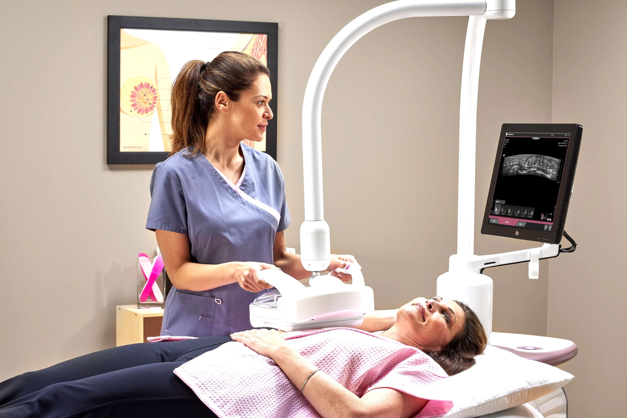
What Is a Breast Ultrasound?
A breast ultrasound uses high-frequency sound waves to create real-time images of the breast tissue. A radiologist or sonographer applies gel to the breast and moves a handheld transducer across the skin. The sound waves bounce off internal structures and return to the transducer, which converts them into images displayed on a monitor. The procedure is painless, involves no radiation, and takes 15 to 30 minutes depending on the area being examined.
Breast ultrasound excels at distinguishing between fluid-filled cysts and solid masses. When a mammogram or physical exam detects a lump, ultrasound is typically the next step to characterize it. Simple cysts (fluid-filled sacs) appear as well-defined dark areas on ultrasound and are almost always benign. Solid masses require further evaluation and may need a biopsy, which can also be guided by ultrasound in real time. For a complete guide to this test, see our article on breast ultrasound in Dubai.
Unlike mammography, ultrasound does not use ionizing radiation, making it safe for pregnant women, younger patients, and those who require frequent follow-up imaging. However, it cannot reliably detect microcalcifications and is not recommended as a standalone screening tool for breast cancer.
Key Differences: Accuracy and Detection
The accuracy of each test depends on what is being looked for and the patient's breast composition. Understanding these differences is essential for choosing the right approach. For a broader overview of screening recommendations, refer to our breast cancer screening recommendations guide.
Cancer Detection Rates
Mammography has a sensitivity of approximately 80-87% for breast cancer detection in the general population, according to the American Cancer Society. However, this drops to 48-64% in women with extremely dense breast tissue (BI-RADS density D). Breast ultrasound has lower overall sensitivity for cancer screening (approximately 50-80%), but it detects an additional 2-4 cancers per 1,000 women when used as a supplement to mammography in dense-breast populations. When both tests are combined, overall detection rates improve significantly.
False Positive Rates
Both mammograms and breast ultrasounds can produce false positive results -- findings that look suspicious but turn out to be benign after further evaluation. Mammograms have a recall rate of about 10% on a first screening (lower on subsequent screenings). Supplemental breast ultrasound tends to increase the false positive rate, which is why it is recommended as an adjunct to mammography rather than a replacement. The trade-off is that more cancers are found, but more benign findings also require follow-up.
Microcalcifications vs Soft Tissue Masses
This is the most important distinction. Mammography is the only imaging modality that reliably detects microcalcifications, which can represent DCIS (the earliest form of breast cancer). Ultrasound cannot see these tiny calcium deposits. Conversely, ultrasound provides superior characterization of soft tissue masses, determining whether a lump is a simple cyst, a complex cyst, or a solid mass requiring biopsy. The two tests are therefore complementary, not interchangeable.
When a Mammogram Is the Better Choice
- Routine breast cancer screening: For women aged 40 and over with no symptoms, mammography is the primary screening tool recommended by the American Cancer Society, WHO, and the UAE Ministry of Health (MOHAP)
- Detecting microcalcifications: Only mammography can identify the tiny calcium deposits that may indicate DCIS or early invasive cancer
- Baseline breast imaging: A first mammogram (baseline) establishes a reference for comparison on future screenings, making subtle changes easier to detect over time
- Post-menopausal women: Breast tissue becomes less dense after menopause, improving mammographic sensitivity and making it even more effective as a screening tool
- Follow-up of previously detected calcifications: Mammography tracks changes in known calcification clusters that may indicate progression
- Insurance-covered annual screening: Most insurance plans in Dubai cover annual mammography for women over 40. For pricing details, see our mammogram cost guide
When Breast Ultrasound Is the Better Choice
- Evaluating a palpable lump: When you or your doctor can feel a breast lump, ultrasound is the best first-line test to determine whether it is a cyst or a solid mass
- Dense breast tissue supplementation: Women with heterogeneously dense or extremely dense breasts (BI-RADS C or D) benefit from supplemental ultrasound because mammography alone may miss cancers hidden by dense tissue
- Women under 30: Younger women typically have very dense breast tissue, which limits mammographic accuracy. Ultrasound is the preferred initial imaging test for breast concerns in this age group
- Pregnancy and breastfeeding: Ultrasound uses no radiation and is safe during pregnancy and lactation, making it the imaging modality of choice for pregnant or nursing women with breast symptoms
- Guiding needle biopsies: When a biopsy is needed, ultrasound provides real-time visualization to guide the needle precisely into the target area
- Monitoring known cysts: Ultrasound tracks changes in previously identified cysts without radiation exposure
- Evaluating breast implants: Ultrasound can assess the integrity of breast implants and detect implant-related complications
Can You Get Both a Mammogram and Breast Ultrasound?
Yes, and in many clinical scenarios, getting both tests provides the most comprehensive breast evaluation. In fact, combined mammography and ultrasound is increasingly considered the standard of care for women with dense breast tissue. Here are the most common situations where both tests are ordered together:
- Dense breast supplemental screening: After a mammogram shows dense tissue (BI-RADS C or D), ultrasound is added to search for cancers that may be hidden by the density
- Abnormal mammogram finding: When a mammogram detects a mass or area of concern, ultrasound is used to further characterize the finding and guide management
- Palpable lump with screening due: If a patient presents with a lump and is also due for screening, both a diagnostic mammogram and targeted ultrasound are performed in the same visit
- High-risk patients: Women with a strong family history of breast cancer or known BRCA gene mutations may undergo mammography plus ultrasound (and sometimes breast MRI) as part of an enhanced screening protocol
- Pre-biopsy planning: Both tests may be performed to fully characterize a lesion before deciding on the biopsy approach
At DCDC, both mammography and breast ultrasound can be performed during a single visit. Our female radiographers ensure your comfort throughout the process, and a subspecialty radiologist reviews both studies together for an integrated interpretation.
Book Your Breast Imaging at DCDC
Schedule a mammogram or breast ultrasound at Doctors Clinic Diagnostic Center in Dubai Healthcare City. Female radiographers, subspecialty reads, and results within 18-24 hours.
Digital mammography from AED 250 | Direct billing with 20+ insurers
Breast Density: Why It Changes the Equation
Breast density is one of the most important factors in determining which imaging approach is right for you. Dense breast tissue appears white on a mammogram -- and so do cancers. This overlap means that a cancer can be hidden within dense tissue on a mammogram, like trying to spot a snowball in a snowstorm. The American College of Radiology (ACR) classifies breast density into four categories using the BI-RADS system:
- BI-RADS A -- Almost entirely fatty: Mammography is highly effective. Cancer detection rates exceed 90%. Approximately 10% of women fall into this category
- BI-RADS B -- Scattered fibroglandular density: Mammography is still effective. Approximately 40% of women have this breast pattern
- BI-RADS C -- Heterogeneously dense: Mammographic sensitivity drops significantly. Supplemental ultrasound is recommended. Approximately 40% of women have this pattern
- BI-RADS D -- Extremely dense: Mammographic sensitivity is lowest. Supplemental imaging (ultrasound or MRI) is strongly recommended. Approximately 10% of women have this density
At DCDC, breast density is assessed and reported with every mammogram. If your mammogram shows dense tissue, your radiologist may recommend adding a breast ultrasound to improve cancer detection. This is part of our commitment to personalized breast care. To learn about early signs of breast cancer that you should be aware of between screenings, see our dedicated guide.
Age and Risk Guidelines: Which Test and When
Breast imaging recommendations vary by age, risk level, and the guidelines your doctor follows. Here is a summary of the major guideline recommendations and how mammography and ultrasound fit into each stage of life.
| Age / Risk Group | Recommended Imaging | Guideline Source |
|---|---|---|
| Under 25, average risk | No routine screening. Ultrasound for palpable lumps only | ACR, ACS |
| 25-29, average risk | No routine screening. Ultrasound for symptoms or clinical concerns | ACR, ACS |
| 30-39, average risk | Clinical breast exam. Consider baseline mammogram at 35-39. Ultrasound for symptoms | ACR, MOHAP |
| 40-49, average risk | Annual mammogram recommended (ACS: optional from 40, mandatory from 45) | ACR, ACS, MOHAP |
| 50-74, average risk | Mammogram every 1-2 years. Add ultrasound if dense breasts | ACR, WHO, NHS, MOHAP |
| 75+, average risk | Continue mammography if life expectancy > 10 years and good health | ACR, ACS |
| Any age, high risk (BRCA, family history) | Annual mammogram + breast MRI starting at 25-30. Ultrasound if MRI unavailable | ACR, ACS, NCCN |
| Any age, dense breasts (C or D) | Mammogram + supplemental ultrasound or MRI | ACR |
Screening guidelines vary between organizations. Discuss your personal risk factors with your doctor to determine the right screening schedule for you.
The UAE Ministry of Health and Prevention (MOHAP) recommends mammography screening for women aged 40 and above. In Dubai, insurance plans typically cover annual screening mammography from age 40, making early detection accessible and affordable.
Cost Comparison: Mammogram vs Breast Ultrasound in Dubai
Cost is an important practical consideration when planning breast imaging. Here is how mammogram and breast ultrasound costs compare across Dubai, with DCDC pricing shown for reference.
| Test | DCDC Price (AED) | Dubai Market Range (AED) | Insurance Coverage |
|---|---|---|---|
| 2D Digital Mammogram (bilateral) | From AED 250 | AED 250-600 | Covered annually from age 40 by most insurers |
| 3D Mammogram / Tomosynthesis | From AED 400 | AED 400-900 | Covered by select insurers; may require pre-authorization |
| Breast Ultrasound (bilateral) | From AED 300 | AED 300-700 | Covered when medically indicated (lump, dense tissue, abnormal mammogram) |
| Mammogram + Ultrasound (combined) | From AED 500 | AED 500-1,200 | Often covered as a single diagnostic encounter |
| Breast MRI | From AED 2,000 | AED 2,000-5,000 | Covered for high-risk patients; requires pre-authorization |
Prices are approximate and subject to change. DCDC offers direct billing with 20+ insurance providers. Contact us to verify your specific coverage.
DCDC provides direct billing with over 20 insurance providers, so most patients pay nothing out of pocket for covered breast imaging services. For a detailed breakdown of mammography pricing, see our mammogram cost guide for Dubai.
What to Expect at DCDC: Your Breast Imaging Visit
Knowing what to expect can ease anxiety about breast imaging. Here is a step-by-step overview of the patient experience at Doctors Clinic Diagnostic Center in Dubai Healthcare City.
Before Your Appointment
- Schedule your mammogram for the week after your period ends, when breasts are least tender
- Avoid applying deodorant, powder, or cream to your underarms or chest on the day of the exam (these can appear as artifacts on the images)
- Wear a two-piece outfit so you only need to remove your top
- Bring any prior mammogram images or reports from other facilities for comparison
- Bring your Emirates ID and insurance card
During the Mammogram
At DCDC, all mammograms are performed by experienced female radiographers and technologists in a private, comfortable room. Your breast is positioned on the mammography unit and gently compressed for a few seconds while the image is captured. Compression may feel uncomfortable but should not be painful. Four images are taken -- two of each breast. The entire process takes approximately 10-15 minutes.
During the Breast Ultrasound
You will lie on your back with your arm raised above your head on the side being examined. The sonographer or radiologist applies warm gel and moves the ultrasound probe across the breast and underarm area. The exam is painless and takes 15-30 minutes. If both a mammogram and ultrasound are ordered, they are typically performed during the same visit.
After Your Exam
A subspecialty radiologist interprets your images and prepares a detailed report. At DCDC, results are available within 18-24 hours, with same-day reporting available for urgent cases. Your report is sent directly to your referring physician and can also be accessed through our patient portal. If additional imaging or a biopsy is needed, our team will coordinate the next steps promptly.
"As a subspecialty radiologist, I review every mammogram alongside the ultrasound when both are performed. This integrated reading approach is critical because a finding seen on one modality may only be fully understood in the context of the other. Subspecialty radiologists catch findings that generalists might miss, particularly in dense breasts where subtle asymmetries or architectural distortions require experienced eyes," explains Dr. Osama Elzamzami, Diagnostic Radiologist at DCDC.
Which Test Is Right for You? A Decision Guide
Choosing between a mammogram and a breast ultrasound depends on several factors. Use this guide to understand the most common clinical scenarios and the recommended imaging approach for each.
- You are over 40 with no symptoms: Start with a screening mammogram. This is the standard of care for early detection
- You are over 40 with dense breasts: Mammogram plus supplemental breast ultrasound. The combination improves detection in dense tissue
- You are under 30 with a breast lump: Start with breast ultrasound. Mammography is less effective in very young, dense breast tissue
- You are 30-39 with a breast lump: Diagnostic mammogram plus targeted ultrasound of the lump
- You are pregnant or breastfeeding with a concern: Breast ultrasound first (no radiation). Mammogram may be added if ultrasound findings warrant further evaluation
- You have a family history of breast cancer: Discuss enhanced screening with your doctor. This may include annual mammogram starting at 30, plus breast MRI or ultrasound
- Your mammogram showed something that needs further evaluation: Diagnostic ultrasound is typically the next step to characterize the finding
- You have breast implants: Specialized mammogram (implant-displaced views) plus ultrasound for complete evaluation
When in doubt, consult with your doctor or speak to our breast imaging team at DCDC. We will recommend the right combination of tests based on your individual risk factors and clinical history. For further information on when breast MRI may be needed, see our detailed guide.
Comprehensive Breast Imaging at DCDC Dubai Healthcare City
At Doctors Clinic Diagnostic Center, we offer digital mammography, breast ultrasound, and breast MRI with subspecialty radiologist interpretation. MOHAP-licensed, 4.8/5 Google rating from 1,000+ reviews, and a 98% patient satisfaction rate.
Building 64, Block A, DHCC | Free parking | Sat-Thu 8AM-10:30PM, Fri 9AM-10PM
Why Subspecialty Radiologist Reads Matter
Not all radiology readings are equal. A subspecialty radiologist -- a radiologist with additional training and focused experience in a specific area such as breast imaging -- interprets studies with a higher level of expertise than a general radiologist. Research published in the journal Radiology has shown that high-volume, fellowship-trained breast radiologists have higher cancer detection rates and lower recall rates compared to general radiologists.
At DCDC, every breast imaging study is interpreted by a subspecialty radiologist. This means your mammogram or ultrasound is read by a doctor whose daily practice is focused on diagnostic imaging, not someone who reads breast images occasionally between other unrelated studies. This expertise is particularly valuable for detecting subtle findings in dense breasts, characterizing complex cysts, and identifying architectural distortions that can be the earliest sign of breast cancer.
Frequently Asked Questions
Related Services at DCDC
Expert care and advanced diagnostics at Dubai Healthcare City
Digital Mammography
2D and 3D mammogram screening with female radiographers and same-day results.
Book AppointmentBreast Ultrasound
Radiation-free breast imaging ideal for dense tissue and lump evaluation.
Book AppointmentBreast MRI
High-sensitivity breast MRI for high-risk screening and detailed tissue evaluation.
Book AppointmentFrequently Asked Questions
Final Thoughts
Mammograms and breast ultrasounds are both essential tools in breast health, but they serve different purposes. Mammography is the gold standard for routine screening and the only test that detects microcalcifications -- early markers of breast cancer that no other imaging modality can reliably identify. Breast ultrasound is the gold standard for evaluating palpable lumps, characterizing cysts, and supplementing mammography in women with dense breast tissue. For many women, especially those with dense breasts, the most thorough approach combines both tests.
The most important step you can take for your breast health is to follow recommended screening guidelines and not delay imaging when symptoms arise. Early detection through regular screening remains the single most effective strategy for improving breast cancer outcomes. Whether you need a mammogram, a breast ultrasound, or both, having these tests performed by experienced professionals with subspecialty expertise ensures the most accurate results.
At Doctors Clinic Diagnostic Center in Dubai Healthcare City, we provide comprehensive breast imaging with digital mammography from AED 250, female radiographers, subspecialty radiologist interpretation, and results within 18-24 hours. Our MOHAP-licensed facility offers direct billing with 20+ insurance providers, free parking, and extended hours (Sat-Thu 8AM-10:30PM, Fri 9AM-10PM) to make breast screening accessible and convenient.
Sources & References
This article was reviewed by our medical team and references the following sources:
- American Cancer Society -- Breast Cancer Screening Guidelines
- American College of Radiology -- ACR Appropriateness Criteria: Breast Cancer Screening
- World Health Organization -- Breast Cancer Early Diagnosis and Screening
- Mayo Clinic -- Mammogram: What You Can Expect
- NHS -- Breast Cancer Screening
- Radiology Journal -- Performance of Screening Breast Ultrasound as an Adjunct to Mammography
Medical content on this site is reviewed by DHA-licensed physicians. See our editorial policy for more information.

Written by
Dr. Osama Elzamzami
Diagnostic Radiologist
MD, FRCR
Dr. Osama Elzamzami is a Diagnostic Radiologist at Doctors Clinic Diagnostic Center (DCDC) in Dubai Healthcare City.
Related Articles
Mammogram Cost Dubai: AED 250–900 (2026)

Breast Cancer Screening: Mammogram & BRCA Guide
Breast Ultrasound Dubai: Cost & Guide
10 Early Signs of Breast Cancer to Watch For

Women's Health Screening Dubai: Tests by Age
More in Women's Health

Perimenopause vs Menopause Dubai: Signs (2026)
Read More
Mother & Baby Home Care Dubai: Guide (2026)
Read More
Gestational Diabetes Dubai: Screening (2026)
Read More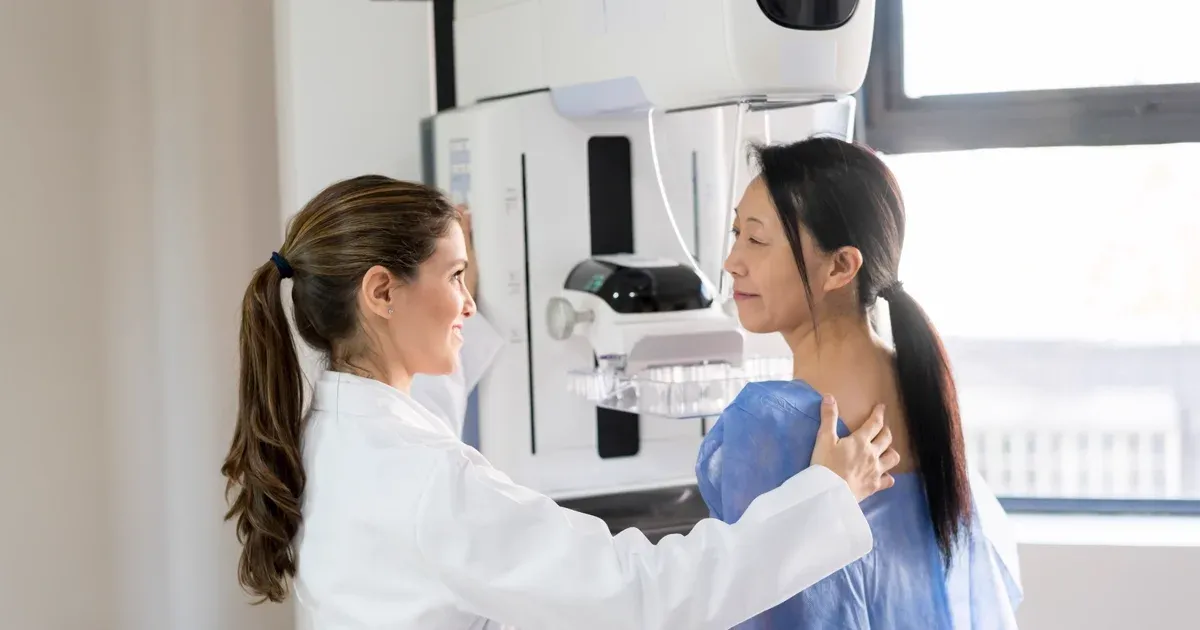
Mammogram Preparation Dubai: What to Expect (2026)
Read More
CA-125 Test Dubai: Ovarian Cancer Marker (2026)
Read More
Endometriosis Dubai: Symptoms & Diagnosis (2026)
Read More© 2026 Doctors Clinic Diagnostic Center (DCDC), Dubai Healthcare City. Originally published at https://doctorsclinicdubai.ae/blog/mammogram-vs-breast-ultrasound-dubai. All rights reserved. Unauthorized reproduction is prohibited.