
मुख्य बातें
- स्तन कैंसर UAE में महिलाओं में सबसे आम कैंसर है, लेकिन उचित उपचार के साथ 99% प्रारंभिक-चरण के मामले ठीक हो सकते हैं
- 40 वर्ष की आयु से वार्षिक मैमोग्राम शुरू करें, या जोखिम कारक होने पर पहले (पारिवारिक इतिहास, BRCA जीन, घनी स्तन ऊतक)
- लगभग 85% स्तन कैंसर बिना पारिवारिक इतिहास वाली महिलाओं में होते हैं, इसलिए हर महिला को नियमित स्क्रीनिंग की आवश्यकता है
- मैमोग्राम (400-700 AED) स्वर्ण मानक है; अल्ट्रासाउंड (400-600 AED) घने स्तन या गांठ के मूल्यांकन के लिए जोड़ा जाता है
- अधिकांश स्तन गांठें सौम्य (कैंसर नहीं) होती हैं, लेकिन मन की शांति के लिए किसी भी नई गांठ का 1-2 सप्ताह के भीतर मूल्यांकन करवाएं
- 3D मैमोग्राफी 2D की तुलना में 20-40% अधिक कैंसर का पता लगाती है, विशेष रूप से घने स्तन ऊतक के लिए मूल्यवान
मुझे एक रेडियोलॉजिस्ट के रूप में कुछ बात साझा करनी है जो मुझे चिंतित करती है: मैं नियमित रूप से उन महिलाओं में स्तन कैंसर का निदान करता हूं जो "ठीक महसूस कर रही थीं" और "कोई लक्षण नहीं थे।" जब तक कैंसर ऐसे लक्षण पैदा करता है जो आप महसूस कर सकती हैं (गांठ, त्वचा में बदलाव, निप्पल से स्राव), यह पहले से ही हमारी इच्छा से बड़ा हो चुका होता है। स्क्रीनिंग का पूरा उद्देश्य कैंसर को महसूस करने से पहले खोजना है - जब यह छोटा और अत्यधिक उपचार योग्य हो।
फिर भी कई महिलाएं स्क्रीनिंग में देरी करती हैं। कुछ डरती हैं कि हम क्या पा सकते हैं। अन्य काम और परिवार में व्यस्त हैं। कुछ मानती हैं कि स्तन कैंसर केवल पारिवारिक इतिहास वाली महिलाओं को होता है (यह सच नहीं है - 85% मामलों में कोई पारिवारिक इतिहास नहीं होता)। कुछ ने वर्षों पहले असुविधाजनक मैमोग्राम कराया और कभी नहीं लौटीं। यहां वास्तविकता है: प्रारंभिक चरण के स्तन कैंसर में पांच वर्ष की जीवित रहने की दर 99% है। उन्नत स्तन कैंसर में जीवित रहने की दर बहुत कम होती है। स्क्रीनिंग ही अंतर पैदा करती है। यह गाइड स्पष्ट रूप से बताएगी कि स्क्रीनिंग में क्या शामिल है, आपको इसकी कब आवश्यकता है, और अगर हम कुछ पाते हैं तो क्या होता है।
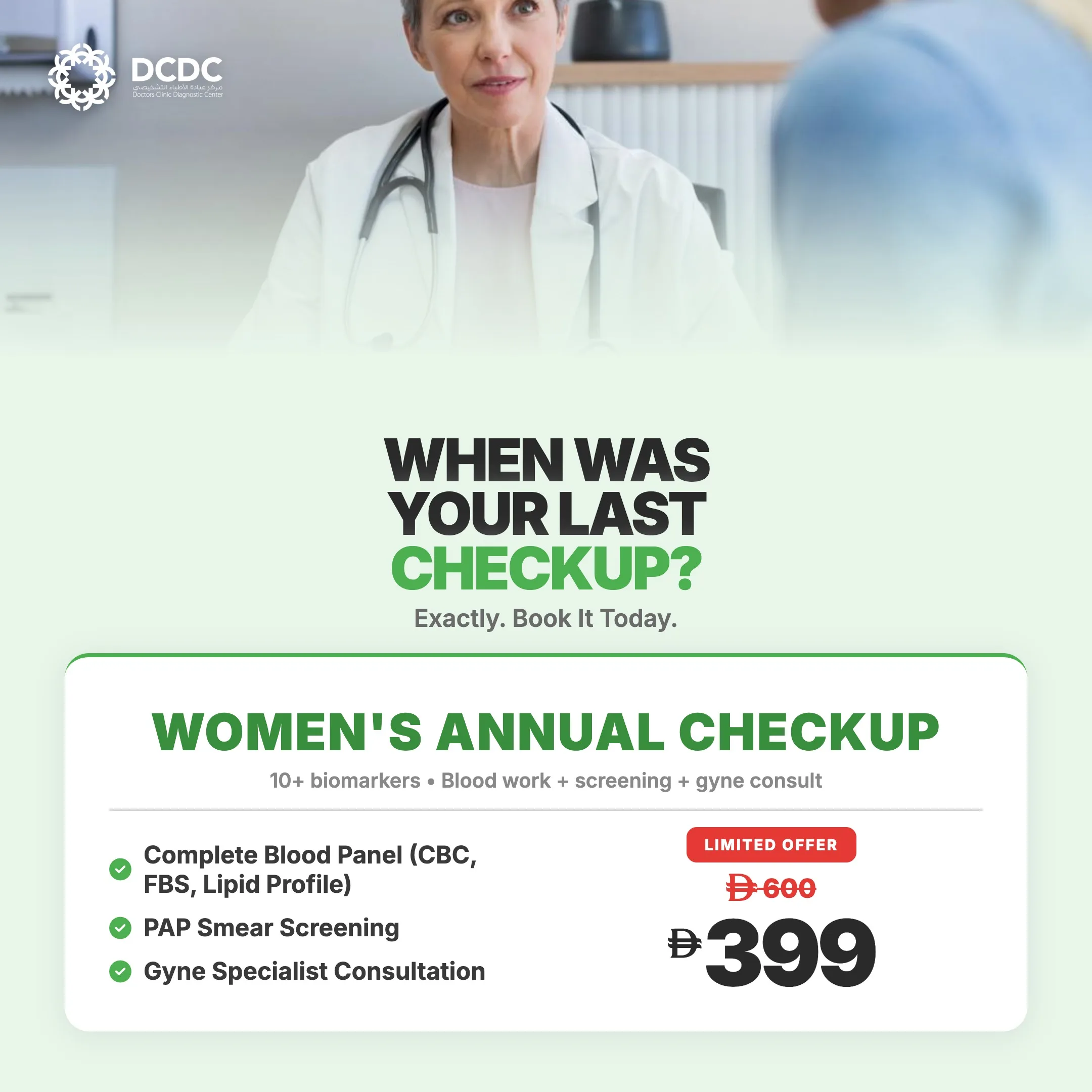
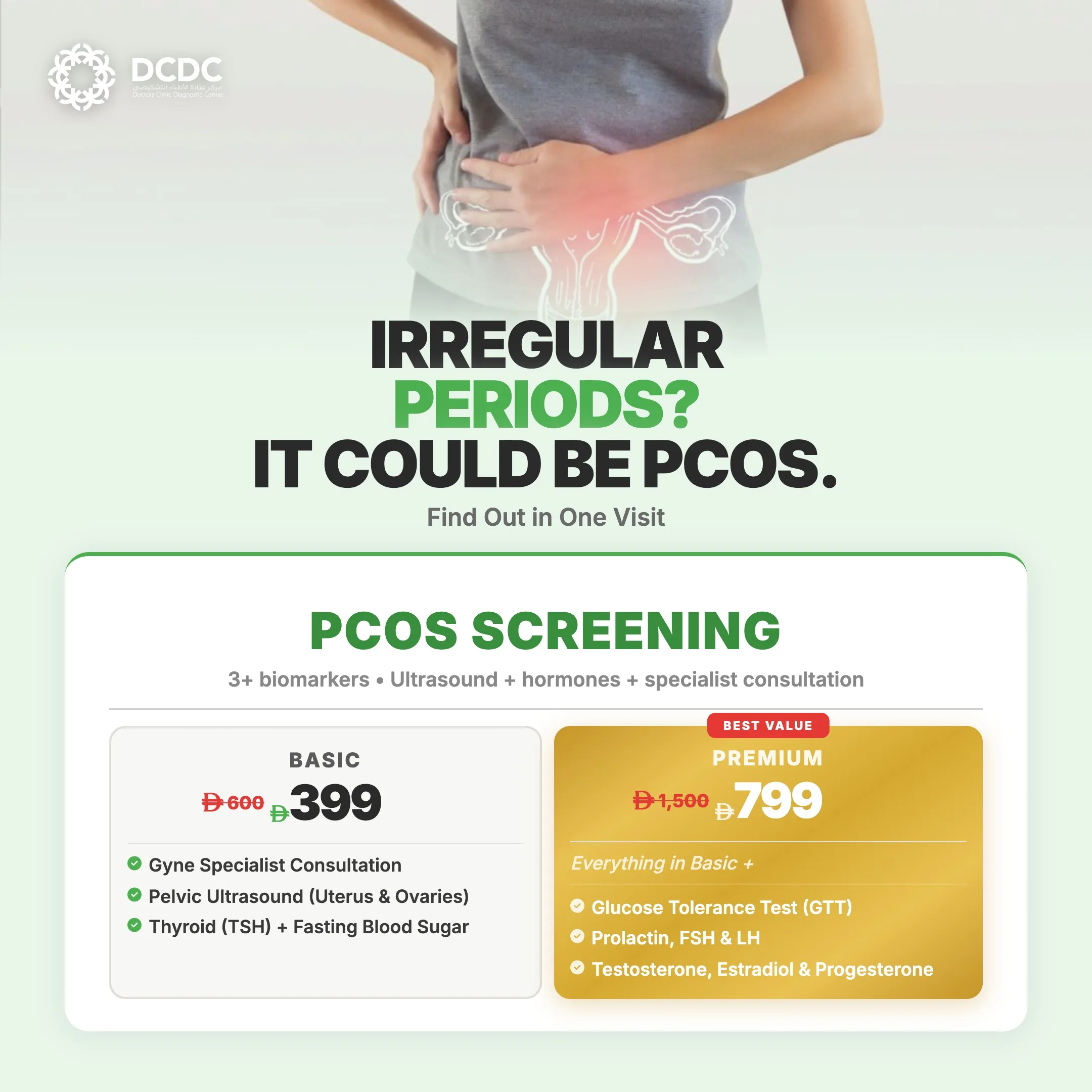
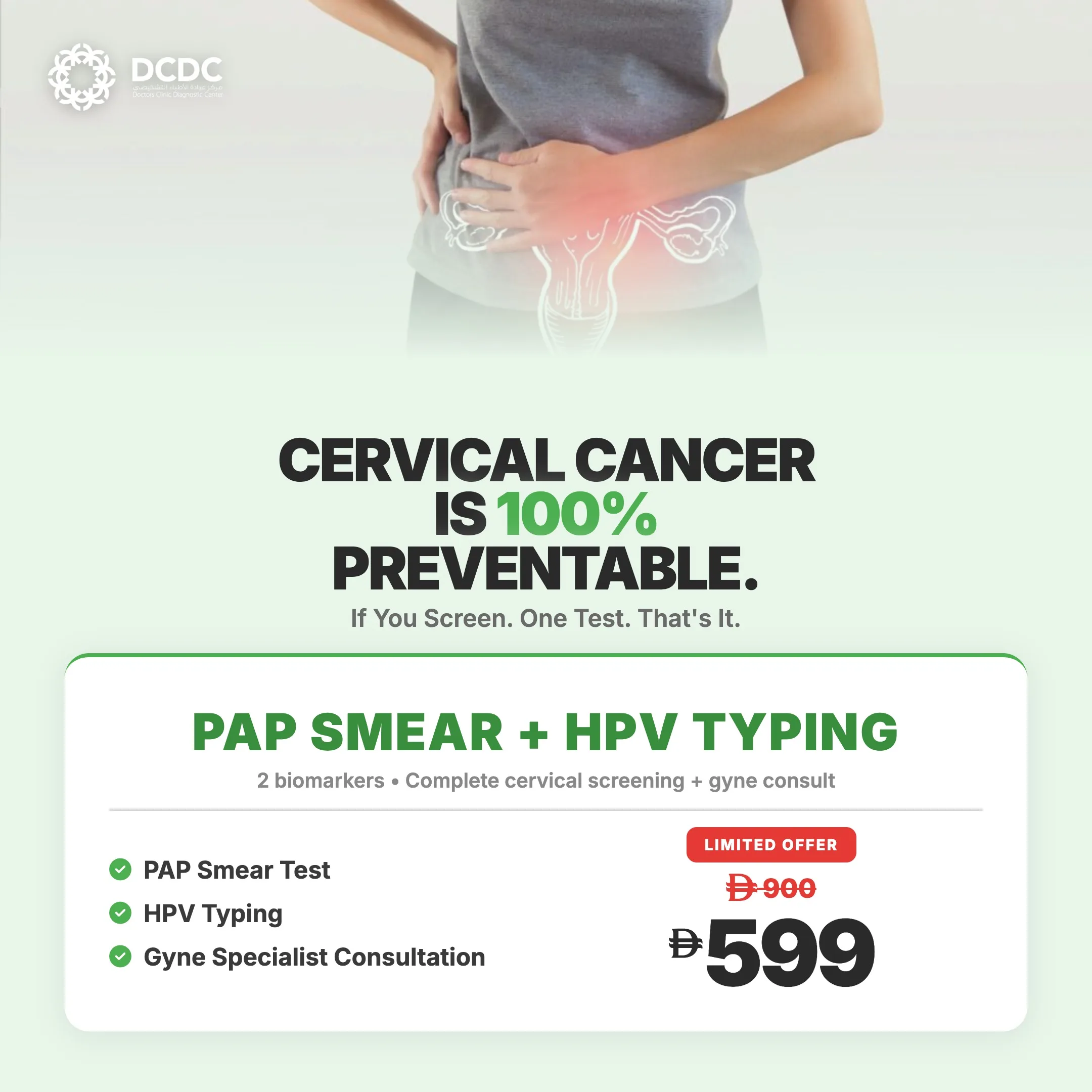
Health Screening Packages
Save with our bundled screening packages — specialist consultation included


दुबई में स्तन कैंसर के जोखिम को समझना
स्तन कैंसर UAE और पूरे खाड़ी क्षेत्र में महिलाओं में सबसे आम कैंसर है। जबकि जागरूकता में काफी सुधार हुआ है, स्क्रीनिंग दरें अभी भी इष्टतम स्तर से पीछे हैं। कई महिलाएं बाद के चरण की बीमारी के साथ आती हैं जो पहले पकड़ी जा सकती थी।
कई कारक आपके व्यक्तिगत जोखिम को प्रभावित करते हैं:
जोखिम बढ़ाने वाले कारक
- आयु: उम्र बढ़ने के साथ जोखिम बढ़ता है, और अधिकांश स्तन कैंसर 50 वर्ष की आयु के बाद होते हैं
- पारिवारिक इतिहास: माँ, बहन या बेटी को स्तन कैंसर होने से जोखिम काफी बढ़ जाता है
- आनुवंशिक उत्परिवर्तन: BRCA1/BRCA2 जीन जोखिम को नाटकीय रूप से बढ़ाते हैं (85% तक जीवनकाल जोखिम)
- घने स्तन ऊतक: थोड़ा अधिक जोखिम, साथ ही मैमोग्राम पर कैंसर का पता लगाना कठिन
- पिछली स्तन असामान्यताएं: एटिपिकल हाइपरप्लासिया या लोब्युलर कार्सिनोमा इन सिटू
- हार्मोनल कारक: जल्दी मासिक धर्म, देर से रजोनिवृत्ति, कोई गर्भधारण नहीं, या 30 के बाद पहली गर्भावस्था
- हार्मोन रिप्लेसमेंट थेरेपी: संयुक्त एस्ट्रोजन-प्रोजेस्टेरोन थेरेपी जोखिम बढ़ाती है
- पिछला छाती विकिरण: विशेष रूप से किशोरावस्था या युवा वयस्कता के दौरान
- रजोनिवृत्ति के बाद मोटापा: एस्ट्रोजन के स्तर और कैंसर के जोखिम को बढ़ाता है
- शराब का सेवन: मध्यम मात्रा में पीना भी जोखिम बढ़ाता है
महत्वपूर्ण: अधिकांश मामलों में कोई स्पष्ट जोखिम कारक नहीं होते
यहां वह है जो मैं चाहता हूं कि हर महिला समझे: लगभग 85% स्तन कैंसर बिना रोग के पारिवारिक इतिहास वाली महिलाओं में होते हैं। अधिकांश मामले वंशानुगत नहीं हैं। वे यादृच्छिक आनुवंशिक उत्परिवर्तन से होते हैं जो उम्र और जीवन के अनुभवों के साथ जमा होते हैं। कोई जोखिम कारक न होने का मतलब यह नहीं है कि आप सुरक्षित हैं। इसका मतलब है कि आप औसत जोखिम में हैं, और आपको अभी भी नियमित स्क्रीनिंग की आवश्यकता है।
मैमोग्राफी: स्क्रीनिंग के लिए स्वर्ण मानक
मैमोग्राफी स्तन कैंसर के लिए सबसे प्रभावी स्क्रीनिंग उपकरण बनी हुई है। यह एकमात्र इमेजिंग विधि है जो नियमित स्क्रीनिंग के लिए उपयोग किए जाने पर स्तन कैंसर मृत्यु दर को कम करने के लिए सिद्ध है। मुझे बताने दें कि इसमें क्या शामिल है और यह क्यों काम करती है।
मैमोग्राफी कैसे काम करती है
मैमोग्राम स्तन का कम-खुराक एक्स-रे है। प्रत्येक स्तन को एक समतल सतह पर रखा जाता है और एक पारदर्शी पैडल द्वारा धीरे से दबाया जाता है। यह संपीड़न आवश्यक है क्योंकि यह स्तन ऊतक को फैलाता है ताकि हम सभी परतों के माध्यम से देख सकें, गति धुंधलापन कम करता है, और विकिरण खुराक को कम करता है।
हम आमतौर पर प्रत्येक स्तन की दो छवियां लेते हैं: ऊपर से (क्रैनियोकॉडल व्यू) और एक कोण से (मीडियोलेटरल ऑब्लिक व्यू)। पूरी प्रक्रिया में लगभग 15-20 मिनट लगते हैं।
मैमोग्राम क्या पता लगाते हैं
- माइक्रोकैल्सीफिकेशन: छोटे कैल्शियम जमाव जो प्रारंभिक कैंसर या पूर्व-कैंसर परिवर्तनों का संकेत दे सकते हैं
- द्रव्यमान: ठोस या सिस्टिक गांठें जिन्हें आगे मूल्यांकन की आवश्यकता हो सकती है
- वास्तुशिल्प विकृति: असामान्य ऊतक पैटर्न जो कैंसर का संकेत दे सकते हैं
- असममितताएं: ऐसे क्षेत्र जो दोनों स्तनों के बीच अलग दिखते हैं
महत्वपूर्ण बात यह है कि मैमोग्राम गांठ महसूस होने से 1-3 साल पहले कैंसर का पता लगा सकते हैं। यह प्रारंभिक पहचान ही जीवन बचाती है।
3D मैमोग्राफी (टोमोसिंथेसिस)
3D मैमोग्राफी एक उन्नत तकनीक है जो विभिन्न कोणों से कई छवियां लेती है, स्तन का त्रि-आयामी दृश्य बनाती है। इसे ब्रेड काटने जैसा समझें: सभी स्लाइस को एक साथ संपीड़ित देखने के बजाय, आप प्रत्येक स्लाइस को अलग-अलग देख सकते हैं।
3D मैमोग्राफी के लाभ:
- मानक 2D मैमोग्राफी की तुलना में 20-40% अधिक कैंसर पाती है
- घने स्तनों के लिए विशेष रूप से मूल्यवान जहां ओवरलैपिंग ऊतक कैंसर छिपा सकते हैं
- गलत सकारात्मक (कॉलबैक) को लगभग 15-40% कम करती है
- मानक मैमोग्राफी के समान विकिरण खुराक
यदि आपके पास घने स्तन हैं या सबसे गहन उपलब्ध स्क्रीनिंग चाहती हैं, तो अपॉइंटमेंट शेड्यूल करते समय 3D मैमोग्राफी के बारे में पूछें।
| मैमोग्राफी सेवा | लागत (AED) |
|---|---|
| स्क्रीनिंग मैमोग्राम | 400-700 |
| डायग्नोस्टिक मैमोग्राम (अतिरिक्त व्यू के साथ) | 500-900 |
| 3D मैमोग्राफी/टोमोसिंथेसिस | 600-1,000 |
अधिकांश बीमा योजनाएं 40+ महिलाओं के लिए स्क्रीनिंग मैमोग्राम को निवारक देखभाल के रूप में कवर करती हैं। परिणाम आमतौर पर उसी दिन या 24 घंटे के भीतर उपलब्ध होते हैं।
स्तन अल्ट्रासाउंड: सही पूरक
अल्ट्रासाउंड स्तन ऊतक की छवियां बनाने के लिए ध्वनि तरंगों (कोई विकिरण नहीं) का उपयोग करता है। यह मैमोग्राफी से अलग उद्देश्यों की पूर्ति करता है और अक्सर इसके साथ उपयोग किया जाता है, विकल्प के रूप में नहीं।
अल्ट्रासाउंड कब उपयोग किया जाता है
- गांठों का मूल्यांकन: अल्ट्रासाउंड ठोस द्रव्यमान को तरल-भरी सिस्ट से अलग करने में उत्कृष्ट है - एक साधारण सिस्ट लगभग हमेशा सौम्य होती है और अक्सर उपचार की आवश्यकता नहीं होती।
- घने स्तन स्क्रीनिंग: घने ऊतक वाली महिलाओं के लिए मैमोग्राफी में जोड़ा जाता है जहां मैमोग्राम असामान्यताएं छोड़ सकते हैं
- युवा महिलाएं: 30 वर्ष से कम उम्र की महिलाओं के लिए प्राथमिक इमेजिंग (घने ऊतक मैमोग्राम को कम उपयोगी बनाते हैं, और छोटी ऊतक विकिरण के प्रति अधिक संवेदनशील होती है)
- गर्भावस्था और स्तनपान: गर्भावस्था के दौरान विकिरण की कोई चिंता नहीं; स्तनपान कराने वाली माताओं में स्तन समस्याओं का मूल्यांकन कर सकता है
- बायोप्सी का मार्गदर्शन: कोर बायोप्सी के दौरान सुई प्लेसमेंट के लिए रीयल-टाइम इमेजिंग
- मैमोग्राम निष्कर्षों की फॉलो-अप: मैमोग्राफी पर असामान्य दिखने वाले क्षेत्रों का आगे मूल्यांकन
अल्ट्रासाउंड की सीमाएं
अल्ट्रासाउंड माइक्रोकैल्सीफिकेशन का पता नहीं लगा सकता, जो अक्सर स्तन कैंसर का सबसे पहला संकेत होता है। यह मैमोग्राफी की तुलना में अधिक ऑपरेटर-निर्भर भी है और छोटे कैंसर छोड़ सकता है। इसलिए यह स्क्रीनिंग के लिए मैमोग्राफी का पूरक है, विकल्प नहीं।
स्तन एमआरआई: उच्च-जोखिम स्क्रीनिंग के लिए
एमआरआई स्तन कैंसर के लिए सबसे संवेदनशील इमेजिंग विधि है, मैमोग्राफी या अल्ट्रासाउंड की तुलना में अधिक कैंसर का पता लगाती है। हालांकि, यह अधिक महंगी भी है, अधिक समय लेती है, कंट्रास्ट इंजेक्शन की आवश्यकता होती है, और उच्च गलत-सकारात्मक दरें हैं। इन कारणों से, यह विशिष्ट स्थितियों के लिए आरक्षित है।
किसे स्तन एमआरआई करवानी चाहिए
- BRCA जीन उत्परिवर्तन वाहक: 25-30 वर्ष की आयु से वार्षिक एमआरआई, मैमोग्राफी के साथ बारी-बारी से
- बहुत उच्च जीवनकाल जोखिम (20-25% से अधिक): पारिवारिक इतिहास या अन्य कारकों के आधार पर
- 30 वर्ष की आयु से पहले पिछला छाती विकिरण: हॉजकिन लिम्फोमा जैसी स्थितियों के लिए
- नव निदान स्तन कैंसर: सर्जरी से पहले रोग की पूरी सीमा का मूल्यांकन करने के लिए
- इम्प्लांट अखंडता की निगरानी: सिलिकॉन इम्प्लांट के फटने की जांच
- छिपा प्राथमिक: जब लिम्फ नोड्स में कैंसर पाया जाता है लेकिन मैमोग्राम/अल्ट्रासाउंड स्तन स्रोत नहीं दिखाते
औसत-जोखिम वाली महिलाओं के लिए स्तन एमआरआई की सिफारिश नहीं की जाती क्योंकि उच्च गलत-सकारात्मक दर अनावश्यक बायोप्सी और चिंता की ओर ले जाती है।
स्क्रीनिंग कब शुरू करें: आयु-आधारित दिशानिर्देश
स्क्रीनिंग सिफारिशें आपके जोखिम स्तर पर निर्भर करती हैं। यहां एक व्यावहारिक गाइड है:
| आयु समूह | स्क्रीनिंग सिफारिश |
|---|---|
| आयु 20-39 (औसत जोखिम) | स्तन जागरूकता (अपना सामान्य जानें); हर 1-3 साल में क्लीनिकल स्तन परीक्षा; मैमोग्राम नियमित रूप से अनुशंसित नहीं |
| आयु 40-49 (औसत जोखिम) | वार्षिक मैमोग्राम अनुशंसित; घने स्तन होने पर अल्ट्रासाउंड पर चर्चा करें |
| आयु 50-74 (औसत जोखिम) | वार्षिक या द्विवार्षिक मैमोग्राम; घने स्तन होने पर अल्ट्रासाउंड जारी रखें |
| आयु 75+ (औसत जोखिम) | अच्छे स्वास्थ्य और 10 वर्ष से अधिक जीवन प्रत्याशा होने पर स्क्रीनिंग जारी रखें; अपने डॉक्टर से चर्चा करें |
| उच्च जोखिम (कोई भी आयु) | पहले और अधिक गहन स्क्रीनिंग (एमआरआई + मैमोग्राम), आनुवंशिक परामर्श, जोखिम-कम करने की रणनीतियों की आवश्यकता हो सकती है; विशेषज्ञ से चर्चा करें |
अगर हम कुछ पाते हैं तो क्या होता है
मैमोग्राम के बाद वापस बुलाया जाना चिंता पैदा करता है, लेकिन आंकड़े समझें: लगभग 10% महिलाओं को अतिरिक्त इमेजिंग के लिए वापस बुलाया जाता है, और उनमें से केवल लगभग 10% (सभी स्क्रीन की गई का 1%) को कैंसर होगा। अधिकांश कॉलबैक सौम्य निष्कर्षों के लिए होते हैं।
सामान्य कॉलबैक प्रक्रिया
- अतिरिक्त मैमोग्राम व्यू: चिंता के क्षेत्र को बेहतर देखने के लिए स्पॉट कंप्रेशन या मैग्निफिकेशन व्यू
- स्तन अल्ट्रासाउंड: यह निर्धारित करने के लिए कि निष्कर्ष ठोस है या सिस्टिक
- BI-RADS मूल्यांकन: आपका रेडियोलॉजिस्ट कैंसर की संभावना दर्शाने वाली श्रेणी निर्धारित करता है: 1 = सामान्य, 2 = सौम्य, 3 = संभवतः सौम्य, 4 = संदिग्ध, 5 = कैंसर का अत्यधिक संकेत
- आवश्यकता होने पर बायोप्सी: संदिग्ध निष्कर्षों के लिए, हम ऊतक नमूनाकरण की सिफारिश करते हैं
स्तन बायोप्सी को समझना
यदि बायोप्सी की सिफारिश की जाती है, तो यह आमतौर पर कोर नीडल बायोप्सी होती है - अल्ट्रासाउंड मार्गदर्शन का उपयोग करके स्थानीय एनेस्थीसिया के तहत किया जाने वाला न्यूनतम इनवेसिव प्रक्रिया। एक छोटी सुई सूक्ष्म विश्लेषण के लिए ऊतक के नमूने निकालती है। प्रक्रिया में लगभग 30 मिनट लगते हैं, और आप उसी दिन सामान्य गतिविधियों में वापस आ सकती हैं।
बायोप्सी परिणाम आमतौर पर 2-5 दिन लेते हैं। यदि कैंसर की पुष्टि होती है, तो हम आपको उपचार विकल्पों पर चर्चा करने के लिए स्तन सर्जन और ऑन्कोलॉजिस्ट से जोड़ते हैं।
अपने मैमोग्राम की तैयारी
अपने मैमोग्राम को यथासंभव आरामदायक और प्रभावी बनाने के लिए कुछ सुझाव:
- समय निर्धारण: अपने मासिक धर्म के बाद के सप्ताह के लिए बुक करें जब स्तन कम संवेदनशील होते हैं
- डिओडोरेंट से बचें: अपनी बाहों के नीचे या स्तनों पर डिओडोरेंट, एंटीपर्सपिरेंट, पाउडर या लोशन का उपयोग न करें क्योंकि ये छवियों पर कलाकृतियों के रूप में दिखाई दे सकते हैं
- सुविधाजनक कपड़े पहनें: दो-टुकड़ों वाली पोशाक आसान बनाती है क्योंकि आप कमर से ऊपर कपड़े उतारेंगी
- पिछली इमेजिंग लाएं: यदि आपने कहीं और मैमोग्राम कराया है, तो इसे लाएं या भिजवाएं ताकि हम तुलना कर सकें
- आवश्यकता होने पर दर्द निवारक लें: परीक्षा से पहले इबुप्रोफेन या एसिटामिनोफेन मदद कर सकता है यदि आप विशेष रूप से संवेदनशील हैं
- कैफीन सीमित करें: कुछ महिलाओं को लगता है कि कैफीन स्तन कोमलता बढ़ाता है
मुझे गांठ मिली: अब क्या?
पहले, गहरी सांस लें। गांठ मिलना डरावना है, लेकिन याद रखें कि लगभग 80% स्तन गांठें सौम्य होती हैं। सामान्य गैर-कैंसर कारणों में शामिल हैं:
- सिस्ट: तरल-भरी थैलियां, बहुत आम, विशेष रूप से रजोनिवृत्ति से पहले
- फाइब्रोएडेनोमा: ठोस, रबड़ जैसी, चलने योग्य गांठें, युवा महिलाओं में आम
- फाइब्रोसिस्टिक परिवर्तन: गांठदार, रस्सी जैसे स्तन ऊतक जो आपके चक्र के साथ बदलते हैं
- वसा परिगलन: क्षतिग्रस्त वसायुक्त ऊतक से मजबूत गांठें, अक्सर आघात या सर्जरी के बाद
हालांकि, आपको किसी भी नई गांठ का मूल्यांकन करवाना चाहिए। "देखें कि यह चली जाती है या नहीं" की प्रतीक्षा न करें। DCDC में, हम आमतौर पर आपको 1-2 दिनों के भीतर स्तन अल्ट्रासाउंड के लिए देख सकते हैं, जो अक्सर तत्काल आश्वासन या अगले कदमों का मार्गदर्शन प्रदान कर सकता है।
कब शीघ्र मूल्यांकन लें
- स्तन या बगल में नई गांठ या मोटाई
- स्तन के आकार या आकृति में बदलाव
- त्वचा में परिवर्तन: गड्ढे, सिकुड़न, लालिमा, या संतरे के छिलके जैसी बनावट
- निप्पल में परिवर्तन जैसे उलटाव, स्राव (विशेष रूप से खूनी), या पपड़ी
- एक क्षेत्र में लगातार स्तन दर्द
DCDC दुबई हेल्थकेयर सिटी में स्तन स्क्रीनिंग
हमारे दुबई हेल्थकेयर सिटी क्लिनिक में, हम उसी दिन या अगले दिन की अपॉइंटमेंट उपलब्ध होने के साथ व्यापक स्तन इमेजिंग सेवाएं प्रदान करते हैं:
- डिजिटल मैमोग्राफी अनुभवी महिला तकनीशियनों के साथ
- 3D मैमोग्राफी (टोमोसिंथेसिस) बेहतर पहचान के लिए
- स्तन अल्ट्रासाउंड गांठ मूल्यांकन और घने स्तन स्क्रीनिंग के लिए
- अल्ट्रासाउंड-गाइडेड बायोप्सी जब ऊतक नमूनाकरण की आवश्यकता हो
- स्तन सर्जनों के साथ समन्वय यदि उपचार की आवश्यकता हो
परिणामों की अनुभवी रेडियोलॉजिस्ट द्वारा समीक्षा की जाती है और आमतौर पर 24 घंटे के भीतर उपलब्ध होते हैं। चिंताजनक निष्कर्षों के लिए, हम आपसे सीधे संपर्क करते हैं और अगले कदमों को तेज करते हैं।
आज ही अपनी स्तन स्क्रीनिंग बुक करें
प्रारंभिक पहचान आपके नियंत्रण में है। नियमित स्क्रीनिंग कैंसर को तब पाती है जब यह सबसे अधिक उपचार योग्य होता है। दुबई हेल्थकेयर सिटी में डॉक्टर्स क्लिनिक डायग्नोस्टिक सेंटर में, हमारे अनुभवी रेडियोलॉजिस्ट मैमोग्राफी, स्तन अल्ट्रासाउंड और बायोप्सी सेवाओं सहित व्यापक स्तन इमेजिंग सेवाएं प्रदान करते हैं।
DCDC में संबंधित सेवाएं
दुबई हेल्थकेयर सिटी में विशेषज्ञ देखभाल और उन्नत निदान
Frequently Asked Questions
अपने स्तन स्वास्थ्य पर नियंत्रण रखें
स्तन कैंसर के खिलाफ प्रारंभिक पहचान सबसे शक्तिशाली उपकरण है। जब स्तन कैंसर जल्दी पाया जाता है, फैलने से पहले, जीवित रहने की दर 99% है। यही कारण है कि नियमित स्क्रीनिंग इतनी महत्वपूर्ण है, भले ही आप पूरी तरह से स्वस्थ महसूस करें और कोई लक्षण न हों।
डर, व्यस्त कार्यक्रम, या इस बारे में गलतफहमियां कि किसे स्तन कैंसर होता है, को अपनी स्क्रीनिंग में देरी न करने दें। हर महिला को, पारिवारिक इतिहास की परवाह किए बिना, 40 वर्ष की आयु से (या जोखिम कारक होने पर पहले) नियमित स्तन कैंसर स्क्रीनिंग की आवश्यकता है। अपनी स्तन स्क्रीनिंग बुक करें हमारे दुबई हेल्थकेयर सिटी क्लिनिक में आज।
स्रोत एवं संदर्भ
यह लेख हमारी चिकित्सा टीम द्वारा समीक्षित है और निम्नलिखित स्रोतों का संदर्भ देता है:
- दुबई स्वास्थ्य प्राधिकरण - पिंक कारवां स्तन कैंसर कार्यक्रम
- UAE स्वास्थ्य और रोकथाम मंत्रालय - राष्ट्रीय कैंसर स्क्रीनिंग दिशानिर्देश
- फ्रेंड्स ऑफ कैंसर पेशेंट्स UAE - स्तन कैंसर जागरूकता
- अमेरिकन कैंसर सोसाइटी - स्तन कैंसर स्क्रीनिंग दिशानिर्देश
- RadiologyInfo.org - मैमोग्राफी और स्तन इमेजिंग
इस साइट पर चिकित्सा सामग्री DHA-लाइसेंस प्राप्त चिकित्सकों द्वारा समीक्षित है। हमारी देखें संपादकीय नीति अधिक जानकारी के लिए।

लेखक
डॉ. उसामा अल-ज़मज़मी
सलाहकार रेडियोलॉजिस्ट
MBBS, FRCR, सलाहकार रेडियोलॉजिस्ट
डॉ. उसामा अल-ज़मज़मी DCDC दुबई हेल्थकेयर सिटी में एक सलाहकार रेडियोलॉजिस्ट हैं जिनके पास मैमोग्राफी, स्तन अल्ट्रासाउंड और इमेज-गाइडेड बायोप्सी सहित स्तन इमेजिंग में व्यापक अनुभव है। वे प्रारंभिक पहचान और सटीक निदान के लिए प्रतिबद्ध हैं, व्यापक स्क्रीनिंग कार्यक्रमों के माध्यम से दुबई में महिलाओं को अपने स्तन स्वास्थ्य को बनाए रखने में मदद करते हैं।
संबंधित लेख

महिला स्वास्थ्य स्क्रीनिंग गाइड
दुबई में पैप स्मीयर टेस्ट
दुबई में प्रसूति एवं स्त्री रोग सेवाएं
More in Women's Health

Perimenopause vs Menopause Dubai: Signs (2026)
और पढ़ें
Mother & Baby Home Care Dubai: Guide (2026)
और पढ़ें
Gestational Diabetes Dubai: Screening (2026)
और पढ़ें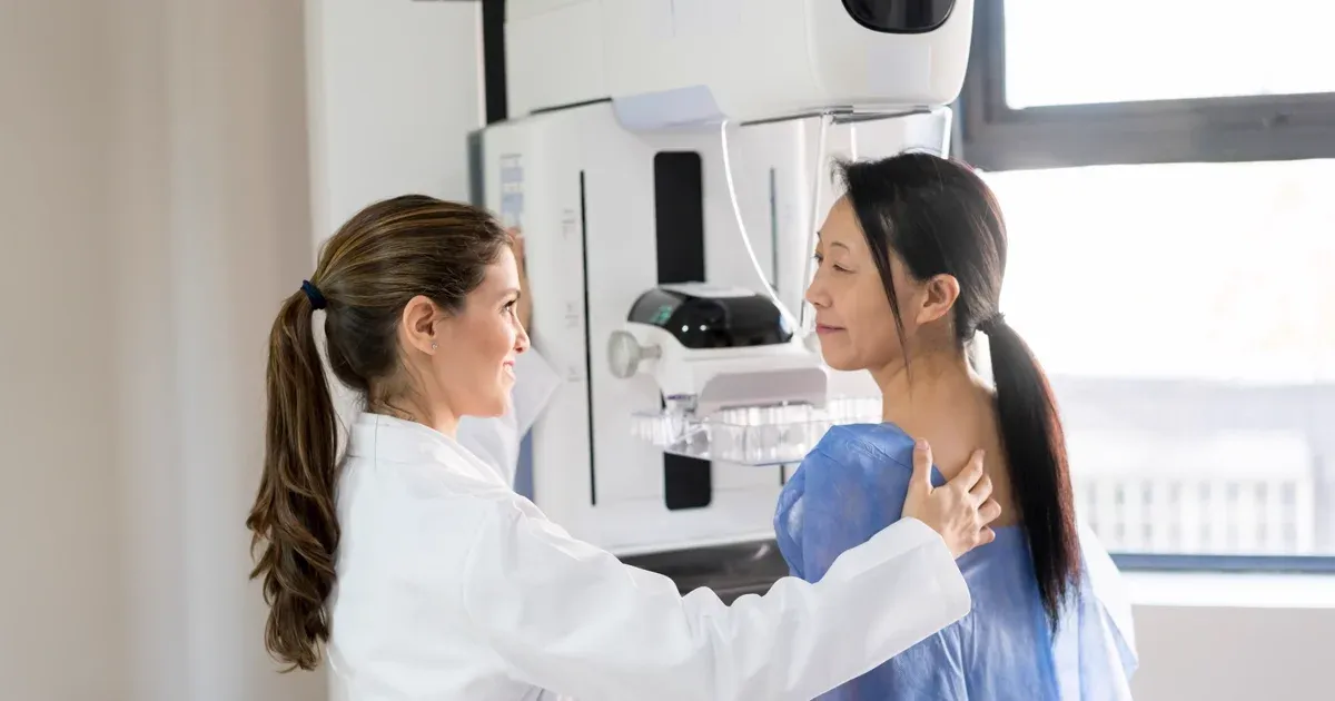
Mammogram Preparation Dubai: What to Expect (2026)
और पढ़ें
CA-125 Test Dubai: Ovarian Cancer Marker (2026)
और पढ़ेंMammogram vs Breast Ultrasound Dubai (2026)
और पढ़ें© 2026 Doctors Clinic Diagnostic Center (DCDC), Dubai Healthcare City. Originally published at https://doctorsclinicdubai.ae/blog/breast-cancer-screening-guide. All rights reserved. Unauthorized reproduction is prohibited.