
Wichtigste Erkenntnisse
- Brustkrebs ist die haeufigste Krebsart bei Frauen in den VAE, aber 99% der Fruehstadien sind bei richtiger Behandlung heilbar
- Beginnen Sie mit jaehrlichen Mammographien ab 40 Jahren, oder frueher bei Risikofaktoren (Familiengeschichte, BRCA-Gene, dichtes Brustgewebe)
- Etwa 85% der Brustkrebsfaelle treten bei Frauen OHNE Familiengeschichte auf, daher braucht jede Frau regelmaessige Vorsorge
- Mammographie (400-700 AED) ist der Goldstandard; Ultraschall (400-600 AED) wird bei dichtem Brustgewebe oder zur Beurteilung von Knoten ergaenzt
- Die meisten Brustknoten sind gutartig (kein Krebs), aber lassen Sie jeden neuen Knoten innerhalb von 1-2 Wochen untersuchen
- 3D-Mammographie erkennt 20-40% mehr Krebsfaelle als 2D, besonders wertvoll bei dichtem Brustgewebe
Lassen Sie mich etwas teilen, das mich als Radiologe beunruhigt: Ich diagnostiziere regelmaessig Brustkrebs bei Frauen, die sich "gut fuehlten" und "keine Symptome hatten". Wenn der Krebs Symptome verursacht, die Sie spueren koennen (einen Knoten, Hautveraenderungen, Brustwarzenausfluss), ist er bereits groesser gewachsen, als uns lieb ist. Der ganze Sinn der Vorsorge besteht darin, Krebs zu finden, bevor Sie ihn spueren koennen - wenn er klein und sehr gut behandelbar ist.
Dennoch verschieben viele Frauen die Vorsorge. Einige haben Angst vor dem, was wir finden koennten. Andere sind mit Arbeit und Familie beschaeftigt. Manche gehen davon aus, dass Brustkrebs nur Frauen mit Familiengeschichte trifft (das stimmt nicht - 85% der Faelle haben keine Familiengeschichte). Einige hatten vor Jahren eine unangenehme Mammographie und kamen nie zurueck. Hier ist die Realitaet: Brustkrebs im Fruehstadium hat eine 99%ige Fuenf-Jahres-Ueberlebensrate. Fortgeschrittener Brustkrebs hat eine viel niedrigere Ueberlebensrate. Die Vorsorge macht den Unterschied. Dieser Leitfaden erklaert genau, was die Vorsorge beinhaltet, wann Sie sie brauchen und was passiert, wenn wir etwas finden.
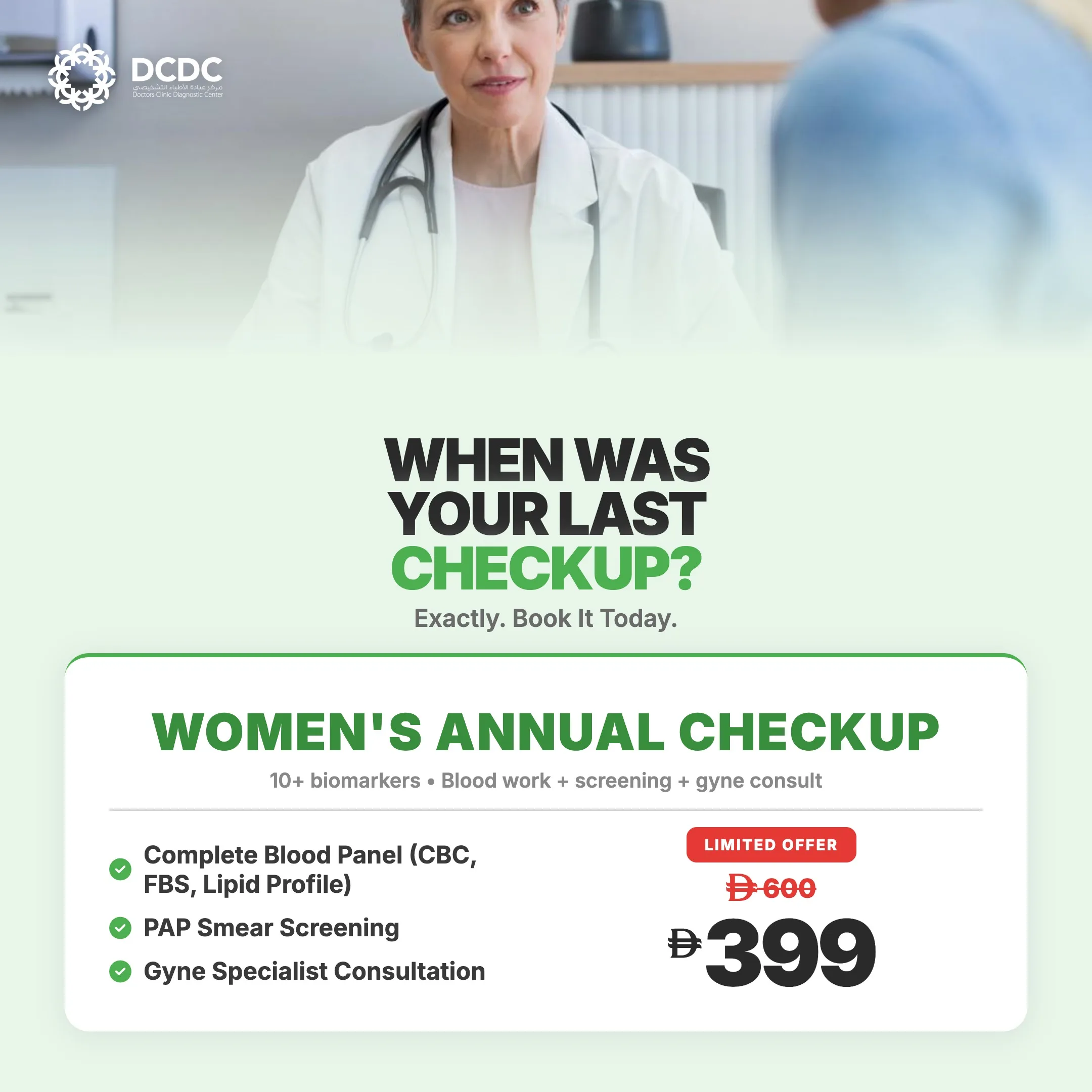
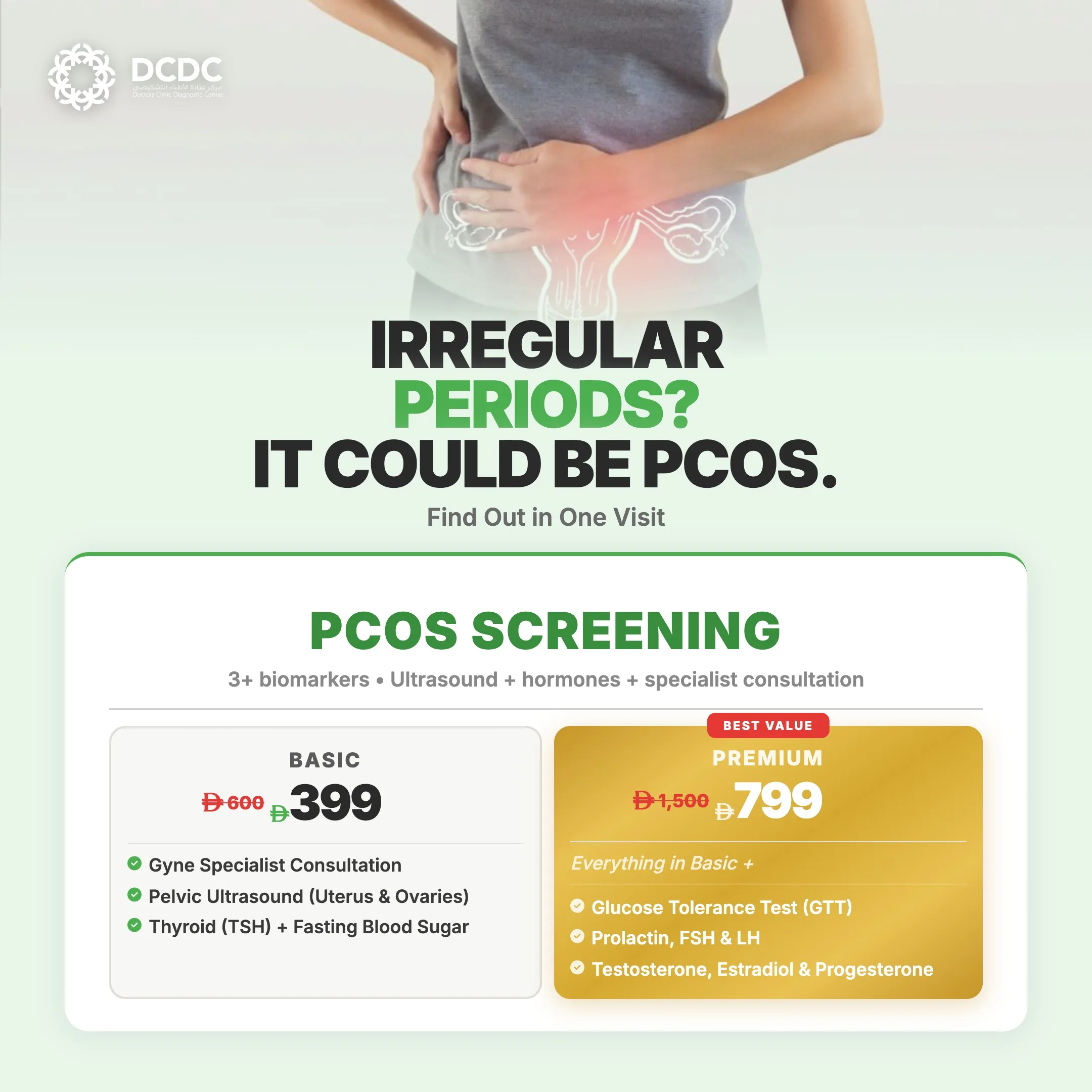
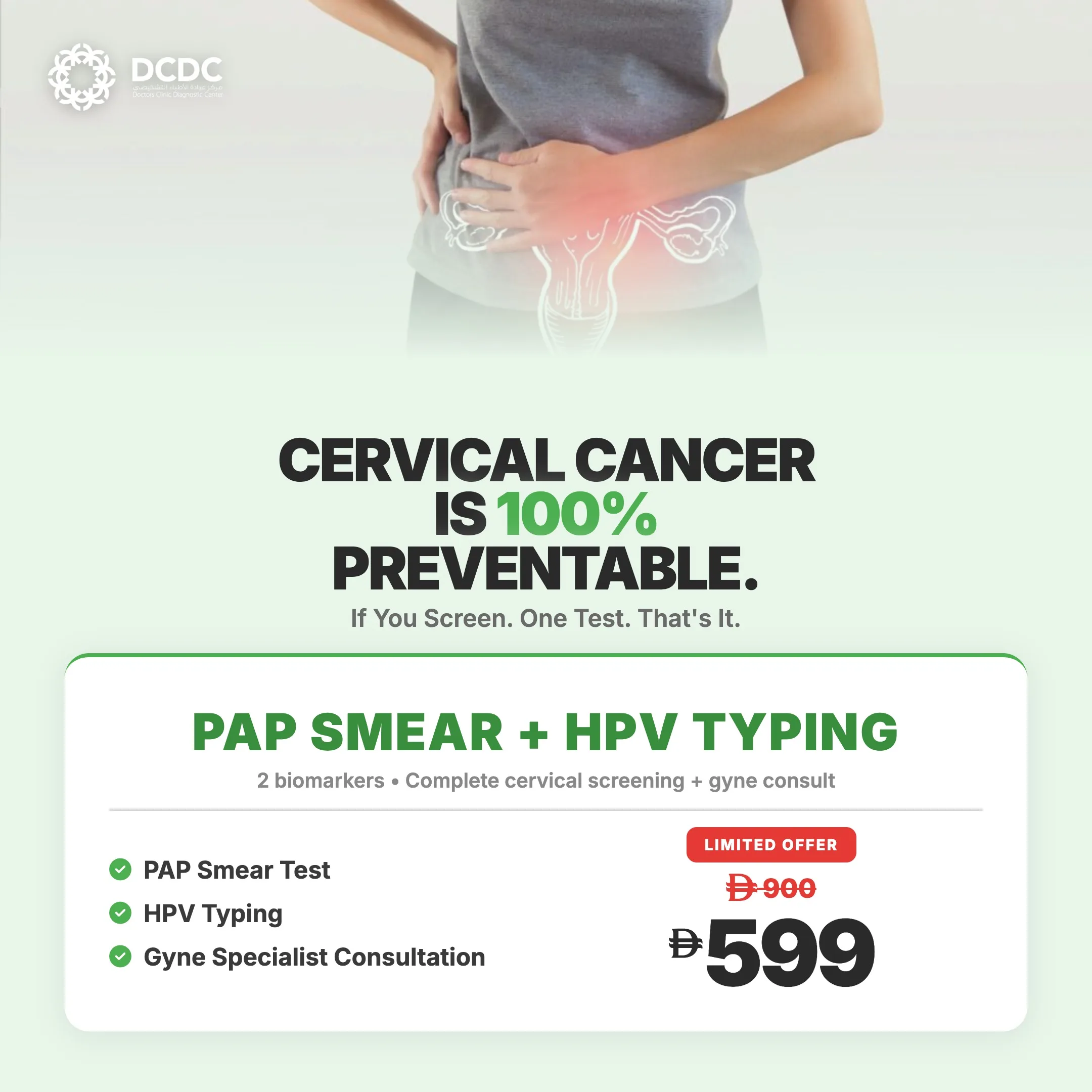
Health Screening Packages
Save with our bundled screening packages — specialist consultation included


Brustkrebsrisiko in Dubai verstehen
Brustkrebs ist die haeufigste Krebsart bei Frauen in den VAE und in der gesamten Golfregion. Waehrend das Bewusstsein sich deutlich verbessert hat, liegen die Vorsorgeraten immer noch unter dem optimalen Niveau. Viele Frauen stellen sich mit Erkrankungen in spaeteren Stadien vor, die frueher haetten erkannt werden koennen.
Mehrere Faktoren beeinflussen Ihr persoenliches Risiko:
Faktoren, die das Risiko erhoehen
- Alter: Das Risiko steigt mit dem Alter, und die meisten Brustkrebsfaelle treten nach dem 50. Lebensjahr auf
- Familiengeschichte: Mutter, Schwester oder Tochter mit Brustkrebs erhoeht das Risiko erheblich
- Genetische Mutationen: BRCA1/BRCA2-Gene erhoehen das Risiko dramatisch (bis zu 85% Lebenszeit-Risiko)
- Dichtes Brustgewebe: Leicht erhoehtes Risiko, ausserdem schwerer Krebs in der Mammographie zu erkennen
- Fruehere Brustauffaelligkeiten: Atypische Hyperplasie oder lobulaeres Karzinom in situ
- Hormonelle Faktoren: Fruehe Menstruation, spaete Menopause, keine Schwangerschaften oder erste Schwangerschaft nach 30
- Hormonersatztherapie: Kombinierte Oestrogen-Progesteron-Therapie erhoeht das Risiko
- Fruehere Brustbestrahlung: Besonders waehrend der Teenagerjahre oder im jungen Erwachsenenalter
- Fettleibigkeit nach der Menopause: Erhoeht Oestrogenspiegel und Krebsrisiko
- Alkoholkonsum: Selbst maessiger Konsum erhoeht das Risiko
Wichtig: Die meisten Faelle haben keine klaren Risikofaktoren
Hier ist, was ich moechte, dass jede Frau versteht: Etwa 85% der Brustkrebsfaelle treten bei Frauen ohne Familiengeschichte der Erkrankung auf. Die meisten Faelle sind nicht erblich. Sie resultieren aus zufaelligen genetischen Mutationen, die sich mit dem Alter und Lebenserfahrungen ansammeln. Keine Risikofaktoren zu haben bedeutet nicht, dass Sie sicher sind. Es bedeutet, dass Sie ein durchschnittliches Risiko haben, und Sie brauchen trotzdem regelmaessige Vorsorge.
Mammographie: Der Goldstandard der Vorsorge
Die Mammographie bleibt das wirksamste Vorsorgeinstrument fuer Brustkrebs. Sie ist die einzige Bildgebungsmodalitaet, die nachweislich die Brustkrebssterblichkeit reduziert, wenn sie fuer Routinevorsorge verwendet wird. Lassen Sie mich erklaeren, was sie beinhaltet und warum sie funktioniert.
Wie Mammographie funktioniert
Eine Mammographie ist eine niedrigdosierte Roentgenaufnahme der Brust. Jede Brust wird auf einer flachen Oberflaeche positioniert und sanft durch eine durchsichtige Platte komprimiert. Diese Kompression ist wichtig, weil sie das Brustgewebe verteilt, sodass wir durch alle Schichten sehen koennen, Bewegungsunschaerfe reduziert und die Strahlendosis minimiert.
Wir nehmen typischerweise zwei Bilder von jeder Brust auf: von oben (craniocaudale Ansicht) und von einem Winkel (mediolateral oblique Ansicht). Der gesamte Vorgang dauert etwa 15-20 Minuten.
Was Mammographien erkennen
- Mikroverkalkungen: Winzige Kalziumablagerungen, die auf fruehen Krebs oder praekanzeroese Veraenderungen hinweisen koennen
- Raumforderungen: Feste oder zystische Knoten, die weitere Untersuchung erfordern koennen
- Architektonische Verzerrung: Abnormale Gewebemuster, die auf Krebs hinweisen koennen
- Asymmetrien: Bereiche, die zwischen den beiden Bruesten unterschiedlich aussehen
Wichtig ist, dass Mammographien Krebs 1-3 Jahre erkennen koennen, bevor Sie einen Knoten spueren wuerden. Diese Frueherkennung rettet Leben.
3D-Mammographie (Tomosynthese)
3D-Mammographie ist eine fortschrittliche Technologie, die mehrere Bilder aus verschiedenen Winkeln aufnimmt und eine dreidimensionale Ansicht der Brust erstellt. Stellen Sie es sich vor wie Brot schneiden: Anstatt alle Scheiben zusammengedrueckt zu sehen, koennen Sie jede Scheibe einzeln betrachten.
Vorteile der 3D-Mammographie:
- Findet 20-40% mehr Krebsfaelle als Standard-2D-Mammographie
- Besonders wertvoll bei dichtem Brustgewebe, wo ueberlappendes Gewebe Krebs verbergen kann
- Reduziert falsch-positive Ergebnisse (Rueckrufe) um etwa 15-40%
- Aehnliche Strahlendosis wie Standard-Mammographie
Wenn Sie dichtes Brustgewebe haben oder die gruendlichste verfuegbare Vorsorge wuenschen, fragen Sie bei der Terminvereinbarung nach 3D-Mammographie.
| Mammographie-Leistung | Kosten (AED) |
|---|---|
| Vorsorge-Mammographie | 400-700 |
| Diagnostische Mammographie (mit zusaetzlichen Aufnahmen) | 500-900 |
| 3D-Mammographie/Tomosynthese | 600-1.000 |
Die meisten Versicherungstarife decken Vorsorge-Mammographien fuer Frauen ab 40 als Praevention ab. Ergebnisse sind typischerweise am selben Tag oder innerhalb von 24 Stunden verfuegbar.
Brustultraschall: Die perfekte Ergaenzung
Ultraschall verwendet Schallwellen (keine Strahlung), um Bilder des Brustgewebes zu erstellen. Er dient anderen Zwecken als die Mammographie und wird oft ergaenzend eingesetzt, nicht als Ersatz.
Wann Ultraschall eingesetzt wird
- Beurteilung von Knoten: Ultraschall ist hervorragend geeignet, feste Raumforderungen von fluessigkeitsgefuellten Zysten zu unterscheiden - eine einfache Zyste ist fast immer gutartig und benoetigt oft keine Behandlung.
- Vorsorge bei dichtem Brustgewebe: Ergaenzend zur Mammographie bei Frauen mit dichtem Gewebe, wo Mammographien Auffaelligkeiten uebersehen koennen
- Junge Frauen: Primaere Bildgebung fuer Frauen unter 30 (dichtes Gewebe macht Mammographien weniger nuetzlich, und juengeres Gewebe ist strahlungsempfindlicher)
- Schwangerschaft und Stillzeit: Keine Strahlungsbedenken waehrend der Schwangerschaft; kann Brustprobleme bei stillenden Muettern beurteilen
- Fuehrung von Biopsien: Echtzeit-Bildgebung fuer Nadelplatzierung bei Stanzbiopsien
- Nachverfolgung von Mammographie-Befunden: Weitere Beurteilung von Bereichen, die in der Mammographie auffaellig erscheinen
Einschraenkungen des Ultraschalls
Ultraschall kann keine Mikroverkalkungen erkennen, die oft das frueheste Zeichen von Brustkrebs sind. Er ist auch untersucherabhaengiger als die Mammographie und kann kleine Krebsfaelle uebersehen. Deshalb ergaenzt er die Mammographie bei der Vorsorge, anstatt sie zu ersetzen.
Brust-MRT: Fuer Hochrisiko-Vorsorge
MRT ist die sensitivste Bildgebungsmodalitaet fuer Brustkrebs und erkennt mehr Krebsfaelle als Mammographie oder Ultraschall. Sie ist jedoch auch teurer, dauert laenger, erfordert eine Kontrastmittelinjektion und hat hoehere falsch-positive Raten. Aus diesen Gruenden ist sie bestimmten Situationen vorbehalten.
Wer sollte ein Brust-MRT bekommen
- BRCA-Genmutationstraegerinnen: Jaehrliches MRT ab 25-30 Jahren, abwechselnd mit Mammographie
- Sehr hohes Lebenszeit-Risiko (ueber 20-25%): Basierend auf Familiengeschichte oder anderen Faktoren
- Fruehere Brustbestrahlung vor dem 30. Lebensjahr: Bei Erkrankungen wie Hodgkin-Lymphom
- Neu diagnostizierter Brustkrebs: Um das volle Ausmass der Erkrankung vor der Operation zu beurteilen
- Ueberwachung der Implantat-Integritaet: Pruefung auf Silikonimplantat-Ruptur
- Okkulter Primaertumor: Wenn Krebs in Lymphknoten gefunden wird, aber Mammographie/Ultraschall die Brustquelle nicht zeigen
Brust-MRT wird fuer Frauen mit durchschnittlichem Risiko nicht empfohlen, da die hohe falsch-positive Rate zu unnoetigen Biopsien und Angst fuehrt.
Wann mit der Vorsorge beginnen: Altersbasierte Richtlinien
Die Vorsorgeempfehlungen haengen von Ihrem Risikoniveau ab. Hier ist ein praktischer Leitfaden:
| Altersgruppe | Vorsorgeempfehlung |
|---|---|
| Alter 20-39 (Durchschnittliches Risiko) | Brustbewusstsein (kennen Sie Ihren Normalzustand); klinische Brustuntersuchung alle 1-3 Jahre; Mammographie nicht routinemaessig empfohlen |
| Alter 40-49 (Durchschnittliches Risiko) | Jaehrliche Mammographie empfohlen; Ultraschall bei dichtem Brustgewebe besprechen |
| Alter 50-74 (Durchschnittliches Risiko) | Jaehrliche oder zweijaehrliche Mammographie; Ultraschall fortsetzen bei dichtem Brustgewebe |
| Alter 75+ (Durchschnittliches Risiko) | Vorsorge fortsetzen bei guter Gesundheit und Lebenserwartung ueber 10 Jahren; mit Ihrem Arzt besprechen |
| Hohes Risiko (Jedes Alter) | Moeglicherweise fruehere und intensivere Vorsorge erforderlich (MRT + Mammographie), genetische Beratung, risikoreduzierende Strategien; mit Spezialist besprechen |
Was passiert, wenn wir etwas finden
Zur Nachuntersuchung nach einer Mammographie zurueckgerufen zu werden, verursacht Angst, aber verstehen Sie die Statistiken: Etwa 10% der Frauen werden zur zusaetzlichen Bildgebung zurueckgerufen, und nur etwa 10% davon (1% aller Untersuchten) werden Krebs haben. Die ueberwiegende Mehrheit der Rueckrufe gilt gutartigen Befunden.
Der typische Rueckruf-Prozess
- Zusaetzliche Mammographie-Aufnahmen: Punktkompression oder Vergroesserungsaufnahmen, um den betroffenen Bereich besser zu sehen
- Brustultraschall: Um zu charakterisieren, ob ein Befund fest oder zystisch ist
- BI-RADS-Beurteilung: Ihr Radiologe weist eine Kategorie zu, die die Wahrscheinlichkeit von Krebs angibt: 1 = normal, 2 = gutartig, 3 = wahrscheinlich gutartig, 4 = verdaechtig, 5 = hochgradig verdaechtig auf Krebs
- Biopsie bei Bedarf: Bei verdaechtigen Befunden empfehlen wir eine Gewebeprobe
Brustbiopsie verstehen
Wenn eine Biopsie empfohlen wird, handelt es sich in der Regel um eine Stanzbiopsie, ein minimalinvasives Verfahren unter oertlicher Betaeubung mit Ultraschallkontrolle. Eine kleine Nadel entnimmt Gewebeproben zur mikroskopischen Analyse. Der Eingriff dauert etwa 30 Minuten, und Sie koennen am selben Tag normale Aktivitaeten wieder aufnehmen.
Biopsie-Ergebnisse dauern typischerweise 2-5 Tage. Wenn Krebs bestaetigt wird, verbinden wir Sie mit Brustchirurgen und Onkologen, um Behandlungsoptionen zu besprechen.
Vorbereitung auf Ihre Mammographie
Einige Tipps, um Ihre Mammographie so angenehm und effektiv wie moeglich zu gestalten:
- Terminplanung: Vereinbaren Sie den Termin fuer die Woche nach Ihrer Periode, wenn die Brueste am wenigsten empfindlich sind
- Deodorant vermeiden: Verwenden Sie kein Deodorant, Antitranspirant, Puder oder Lotion unter den Armen oder auf den Bruesten, da diese als Artefakte auf den Bildern erscheinen koennen
- Bequeme Kleidung tragen: Ein zweiteiliges Outfit erleichtert es, da Sie sich oberhalb der Taille entkleiden muessen
- Fruehere Bildgebung mitbringen: Wenn Sie anderswo Mammographien hatten, bringen Sie diese mit oder lassen Sie sie schicken, damit wir vergleichen koennen
- Schmerzmittel nehmen bei Bedarf: Ibuprofen oder Paracetamol vor der Untersuchung kann helfen, wenn Sie besonders empfindlich sind
- Koffein einschraenken: Einige Frauen finden, dass Koffein die Brustempfindlichkeit erhoeht
Ich habe einen Knoten gefunden: Was nun?
Erstens, atmen Sie durch. Einen Knoten zu finden ist beaengstigend, aber denken Sie daran, dass etwa 80% der Brustknoten gutartig sind. Haeufige nicht-kanzeroese Ursachen sind:
- Zysten: Fluessigkeitsgefuellte Saecke, sehr haeufig, besonders vor der Menopause
- Fibroadenome: Feste, gummiartige, bewegliche Knoten, haeufig bei jungen Frauen
- Fibrozystische Veraenderungen: Knotiges, strangartiges Brustgewebe, das sich mit Ihrem Zyklus veraendert
- Fettnekrose: Feste Knoten aus geschaedigtem Fettgewebe, oft nach Trauma oder Operation
Dennoch muessen Sie jeden neuen Knoten untersuchen lassen. Warten Sie nicht, um zu sehen, "ob er verschwindet". Bei DCDC koennen wir Sie in der Regel innerhalb von 1-2 Tagen fuer einen Brustultraschall sehen, der oft sofortige Beruhigung oder Anleitung fuer die naechsten Schritte bieten kann.
Wann Sie eine zeitnahe Untersuchung suchen sollten
- Neuer Knoten oder Verdickung in Brust oder Achselhoehle
- Veraenderung der Brustgroesse oder -form
- Hautveraenderungen: Dellenbildung, Faeltchenbildung, Roetung oder Orangenhaut-Textur
- Brustwarzenveraenderungen wie Einziehung, Ausfluss (besonders blutig) oder Schuppung
- Anhaltende Brustschmerzen in einem Bereich
Brustvorsorge bei DCDC Dubai Healthcare City
In unserer Klinik in Dubai Healthcare City bieten wir umfassende Brustbildgebungsdienste mit Terminen am selben oder naechsten Tag an:
- Digitale Mammographie mit erfahrenen weiblichen Fachkraeften
- 3D-Mammographie (Tomosynthese) fuer verbesserte Erkennung
- Brustultraschall zur Knotenbeurteilung und Vorsorge bei dichtem Brustgewebe
- Ultraschallgesteuerte Biopsie wenn Gewebeproben benoetigt werden
- Koordination mit Brustchirurgen wenn Behandlung erforderlich ist
Die Ergebnisse werden von erfahrenen Radiologen beurteilt und sind typischerweise innerhalb von 24 Stunden verfuegbar. Bei besorgniserregenden Befunden kontaktieren wir Sie direkt und beschleunigen die naechsten Schritte.
Buchen Sie noch heute Ihre Brustvorsorge
Frueherkennung liegt in Ihrer Hand. Regelmaessige Vorsorge findet Krebs, wenn er am besten behandelbar ist. Im Doctors Clinic Diagnostic Center in Dubai Healthcare City bieten unsere erfahrenen Radiologen umfassende Brustbildgebungsdienste einschliesslich Mammographie, Brustultraschall und Biopsie-Dienste.
Verwandte Leistungen im DCDC
Fachkundige Betreuung und moderne Diagnostik in Dubai Healthcare City
Frequently Asked Questions
Uebernehmen Sie die Kontrolle ueber Ihre Brustgesundheit
Frueherkennung ist das maechtigste Werkzeug, das wir gegen Brustkrebs haben. Wenn Brustkrebs frueh gefunden wird, bevor er sich ausgebreitet hat, betraegt die Ueberlebensrate 99%. Deshalb ist regelmaessige Vorsorge so wichtig, auch wenn Sie sich voellig gesund fuehlen und keine Symptome haben.
Lassen Sie nicht zu, dass Angst, volle Terminkalender oder Missverstaendnisse darueber, wer Brustkrebs bekommt, Ihre Vorsorge verzoegern. Jede Frau, unabhaengig von der Familiengeschichte, braucht regelmaessige Brustkrebsvorsorge ab 40 Jahren (oder frueher bei Risikofaktoren). Buchen Sie Ihre Brustvorsorge in unserer Klinik in Dubai Healthcare City noch heute.
Quellen und Referenzen
Dieser Artikel wurde von unserem medizinischen Team überprüft und bezieht sich auf folgende Quellen:
- Dubai Health Authority - Pink Caravan Brustkrebsprogramm
- UAE Gesundheitsministerium - Nationale Krebsvorsorge-Richtlinien
- Friends of Cancer Patients UAE - Brustkrebsbewusstsein
- American Cancer Society - Brustkrebsvorsorge-Richtlinien
- RadiologyInfo.org - Mammographie und Brustbildgebung
Medizinische Inhalte auf dieser Website werden von DHA-lizenzierten Ärzten überprüft. Siehe unsere redaktionelle Richtlinien für weitere Informationen.

Verfasst von
Dr. Osama Elzamzami
Facharzt fuer Radiologie
MBBS, FRCR, Facharzt fuer Radiologie
Dr. Osama Elzamzami ist Facharzt fuer Radiologie bei DCDC Dubai Healthcare City mit umfangreicher Erfahrung in der Brustbildgebung, einschliesslich Mammographie, Brustultraschall und bildgesteuerten Biopsien. Er widmet sich der Frueherkennung und genauen Diagnose und hilft Frauen in Dubai, ihre Brustgesundheit durch umfassende Vorsorgeprogramme zu erhalten.
Verwandte Artikel

Leitfaden zur Frauengesundheitsvorsorge
Pap-Abstrich-Test in Dubai
Gynaekologie & Geburtshilfe in Dubai
More in Women's Health

Perimenopause vs Menopause Dubai: Signs (2026)
Weiterlesen
Mother & Baby Home Care Dubai: Guide (2026)
Weiterlesen
Gestational Diabetes Dubai: Screening (2026)
Weiterlesen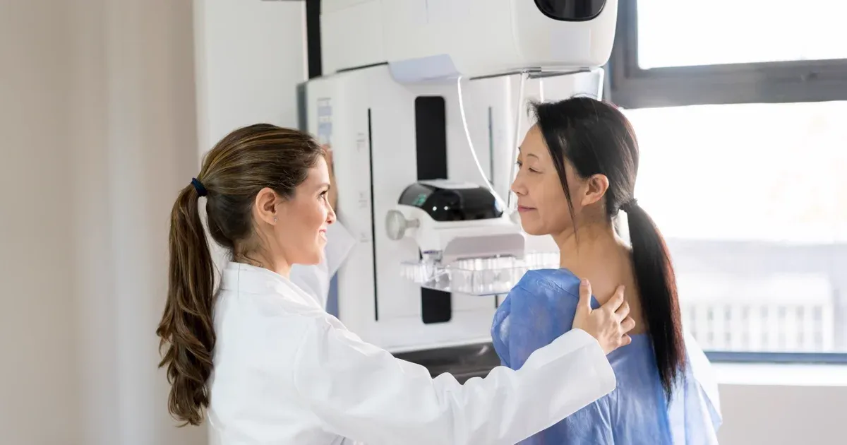
Mammogram Preparation Dubai: What to Expect (2026)
Weiterlesen
CA-125 Test Dubai: Ovarian Cancer Marker (2026)
WeiterlesenMammogram vs Breast Ultrasound Dubai (2026)
Weiterlesen© 2026 Doctors Clinic Diagnostic Center (DCDC), Dubai Healthcare City. Originally published at https://doctorsclinicdubai.ae/blog/breast-cancer-screening-guide. All rights reserved. Unauthorized reproduction is prohibited.