اہم نکات
- MRI is the single most important diagnostic tool for multiple sclerosis — it detects the white matter demyelinating lesions (plaques) that are the hallmark of MS with sensitivity exceeding 95 percent
- The McDonald diagnostic criteria for MS rely heavily on MRI findings, using concepts of dissemination in space (lesions in multiple characteristic brain regions) and dissemination in time (new lesions developing on follow-up scans)
- Gadolinium contrast enhancement on MRI distinguishes active, currently inflamed lesions from older, inactive plaques — a critical distinction for treatment decisions
- Both brain and spinal cord MRI are essential for complete MS evaluation, as approximately 80 to 90 percent of MS patients have brain lesions and 30 to 40 percent have spinal cord lesions at diagnosis
- MS patients typically undergo MRI every 6 to 12 months to monitor disease activity and treatment response, with the goal of achieving no evidence of disease activity (NEDA)
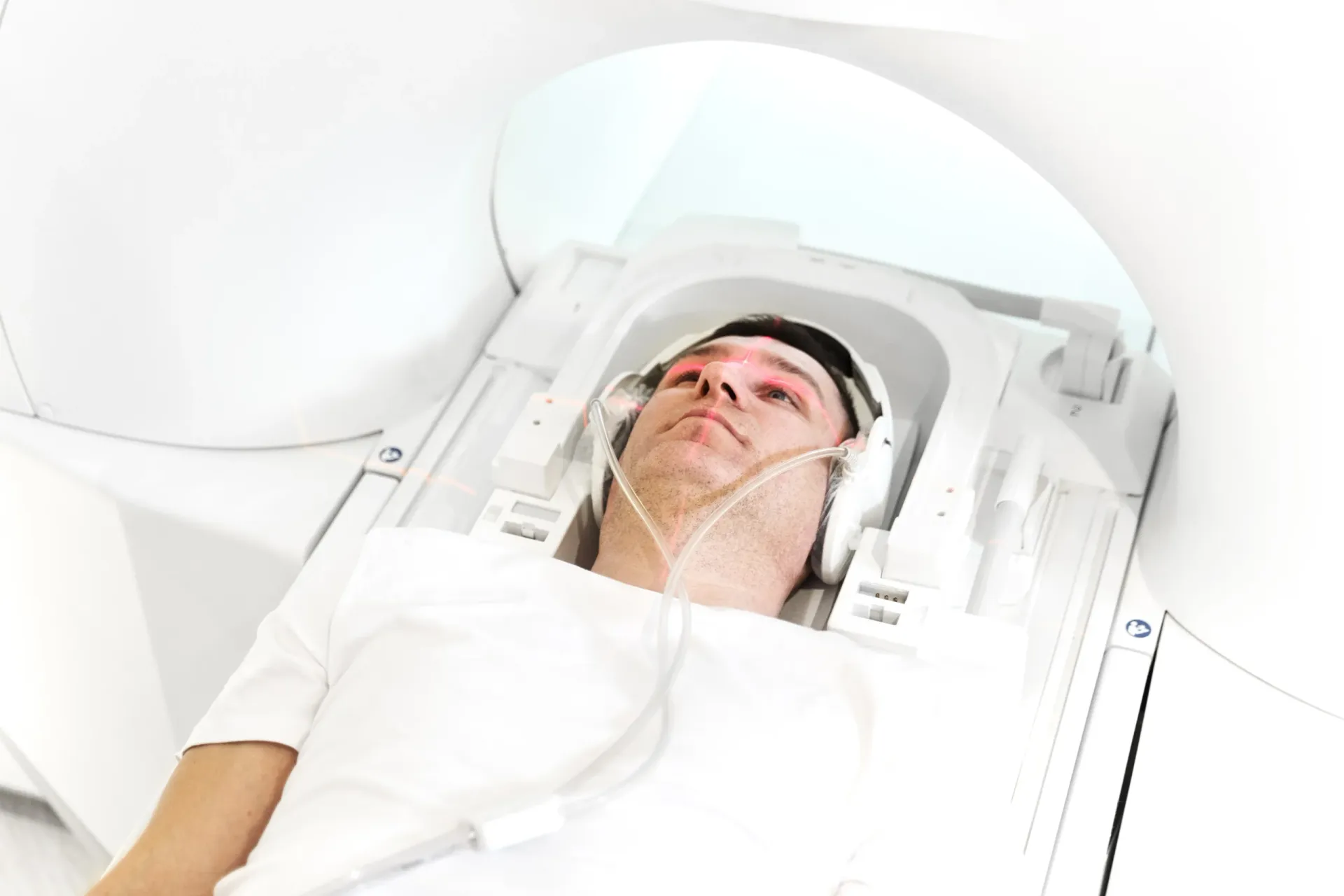
Multiple sclerosis (MS) is a chronic autoimmune disease of the central nervous system in which the immune system attacks the myelin sheath — the protective insulation around nerve fibers in the brain and spinal cord. MRI has revolutionized the diagnosis and management of MS. Before MRI, diagnosing MS required years of clinical observation, multiple neurological episodes, and exclusion of numerous other conditions. Today, MRI can detect MS lesions before symptoms even appear and enables neurologists to monitor disease activity with unprecedented precision.


What Do MS Lesions Look Like on MRI?
Multiple sclerosis lesions appear on MRI as areas of abnormal signal in the white matter of the brain and spinal cord. These lesions represent areas of demyelination — where the immune system has damaged or destroyed the myelin sheath surrounding nerve fibers. Understanding how MS lesions appear on different MRI sequences is essential for diagnosis:
- T2-weighted and FLAIR sequences: MS lesions appear as bright white spots (hyperintense) on T2-weighted and fluid-attenuated inversion recovery (FLAIR) images. FLAIR is particularly valuable because it suppresses the signal from cerebrospinal fluid, making lesions near the ventricles (periventricular lesions) — which are the most characteristic MS lesions — much easier to see. These sequences detect both active and inactive lesions.
- T1-weighted sequences: On standard T1-weighted images, some chronic MS lesions appear as dark spots (hypointense), often called "T1 black holes." These represent areas of more severe tissue destruction where not just myelin but also the underlying nerve fibers (axons) have been damaged. The number and volume of T1 black holes correlate with disability progression.
- Gadolinium-enhanced T1-weighted sequences: When gadolinium contrast dye is administered intravenously, active MS lesions light up (enhance) on T1-weighted images. Enhancement indicates active inflammation and breakdown of the blood-brain barrier — meaning the immune system is currently attacking myelin in that area. Enhancement typically lasts 2 to 8 weeks after a new lesion forms.
- Diffusion-weighted imaging (DWI): Acute MS lesions may show restricted diffusion, helping to differentiate them from other white matter abnormalities such as small vessel disease or other inflammatory conditions.
MS lesions have a characteristic distribution pattern that experienced radiologists recognize. They typically occur in periventricular (adjacent to the brain's ventricles), juxtacortical (at the junction of gray and white matter), infratentorial (brainstem and cerebellum), and spinal cord locations. This distribution pattern is so characteristic that it forms the basis of the McDonald diagnostic criteria for MS.
The McDonald Criteria: How MRI Diagnoses MS
The McDonald criteria, first established in 2001 and most recently revised in 2017, are the internationally accepted diagnostic criteria for multiple sclerosis. These criteria place MRI at the center of MS diagnosis, allowing earlier and more accurate diagnosis than ever before. The two fundamental concepts are:
- Dissemination in space (DIS): MS is characterized by lesions in multiple areas of the central nervous system. The McDonald criteria require at least one T2-hyperintense lesion in at least two of four characteristic locations: periventricular, cortical or juxtacortical, infratentorial, and spinal cord. This demonstrates that the disease is widespread rather than confined to a single area.
- Dissemination in time (DIT): MS is an ongoing disease with new lesions developing over time. DIT can be demonstrated in two ways on MRI: (1) the simultaneous presence of gadolinium-enhancing and non-enhancing lesions on a single scan, which proves that some lesions are new and others are old; or (2) a new T2-hyperintense or gadolinium-enhancing lesion on a follow-up MRI compared to a baseline scan, regardless of the time between scans.
The 2017 McDonald criteria allow MS to be diagnosed on the basis of a single MRI scan if both DIS and DIT criteria are met simultaneously — for example, if the scan shows lesions in at least two characteristic locations and both enhancing and non-enhancing lesions are present. This has dramatically shortened the time to diagnosis, allowing patients to begin disease-modifying therapy earlier when it is most effective.
"The McDonald criteria have transformed MS diagnosis," explains Dr. Osama Elzamzami, Consultant Radiologist at DCDC. "In the past, patients might wait years for a definitive diagnosis, experiencing multiple clinical attacks before MS could be confirmed. Today, with high-quality MRI and an experienced radiologist, we can often make the diagnosis after a single clinical episode and a single MRI scan. Early diagnosis means early treatment, which significantly improves long-term outcomes."
Brain MRI vs Spinal Cord MRI for Multiple Sclerosis
A complete MS evaluation requires imaging of both the brain and spinal cord. Each provides unique and complementary information about the disease:
| Factor | Brain MRI | Spinal Cord MRI |
|---|---|---|
| Lesion prevalence at diagnosis | 85 – 95% of MS patients | 30 – 40% of MS patients |
| Characteristic lesion locations | Periventricular, juxtacortical, infratentorial | Cervical cord (most common), thoracic cord |
| Lesion appearance | Ovoid, perpendicular to ventricles (Dawson fingers) | Short-segment (< 2 vertebral segments), peripheral |
| Role in diagnosis | Primary diagnostic tool, DIS and DIT criteria | Adds DIS location, helps differentiate from NMO |
| Correlation with disability | Moderate (lesion volume, brain atrophy) | Strong — spinal cord lesions correlate closely with physical disability |
| Monitoring frequency | Every 6 – 12 months | Less frequent, typically with symptom changes or baseline |
Both brain and spinal cord MRI are essential for comprehensive MS evaluation.
Brain MRI is the primary diagnostic and monitoring tool because it has the highest sensitivity for detecting MS lesions — approximately 95 percent of MS patients have visible brain lesions on MRI. The classic brain MRI finding in MS is Dawson fingers — ovoid periventricular lesions oriented perpendicular to the lateral ventricles, representing demyelination along the perivenular white matter tracts.
Spinal cord MRI is particularly important for several reasons. First, it adds a fourth location for DIS criteria, potentially enabling diagnosis in patients with fewer brain lesions. Second, spinal cord lesions correlate more strongly with physical disability than brain lesions, making spinal cord imaging crucial for predicting disease outcomes. Third, the pattern of spinal cord lesions helps differentiate MS from neuromyelitis optica spectrum disorder (NMOSD or NMO), which produces longer lesions spanning three or more vertebral segments — a finding atypical for MS.
Book Brain & Spine MRI for MS Evaluation
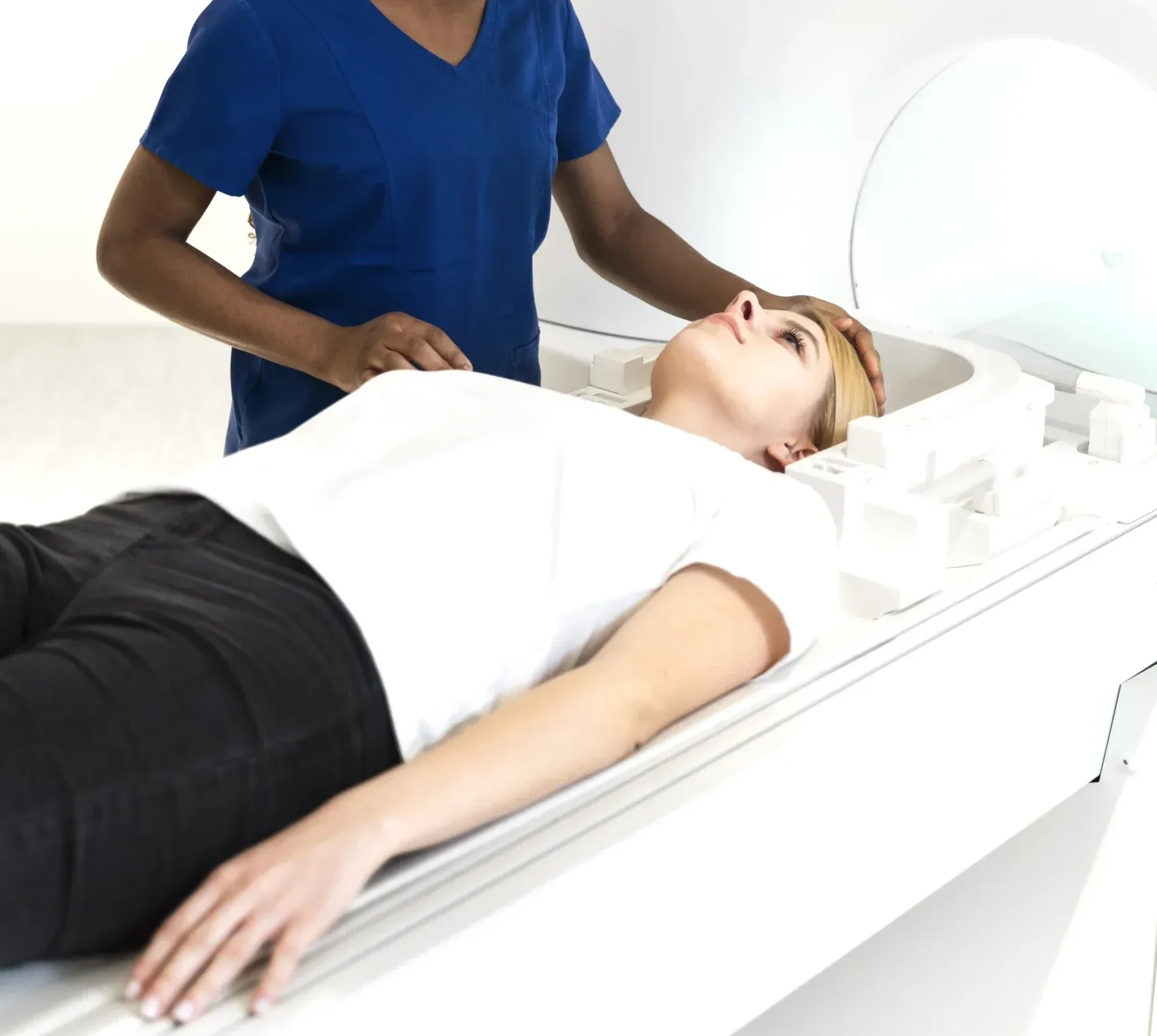
At Doctors Clinic Diagnostic Center in Dubai Healthcare City, we offer comprehensive brain and spinal cord MRI with dedicated MS protocols. Our consultant radiologist provides detailed reports correlating findings with the McDonald criteria.
Gadolinium-Enhancing Lesions: Active vs Old MS Plaques
One of the most clinically important distinctions on MS MRI is between active (enhancing) lesions and inactive (non-enhancing) lesions. This distinction is made possible by gadolinium contrast dye, which is administered intravenously during the MRI examination.
- Active (enhancing) lesions: When a new MS lesion forms, the immune system's inflammatory attack on the myelin sheath causes breakdown of the blood-brain barrier (BBB). Gadolinium, which normally cannot cross an intact BBB, leaks into the area of active inflammation and causes the lesion to appear bright on T1-weighted images. Active enhancement typically persists for 2 to 8 weeks. These lesions represent current disease activity and may or may not be associated with clinical symptoms (many MS lesions are clinically silent).
- Inactive (non-enhancing) lesions: Once the acute inflammatory phase subsides and the BBB repairs itself, the lesion no longer enhances with gadolinium. These older lesions remain visible as bright spots on T2-weighted and FLAIR images but do not enhance on post-contrast T1 images. They represent areas where demyelination has already occurred, though some degree of remyelination (repair) may happen in some lesions over time.
- T1 black holes: Some chronic, non-enhancing lesions evolve into T1 black holes — permanently dark areas on T1-weighted images indicating severe tissue destruction with axonal loss. These represent irreversible damage and correlate with clinical disability. Not all MS lesions become black holes; some resolve or remain isointense on T1.
The presence of gadolinium-enhancing lesions is a key indicator of disease activity that directly influences treatment decisions. If a patient on disease-modifying therapy shows new enhancing lesions, the neurologist may consider escalating to a more potent medication. Conversely, the absence of enhancing lesions on serial MRI scans is one component of the NEDA (No Evidence of Disease Activity) treatment target, which also includes no clinical relapses and no disability progression.
It is worth noting that not every enhancing lesion produces symptoms. MRI is estimated to detect 5 to 10 times more disease activity than clinical assessment alone. This subclinical disease activity — new lesions forming without noticeable symptoms — is one of the primary reasons why regular MRI monitoring is so important in MS management.
How Often Do MS Patients Need MRI Scans?
Regular MRI monitoring is a cornerstone of modern MS management. The frequency of MRI scans depends on the patient's disease stage, treatment status, and clinical stability:
- At diagnosis (baseline): A comprehensive baseline MRI of the brain and spinal cord (with and without gadolinium contrast) is performed. This scan establishes the lesion burden, distribution, and activity level against which all future scans will be compared.
- 3 to 6 months after starting treatment: A follow-up brain MRI is typically performed 3 to 6 months after initiating disease-modifying therapy to establish a new baseline on treatment. This early scan detects any disease activity that may have occurred during the interval before the medication reached full efficacy.
- Every 6 to 12 months during treatment: Routine monitoring MRI is recommended every 6 to 12 months for most MS patients. The more aggressive the disease or the newer the treatment, the more frequently MRI may be performed. Each scan is compared to previous images to detect new or enlarging T2 lesions and any gadolinium-enhancing activity.
- When symptoms worsen or change: Any new neurological symptoms, suspected relapse, or unexplained deterioration should prompt MRI to assess for new disease activity and to distinguish true MS relapses from pseudo-relapses (worsening of existing symptoms due to heat, infection, or stress).
- Less frequent monitoring (long-term stable patients): Patients who have been stable on treatment for several years — with no new lesions, no relapses, and no disability progression — may be able to extend their MRI monitoring interval to every 12 to 24 months, though this should be decided by the neurologist on an individual basis.
The concept of NEDA (No Evidence of Disease Activity) has become the treatment target for modern MS management. NEDA is achieved when a patient has: (1) no clinical relapses, (2) no sustained disability progression, and (3) no new or enlarging T2 lesions and no gadolinium-enhancing lesions on MRI. Achieving NEDA requires regular MRI monitoring as the cornerstone of treatment assessment.
Cost of MRI for Multiple Sclerosis in Dubai
Because MS management requires both brain and spinal cord MRI, often with contrast, and regular monitoring over years to decades, understanding the cost structure is important for patients:
| MRI Protocol | Approximate Cost (AED) | When Used |
|---|---|---|
| Brain MRI with contrast (MS protocol) | 1,500 – 2,000 | Baseline and monitoring scans |
| Cervical spine MRI with contrast | 1,500 – 2,000 | Baseline assessment, symptom changes |
| Brain + cervical spine MRI (combined) | 2,500 – 3,500 | Comprehensive evaluation |
| Brain + full spine MRI (combined) | 3,500 – 4,500 | Complete baseline or diagnostic workup |
Prices are approximate and include radiologist report at DCDC. Insurance typically covers MS-related MRI with neurologist referral.
MS-related MRI scans are covered by virtually all health insurance plans in the UAE when ordered by a neurologist with appropriate documentation. Given the chronic nature of MS and the need for serial imaging, insurance authorization is critical. At Doctors Clinic Diagnostic Center, our administrative team assists with insurance pre-authorization to ensure coverage for your MS monitoring scans.
Expert MS MRI Interpretation at DCDC
Every MS MRI at Doctors Clinic Diagnostic Center is interpreted by a consultant radiologist experienced in neuroradiology. Our structured reports include detailed lesion counts, comparisons with prior scans, and gadolinium enhancement assessment to support your neurologist's treatment decisions. Learn more about our MRI services.
DCDC میں متعلقہ خدمات
دبئی ہیلتھ کیئر سٹی میں ماہرانہ دیکھ بھال اور جدید تشخیص
اکثر پوچھے گئے سوالات
آخری خیالات
MRI is indispensable in every stage of multiple sclerosis management — from initial diagnosis through long-term monitoring of disease activity and treatment response. The ability to visualize demyelinating lesions, distinguish active from inactive disease, and detect subclinical activity that would otherwise go unnoticed makes MRI the most powerful tool in the MS neurologist's arsenal.
At Doctors Clinic Diagnostic Center in Dubai Healthcare City, our consultant radiologists provide expert MS MRI interpretation with detailed lesion tracking, comparison with prior studies, and clear communication of findings to your neurologist. Whether you are undergoing your first diagnostic MRI or a routine monitoring scan, our team delivers the imaging quality and expertise that MS management demands.
ذرائع اور حوالہ جات
یہ مضمون ہماری طبی ٹیم نے جائزہ لیا ہے اور درج ذیل ذرائع کا حوالہ دیتا ہے:
- Thompson AJ et al. - Diagnosis of multiple sclerosis: 2017 revisions of the McDonald criteria, The Lancet Neurology
- National Multiple Sclerosis Society - MRI and MS
- Consortium of MS Centers - MRI Protocol Guidelines
- Radiological Society of North America - MS Imaging
- American Academy of Neurology - Practice Guidelines for MS
اس سائٹ پر طبی مواد کا جائزہ DHA لائسنس یافتہ ڈاکٹرز نے لیا ہے۔ ہماری دیکھیں تحریری پالیسی مزید معلومات کے لیے۔

تحریر
Dr. Osama Elzamzami
Diagnostic Radiology
MD, FRCR
Dr. Osama Elzamzami is a Consultant Radiologist specializing in diagnostic imaging including MRI, CT, and ultrasound at DCDC Dubai Healthcare City.
متعلقہ مضامین
Brain MRI in Dubai: What It Detects, Cost & When You Need One
MRI with Contrast: What to Expect, Safety & Cost
MRI for Headaches: When You Need a Brain Scan for Migraines
Spine MRI in Dubai: Cost, What It Shows & When You Need One
MRI Cost in Dubai: Complete Guide to Types & Pricing (2026)
Diagnostic Imaging میں مزید
CT Scan Cost Dubai: Prices & Types (2026)
مزید پڑھیں
DEXA Scan Dubai: Cost, T-Score & Who Needs It (2026)
مزید پڑھیں![Full Body MRI Cost Dubai: AED 5,000-15,000 [2026]](/wp-media/blog/full-body-mri-scan.webp)
Full Body MRI Cost Dubai: AED 5,000-15,000 [2026]
مزید پڑھیں
MRI Preparation Dubai: Complete Guide (2026)
مزید پڑھیں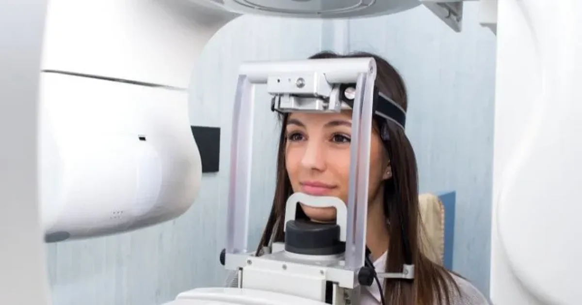
CBCT Scan: 3D Dental Imaging Guide Dubai (2026)
مزید پڑھیں
Ultrasound Preparation Dubai: What to Expect (2026)
مزید پڑھیں© 2026 Doctors Clinic Diagnostic Center (DCDC), Dubai Healthcare City. Originally published at https://doctorsclinicdubai.ae/blog/mri-for-multiple-sclerosis. All rights reserved. Unauthorized reproduction is prohibited.