Ключевые выводы
- An echocardiogram uses ultrasound to create real-time images of the heart, showing valve function, pumping strength (ejection fraction), wall motion, and blood flow
- Four main types exist: TTE (standard), TEE (through esophagus), stress echo, and Doppler echo, each suited to different clinical questions
- The standard TTE takes 20-40 minutes, requires no preparation, is painless, and uses no radiation
- Echocardiogram cost in Dubai starts from approximately AED 850 for a standard TTE with Doppler
- Common reasons for ordering include shortness of breath, heart murmur, chest pain, heart failure monitoring, and pre-surgical cardiac clearance
An echocardiogram (commonly called an "echo test") is a non-invasive ultrasound examination that creates real-time moving images of your heart. It shows how your heart valves open and close, how strongly your heart muscle contracts, how blood flows through the chambers, and whether any structural abnormalities are present. As one of the most important tests in cardiology, an echocardiogram at DCDC provides critical diagnostic information without radiation, injections, or recovery time.
Whether your doctor has detected a heart murmur, you are experiencing unexplained shortness of breath, or you need cardiac clearance before surgery, an echocardiogram is likely the first and most informative imaging test that will be ordered. This guide covers all four types of echo testing, what each reveals about your heart, how to prepare, what the procedure involves, and the cost in Dubai.

What Does an Echocardiogram Show?
An echocardiogram provides a remarkable amount of information about heart structure and function in a single non-invasive test:
- Ejection fraction (EF): The percentage of blood pumped out of the left ventricle with each heartbeat. Normal is 55-70%. Below 40% indicates heart failure. This is the single most important number in cardiology for assessing heart function.
- Valve function: Shows whether heart valves open fully (stenosis if they do not) and close completely (regurgitation if they leak). All four valves (mitral, aortic, tricuspid, pulmonary) are assessed.
- Wall motion: Reveals whether all regions of the heart muscle are contracting normally. Areas that are not moving properly may indicate previous heart attack damage or reduced blood supply.
- Chamber size: Measures the dimensions of all four heart chambers. Enlarged chambers can indicate valve disease, heart failure, or chronic high blood pressure.
- Pericardial effusion: Detects fluid around the heart, which can cause compression (tamponade) if severe.
- Blood flow patterns: Doppler technology shows the speed and direction of blood flow through the heart, detecting abnormal flow patterns that indicate valve disease or congenital defects.
- Pulmonary artery pressure: Estimates pressure in the lung circulation, which is elevated in pulmonary hypertension.
- Congenital defects: Holes between chambers (ASD, VSD), abnormal vessel connections, and other structural birth defects.
Types of Echocardiogram
Transthoracic Echocardiogram (TTE)
TTE is the standard echocardiogram and by far the most commonly performed type. The sonographer places an ultrasound probe (transducer) on different positions on your chest wall to obtain images of the heart from various angles. No instruments enter your body. The test takes 20-40 minutes, requires no preparation, and you can resume all activities immediately afterward.
TTE includes 2D imaging (real-time moving pictures of the heart), M-mode (a single line of ultrasound used for precise measurements), and Doppler assessment (measuring blood flow velocity and direction). Modern echo machines also offer tissue Doppler imaging and strain analysis for more detailed assessment of heart muscle function.
Transesophageal Echocardiogram (TEE)
TEE involves passing a specialized ultrasound probe through the mouth into the esophagus (food pipe), which sits directly behind the heart. Because the esophagus is much closer to the heart than the chest wall, TEE produces significantly higher-resolution images, especially of the posterior (back) structures including the mitral valve, left atrium, and aorta.
TEE is ordered when TTE images are suboptimal (due to body habitus, lung disease, or chest wall anatomy), when detailed valve assessment is needed before surgery, when looking for blood clots in the left atrial appendage (common before cardioversion for atrial fibrillation), or for evaluating endocarditis (infection of heart valves). TEE requires fasting, sedation, and local anesthetic throat spray.
Stress Echocardiogram
A stress echo combines echocardiography with exercise or pharmacological stress to evaluate how the heart performs under increased demand. Echo images are taken at rest and immediately after peak exercise (on a treadmill or stationary bike). If you cannot exercise, a medication called dobutamine is infused through an IV to simulate the heart rate increase of exercise.
The key finding on stress echo is whether any wall segments that moved normally at rest develop abnormal motion during stress, which indicates reduced blood supply to that region (ischemia) from coronary artery disease. Stress echo has a sensitivity of approximately 80-85% and specificity of 85-90% for detecting significant coronary artery disease. For more on cardiac stress testing, see our guide on stress test in Dubai.
Doppler Echocardiogram
Doppler is not a separate test but a component of every standard echocardiogram. It uses the Doppler effect (change in frequency of sound waves reflecting off moving blood cells) to measure the speed and direction of blood flow throughout the heart. Color Doppler overlays a color map on the 2D image, showing blood flowing toward the probe in red and away in blue. Continuous-wave and pulsed-wave Doppler provide precise velocity measurements at specific points.
Doppler is essential for quantifying valve stenosis severity (measuring the pressure gradient across a narrowed valve) and regurgitation severity (measuring the volume and velocity of backward blood flow through a leaking valve).
When Does a Cardiologist Order an Echocardiogram?
Echocardiography is one of the most frequently ordered cardiac tests. Common reasons include:
- Heart murmur detected on examination: To determine whether the murmur represents significant valve disease
- Shortness of breath: To assess heart function (ejection fraction) and rule out heart failure, valve disease, or pericardial effusion
- Chest pain: To check for wall motion abnormalities suggesting coronary artery disease or previous heart attack
- Heart failure monitoring: Regular echos track ejection fraction and guide medication adjustment
- Atrial fibrillation: To assess heart structure and check for clots before cardioversion
- Pre-surgical cardiac clearance: To ensure heart function is adequate before major non-cardiac surgery
- Hypertension assessment: To check for left ventricular hypertrophy (thickened heart muscle from chronic high blood pressure)
- After heart attack: To assess damage, measure ejection fraction, and guide rehabilitation
- Congenital heart disease: To evaluate and monitor structural heart defects
- Follow-up after valve surgery or replacement: To verify prosthetic valve function
Echocardiogram Preparation and Procedure
For Standard TTE
No preparation is required. You can eat, drink, and take your medications normally. You will be asked to remove clothing above the waist and wear a hospital gown. You lie on your left side on an examination bed while the sonographer applies ultrasound gel and moves the probe across different chest positions. You may be asked to change position, hold your breath briefly, or exhale during certain measurements. The test takes 20 to 40 minutes and is completely painless.
For TEE
Fasting for 4-6 hours is required because of the esophageal probe and sedation. You will receive a local anesthetic spray to numb the throat and intravenous sedation for comfort. The probe is guided through the mouth into the esophagus under your conscious cooperation. The procedure takes 20-30 minutes, and you will need 1-2 hours of recovery from sedation. You should not drive for the rest of the day.
For Stress Echo
Wear comfortable exercise clothing and shoes if treadmill exercise is planned. Avoid heavy meals for 2 hours before. Some medications may need to be held (your doctor will specify). The complete test including rest images, exercise, and recovery images takes approximately 45-60 minutes.
Echocardiogram vs ECG vs Cardiac MRI
Patients often ask how an echocardiogram compares to other heart tests. Each provides different but complementary information.
| Feature | Echocardiogram | ECG (Electrocardiogram) | Cardiac MRI |
|---|---|---|---|
| What it assesses | Heart structure, valves, pumping function, blood flow | Heart rhythm, rate, electrical conduction | Heart structure, muscle damage, fibrosis, function |
| Technology | Ultrasound waves | Electrical sensors on skin | Magnetic field and radio waves |
| Duration | 20-40 minutes | 5-10 minutes | 45-60 minutes |
| Radiation | None | None | None |
| Cost in Dubai (AED) | 850 - 2,000 | 200 - 500 | 3,000 - 6,000 |
| Detects valve disease | Yes - primary test | No | Yes - less commonly used for this |
| Detects arrhythmia | No | Yes - primary test | No |
| Measures ejection fraction | Yes - standard measurement | No | Yes - gold standard accuracy |
| Detects heart attack damage | Yes - wall motion abnormalities | Yes - ST changes, Q waves | Yes - with high precision (scar mapping) |
| Availability | Very widely available | Available everywhere | Limited to specialized centers |
ECG and echo are complementary tests. ECG evaluates electrical activity; echo evaluates structural and functional aspects. Cardiac MRI provides the most detailed tissue characterization.
In most clinical scenarios, a cardiologist will order both an ECG and an echocardiogram because they provide different types of information that together give a comprehensive picture of heart health. For the most detailed heart muscle assessment including scar mapping after heart attack, a cardiac MRI may be recommended.
Echocardiogram Cost in Dubai
| Type | Cost Range (AED) | What's Included |
|---|---|---|
| Standard TTE with Doppler | 850 - 2,000 | 2D echo, M-mode, color Doppler, measurements |
| TTE with cardiologist consultation | 1,500 - 3,000 | Echo test plus specialist review and recommendations |
| TEE (transesophageal) | 2,500 - 5,000 | Includes sedation, procedure, and monitoring |
| Stress echocardiogram (exercise) | 2,000 - 4,000 | Resting and exercise echo with interpretation |
| Stress echocardiogram (dobutamine) | 2,500 - 5,000 | Pharmacological stress with echo and monitoring |
| Pediatric echocardiogram | 1,000 - 2,500 | Specialized assessment for congenital heart disease |
Echo testing is typically covered by insurance when ordered by a physician. TTE is the most commonly performed and most affordable type.
Most insurance plans in Dubai cover echocardiography when ordered by a physician, particularly when there is a clear medical indication such as a murmur, symptoms of heart failure, or pre-surgical assessment. A cardiology consultation ensures the appropriate type of echo is ordered and properly interpreted.
"The echocardiogram is the cardiologist's stethoscope of the 21st century. It provides more diagnostic information about heart structure and function in 30 minutes than any other single cardiac test. I recommend it as a baseline for anyone with cardiac symptoms, risk factors, or need for surgical clearance," observes Dr. Osama Elzamzami.
Book Your Echocardiogram in Dubai
Our echocardiogram service provides comprehensive heart assessment with same-day results and expert interpretation.
Cardiac Services at DCDC Dubai Healthcare City
At Doctors Clinic Diagnostic Center, our cardiology team offers complete echo testing including TTE, stress echo, and Doppler assessment. Located in Dubai Healthcare City.
Связанные услуги в DCDC
Квалифицированная помощь и современная диагностика в Dubai Healthcare City
Frequently Asked Questions
Final Thoughts
The echocardiogram is one of the most valuable, versatile, and patient-friendly diagnostic tests in all of cardiology. It provides detailed information about heart structure, valve function, pumping strength, and blood flow patterns without radiation, needles, or recovery time. From detecting subtle valve disease to monitoring heart failure treatment, echo testing is central to cardiac care.
If your doctor has recommended an echocardiogram, the test is a straightforward, painless experience that typically takes less than an hour and provides information that directly guides your care. The cost is modest relative to its diagnostic value, and insurance coverage is broad for medically indicated studies.
For comprehensive echocardiography in Dubai, Doctors Clinic Diagnostic Center in Dubai Healthcare City offers expert cardiac ultrasound with same-day results and integrated cardiology services.
Источники и ссылки
Эта статья проверена нашей медицинской командой и ссылается на следующие источники:
- American Society of Echocardiography - Echo Standards
- European Association of Cardiovascular Imaging
- American College of Cardiology - Appropriate Use Criteria for Echo
- British Society of Echocardiography - Echo Standards
- American Heart Association - Echocardiography
Медицинский контент на этом сайте проверяется врачами, лицензированными DHA. См. нашу редакционную политику для получения дополнительной информации.

Автор
Dr. Osama Elzamzami
Consultant Radiologist
MD, Radiology
Dr. Osama Elzamzami is a Consultant Radiologist at DCDC Dubai Healthcare City with expertise in diagnostic imaging and cardiac assessment.
Related Articles
ECG vs Echocardiogram: What Each Heart Test Tells You
Stress Test Dubai: Types, Cost & Preparation
Cardiac MRI Dubai

Heart Health Prevention Dubai
More in Cardiology

Cardiac Rehabilitation Dubai: Recovery (2026)
Читать далееTroponin Test Dubai: Heart Attack Marker (2026)
Читать далее
Best Cardiologist in Dubai: Dr. Shahoo Mazhari at DCDC (2026)
Читать далееCardiologist in Dubai Healthcare City: Dr. Shahoo Mazhari at DCDC (2026)
Читать далее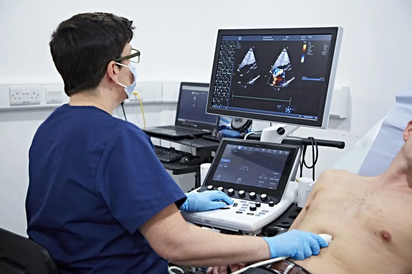
Echocardiogram (Echo) Test in Dubai: Cost, Procedure & What to Expect (2026)
Читать далееIranian Cardiologist in Dubai: Dr. Shahoo Mazhari at DCDC (2026)
Читать далее© 2026 Doctors Clinic Diagnostic Center (DCDC), Dubai Healthcare City. Originally published at https://doctorsclinicdubai.ae/blog/echocardiogram-guide-dubai. All rights reserved. Unauthorized reproduction is prohibited.