मुख्य बातें
- कम हृदय गति बनाए रखने में मदद के लिए अपने सीटी एंजियोग्राम से कम से कम 24-48 घंटे पहले कैफीन से बचें
- उच्च गुणवत्ता वाली छवियों के लिए 65 बीट प्रति मिनट से कम आराम की हृदय गति आदर्श है; यदि आवश्यक हो तो बीटा-ब्लॉकर्स दिए जा सकते हैं
- स्कैन से 4-6 घंटे पहले उपवास करें, लेकिन निर्धारित दवाएं पानी के साथ जारी रखें
- कंट्रास्ट डाई देने से पहले गुर्दे की कार्यक्षमता (क्रिएटिनिन) की जांच होनी चाहिए
- आरामदायक, ढीले-ढाले कपड़े पहनें और स्कैन से पहले सभी धातु के आभूषण हटा दें
सफल सीटी एंजियोग्राम के लिए उचित तैयारी आवश्यक है। कई अन्य इमेजिंग परीक्षणों के विपरीत, कोरोनरी सीटी एंजियोग्राफी के लिए आपकी अपॉइंटमेंट से पहले दिनों और घंटों में विशिष्ट कदमों की आवश्यकता होती है ताकि आपके हृदय की धमनियों की सबसे स्पष्ट छवियां प्राप्त हो सकें। इन दिशानिर्देशों का बारीकी से पालन करना एक सफल डायग्नोस्टिक स्कैन और दोहराए जाने की आवश्यकता वाले स्कैन के बीच का अंतर हो सकता है।
यह चेकलिस्ट आपके सीटी एंजियोग्राम से पहले जानने योग्य हर बात को कवर करती है, आहार प्रतिबंधों और दवा संबंधी विचारों से लेकर क्लिनिक पहुंचने पर क्या होता है तक।

सीटी एंजियोग्राम के लिए तैयारी क्यों महत्वपूर्ण है
कोरोनरी सीटी एंजियोग्राम एक दिल की धड़कन के दौरान आपके हृदय की धमनियों की छवियां कैप्चर करता है। चूंकि हृदय लगातार गति में रहता है, स्कैनर को आपकी हृदय गति धीमी और स्थिर होनी चाहिए ताकि तेज, गति-मुक्त छवियां उत्पन्न हो सकें। कैफीन का सेवन, तनाव, निर्जलीकरण और कुछ दवाएं जैसे कारक आपकी हृदय गति बढ़ा सकते हैं और छवि गुणवत्ता कम कर सकते हैं।
इसके अतिरिक्त, स्कैन में अंतःशिरा कंट्रास्ट डाई शामिल होती है जो छवियों में आपकी धमनियों को उजागर करती है। इस कंट्रास्ट के लिए पर्याप्त गुर्दे की कार्यक्षमता और जलयोजन की आवश्यकता होती है ताकि आपका शरीर इसे सुरक्षित रूप से प्रोसेस कर सके। उचित तैयारी इन सभी कारकों को संबोधित करती है।
आपके सीटी एंजियोग्राम से 48 घंटे पहले
कैफीन को पूरी तरह बंद करें
कैफीन एक उत्तेजक है जो हृदय गति बढ़ाता है और इसे अनियमित बना सकता है। आपको अपने स्कैन से कम से कम 24 से 48 घंटे पहले कैफीन के सभी स्रोतों से बचना चाहिए। इसमें कॉफी, चाय, एनर्जी ड्रिंक, चॉकलेट, कैफीन युक्त सॉफ्ट ड्रिंक और कैफीन युक्त कुछ दवाएं या सप्लीमेंट शामिल हैं।
कई मरीज कम आंकते हैं कि कैफीन उनकी हृदय गति को कितना प्रभावित करता है। यहां तक कि डीकैफिनेटेड कॉफी में भी थोड़ी मात्रा होती है। यदि आप नियमित कैफीन उपभोक्ता हैं, तो स्कैन से दो पूरे दिन पहले प्रतिबंध शुरू करना आपके शरीर को समायोजित होने का समय देता है और विथड्रॉल-संबंधित हृदय गति उतार-चढ़ाव से बचने में मदद करता है।
हाल ही में हमारे पास एक मरीज था जिसने अपने निर्धारित सीटी एंजियोग्राम की सुबह एक कप कॉफी पी ली, यह समझे बिना कि यह उसके स्कैन को कितना प्रभावित करेगा। जब वह DCDC पहुंचा, उसकी हृदय गति 92 बीट प्रति मिनट थी, स्पष्ट छवियों के लिए आवश्यक 65 बीट प्रति मिनट के लक्ष्य से काफी ऊपर। बीटा-ब्लॉकर्स देने के बावजूद, हम उसकी हृदय गति को पर्याप्त रूप से कम नहीं कर सके और स्कैन को अगले सप्ताह के लिए पुनर्निर्धारित करना पड़ा। मरीज ने बाद में हमें बताया कि काश उसने कैफीन प्रतिबंध को अधिक गंभीरता से लिया होता। उसका अनुभव एक अनुस्मारक है कि तैयारी का यह एक कदम निर्धारित कर सकता है कि आपका स्कैन समय पर होगा या फिर से आना पड़ेगा।
"कैफीन सीटी एंजियोग्राम को पुनर्निर्धारित करने का नंबर एक कारण है," DCDC के सलाहकार हृदय रोग विशेषज्ञ डॉ. शाहू मज़हरी कहते हैं। "मरीज अक्सर सोचते हैं कि एक कप कॉफी से फर्क नहीं पड़ेगा, लेकिन थोड़ी सी मात्रा भी हृदय गति को इतना बढ़ा सकती है कि छवि गुणवत्ता से समझौता हो। 48 घंटे का कैफीन-मुक्त अवधि वैकल्पिक नहीं है — यह आवश्यक है।"
अपने डॉक्टर के साथ अपनी दवाओं की समीक्षा करें
कुछ दवाओं को आपके सीटी एंजियोग्राम से पहले समायोजित करने की आवश्यकता हो सकती है। अपनी सभी वर्तमान दवाओं के बारे में अपने हृदय रोग विशेषज्ञ या इमेजिंग सेंटर से चर्चा करें, जिसमें शामिल हैं:
- बीटा-ब्लॉकर्स: यदि आप पहले से बीटा-ब्लॉकर्स ले रहे हैं, तो आपका डॉक्टर 65 बीट प्रति मिनट से कम लक्षित हृदय गति प्राप्त करने में मदद के लिए खुराक समायोजित कर सकता है।
- मेटफॉर्मिन: मेटफॉर्मिन लेने वाले मधुमेह के मरीजों को कंट्रास्ट डाई और गुर्दे की कार्यक्षमता के बीच संभावित इंटरैक्शन के कारण स्कैन के बाद 48 घंटे के लिए इसे बंद करने की सलाह दी जा सकती है।
- स्तंभन दोष की दवाएं: सिल्डेनाफिल (वियाग्रा) जैसी दवाओं को आमतौर पर 48 घंटे पहले बंद कर देना चाहिए, क्योंकि ये नाइट्रोग्लिसरीन के साथ इंटरैक्ट कर सकती हैं जो स्कैन के दौरान उपयोग हो सकती है।
- रक्त पतला करने वाली दवाएं: आमतौर पर सामान्य रूप से जारी रखें, लेकिन टीम को सभी एंटीकोआगुलेंट्स या एंटीप्लेटलेट एजेंट्स के बारे में सूचित करें जो आप ले रहे हैं।
अच्छी तरह हाइड्रेटेड रहें
अच्छा जलयोजन गुर्दे की कार्यक्षमता का समर्थन करता है, जो स्कैन के दौरान उपयोग की जाने वाली कंट्रास्ट डाई को प्रोसेस करने के लिए महत्वपूर्ण है। अपनी अपॉइंटमेंट से पहले दो दिनों में भरपूर पानी पिएं। शराब से बचें, क्योंकि यह निर्जलीकरण का कारण बन सकती है और हृदय ताल को प्रभावित कर सकती है।
आपके सीटी एंजियोग्राम से एक दिन पहले
- कैफीन और शराब से परहेज जारी रखें
- पूरे दिन में कम से कम 1.5 से 2 लीटर पानी पिएं
- धातु की ज़िप, बटन या तार के बिना आरामदायक, ढीले कपड़े तैयार करें
- सभी आभूषण, घड़ियां और पियर्सिंग अलग रखें और घर पर छोड़ दें
- अपनी अपॉइंटमेंट का समय और क्लिनिक के किसी भी विशिष्ट निर्देश की पुष्टि करें
- अच्छी नींद लें; थकान और अपर्याप्त नींद आराम की हृदय गति बढ़ा सकती है
आपके सीटी एंजियोग्राम के दिन
उपवास आवश्यकताएं
आपको अपने निर्धारित स्कैन समय से 4 से 6 घंटे पहले उपवास करना चाहिए। इसका मतलब है सादे पानी के अलावा कोई भोजन या पेय नहीं। उपवास कंट्रास्ट डाई से मतली के जोखिम को कम करता है और स्कैन के दौरान स्थिर शारीरिक स्थितियों को बनाए रखने में मदद करता है।
हालांकि, आपको अपनी निर्धारित दवाएं पानी की छोटी घूंट के साथ जारी रखनी चाहिए जब तक कि आपके डॉक्टर ने विशेष रूप से अन्यथा निर्देश न दिया हो। यह विशेष रूप से रक्तचाप की दवाओं और बीटा-ब्लॉकर्स के लिए महत्वपूर्ण है जो हृदय गति को नियंत्रित रखने में मदद करती हैं।
क्या पहनें और क्या लाएं
- आरामदायक, ढीले कपड़े पहनें (आपको अस्पताल का गाउन दिया जा सकता है)
- सभी धातु की वस्तुएं हटाएं: आभूषण, घड़ियां, बेल्ट, हेयर क्लिप और पियर्सिंग
- अपनी वर्तमान दवाओं और खुराक की सूची लाएं
- हालिया रक्त परीक्षण परिणाम लाएं, विशेष रूप से गुर्दे की कार्यक्षमता (क्रिएटिनिन/eGFR) यदि उपलब्ध हो
- अपना अमीरात आईडी या पासपोर्ट और बीमा कार्ड लाएं
- कोई भी पिछली कार्डियक इमेजिंग रिपोर्ट या रेफरल पत्र लाएं
DCDC में अपने सीटी एंजियोग्राम की तैयारी
दुबई हेल्थकेयर सिटी में डॉक्टर्स क्लिनिक डायग्नोस्टिक सेंटर में, हम हर मरीज को सर्वोत्तम संभव परिणाम सुनिश्चित करने के लिए उचित सीटी एंजियोग्राम की तैयारी में मार्गदर्शन करते हैं।
हृदय गति लक्ष्य: 65 बीपीएम से कम क्यों महत्वपूर्ण है
कोरोनरी सीटी एंजियोग्राम के लिए इष्टतम हृदय गति 65 बीट प्रति मिनट (बीपीएम) से कम है, कुछ केंद्र 60 बीपीएम से कम को प्राथमिकता देते हैं। इन हृदय गतियों पर, हृदय धड़कनों के बीच पर्याप्त समय तक आराम करता है ताकि स्कैनर कोरोनरी धमनियों की स्पष्ट, गति-मुक्त छवियां कैप्चर कर सके।
जब आप क्लिनिक पहुंचेंगे, तो आपकी हृदय गति मापी जाएगी। यदि यह लक्षित सीमा से ऊपर है, तो चिकित्सा टीम इसे कम करने के लिए मुंह से या अंतःशिरा रूप से शॉर्ट-एक्टिंग बीटा-ब्लॉकर (आमतौर पर मेटोप्रोलोल) दे सकती है। यह सीटी एंजियोग्राम की तैयारी का एक नियमित और सुरक्षित हिस्सा है।
जो कारक आपकी हृदय गति बढ़ा सकते हैं और स्कैन के दिन जिनसे बचना चाहिए उनमें कैफीन, धूम्रपान, ज़ोरदार व्यायाम, तनाव और अपॉइंटमेंट के लिए भागदौड़ शामिल हैं। 15 से 20 मिनट पहले पहुंचें ताकि स्कैन से पहले आराम का समय मिले।
"हम DCDC में हर महीने 1,000 से अधिक डायग्नोस्टिक स्कैन करते हैं, और जो मरीज़ सबसे अच्छे परिणाम प्राप्त करते हैं वे वो हैं जो तैयारी के चरणों का सावधानीपूर्वक पालन करते हैं," DCDC के सलाहकार हृदय रोग विशेषज्ञ डॉ. शाहू मज़हरी कहते हैं। "शांत और अच्छी तरह हाइड्रेटेड होकर कम हृदय गति के साथ पहुंचना छवि स्पष्टता में पूरा फर्क डालता है।"
गुर्दे की कार्यक्षमता जांच: क्रिएटिनिन क्यों महत्वपूर्ण है
सीटी एंजियोग्राम के दौरान उपयोग की जाने वाली कंट्रास्ट डाई (आयोडीन-आधारित) गुर्दों द्वारा प्रोसेस और उत्सर्जित की जाती है। स्कैन से पहले, क्रिएटिनिन और अनुमानित ग्लोमेरुलर फिल्ट्रेशन रेट (eGFR) मापने वाला रक्त परीक्षण आवश्यक है ताकि पुष्टि हो सके कि आपके गुर्दे कंट्रास्ट को सुरक्षित रूप से संभालने के लिए पर्याप्त अच्छी तरह काम कर रहे हैं।
यह रक्त परीक्षण आदर्श रूप से आपके स्कैन के 1 से 3 महीने के भीतर किया जाना चाहिए। मधुमेह, उच्च रक्तचाप, ज्ञात गुर्दे की बीमारी वाले मरीज या 60 वर्ष से अधिक आयु वाले मरीज कंट्रास्ट-संबंधित गुर्दे की समस्याओं के उच्च जोखिम में हैं और उनकी अधिक बारीकी से निगरानी की जाएगी।
यदि आपके गुर्दे की कार्यक्षमता सीमारेखा पर है, तो आपका डॉक्टर आपके गुर्दों की सुरक्षा के लिए स्कैन से पहले और बाद में अंतःशिरा तरल पदार्थों से प्री-हाइड्रेशन की सिफारिश कर सकता है। दुर्लभ मामलों में, यदि गुर्दे की कार्यक्षमता काफी कम हो, तो स्कैन स्थगित किया जा सकता है।
एलर्जी संबंधी विचार
यदि आपको आयोडीन-आधारित कंट्रास्ट डाई, शेलफिश से ज्ञात एलर्जी है, या आपको पहले किसी कंट्रास्ट एजेंट से एलर्जिक प्रतिक्रिया हुई है, तो चिकित्सा टीम को पहले से सूचित करें। प्रतिक्रिया के जोखिम को कम करने के लिए एंटीहिस्टामिन और/या स्टेरॉयड के साथ प्री-मेडिकेशन का प्रबंध किया जा सकता है।
कंट्रास्ट इंजेक्शन के दौरान गर्मी का अहसास, धातु जैसा स्वाद या संक्षिप्त मतली जैसी हल्की प्रतिक्रियाएं सामान्य और स्वाभाविक हैं। वास्तविक एलर्जिक प्रतिक्रियाएं दुर्लभ हैं लेकिन जब टीम तैयार हो तो प्रबंधनीय हैं।
सीटी एंजियोग्राम तैयारी चेकलिस्ट
| कब | कार्रवाई |
|---|---|
| 48 घंटे पहले | सभी कैफीन बंद करें (कॉफी, चाय, एनर्जी ड्रिंक, चॉकलेट) |
| 48 घंटे पहले | अपने डॉक्टर से दवाओं पर चर्चा करें (मेटफॉर्मिन, स्तंभन दोष की दवाएं) |
| 48 घंटे पहले | शराब से बचें |
| 2 दिन पहले | प्रतिदिन 1.5-2 लीटर तक पानी का सेवन बढ़ाएं |
| 1 दिन पहले | अपॉइंटमेंट और तैयारी के निर्देशों की पुष्टि करें |
| 1 दिन पहले | बिना धातु के आरामदायक कपड़े तैयार करें |
| रात को | पर्याप्त नींद लें; देर रात का खाना टालें |
| 4-6 घंटे पहले | उपवास शुरू करें (पानी की अनुमति है) |
| स्कैन की सुबह | निर्धारित दवाएं पानी की छोटी घूंट के साथ लें |
| क्लिनिक में | 15-20 मिनट पहले पहुंचें; आईडी, बीमा और दवाओं की सूची लाएं |
अपने सीटी एंजियोग्राम की इष्टतम तैयारी सुनिश्चित करने के लिए इस समयरेखा का पालन करें।
सीटी एंजियोग्राम के दौरान क्या होता है
प्रक्रिया को समझना चिंता कम करने में मदद करता है। जब आप पहुंचेंगे, कंट्रास्ट इंजेक्शन के लिए आपकी बांह में एक IV लाइन लगाई जाएगी। आपके हृदय ताल की निगरानी के लिए ECG लीड आपकी छाती से जोड़े जाएंगे। आप एक संकीर्ण मेज पर लेटेंगे जो CT स्कैनर में स्लाइड होती है, जो एक खुली रिंग के आकार का है (MRI जैसी बंद सुरंग नहीं)।
बेहतर दृश्यता के लिए आपकी कोरोनरी धमनियों को चौड़ा करने के लिए आपकी जीभ के नीचे नाइट्रोग्लिसरीन स्प्रे दिया जा सकता है। स्कैन के दौरान, स्कैनर छवियां कैप्चर करते समय आपसे लगभग 10 से 15 सेकंड के लिए अपनी सांस रोकने को कहा जाएगा। वास्तविक स्कैनिंग का समय आमतौर पर एक मिनट से कम होता है, हालांकि पूरी अपॉइंटमेंट आमतौर पर 30 से 60 मिनट लेती है।
स्कैन के बाद: क्या उम्मीद करें
- कंट्रास्ट डाई को शरीर से बाहर निकालने में मदद के लिए स्कैन के बाद अतिरिक्त पानी पिएं
- स्कैन के तुरंत बाद सामान्य खान-पान फिर से शुरू करें
- यदि आपको बीटा-ब्लॉकर्स मिले हैं, तो आपको हल्की नींद आ सकती है; प्रभावित होने पर 1-2 घंटे ड्राइविंग से बचें
- परिणाम आमतौर पर 24-48 घंटों के भीतर उपलब्ध होते हैं
- यदि आपको त्वचा पर दाने, सूजन या सांस लेने में कठिनाई जैसे असामान्य लक्षण हों तो क्लिनिक से संपर्क करें
DCDC में अपने सीटी एंजियोग्राम की तैयारी कर रहे हैं?
दुबई हेल्थकेयर सिटी में डॉक्टर्स क्लिनिक डायग्नोस्टिक सेंटर में, हम हर मरीज को सर्वोत्तम संभव परिणाम सुनिश्चित करने के लिए उचित सीटी एंजियोग्राम की तैयारी में मार्गदर्शन करते हैं। प्रति माह 1,000 से अधिक डायग्नोस्टिक स्कैन और दुबई हेल्थकेयर सिटी में 13+ वर्षों की सेवा के साथ, हमारी अनुभवी टीम आपकी अपॉइंटमेंट से पहले व्यक्तिगत निर्देशों के साथ आपसे संपर्क करेगी। हम पूरे UAE और अंतरराष्ट्रीय मरीजों का स्वागत करते हैं।
DCDC में संबंधित सेवाएं
दुबई हेल्थकेयर सिटी में विशेषज्ञ देखभाल और उन्नत निदान
CT Angiogram
Detailed CT imaging of your coronary arteries with expert guidance
अपॉइंटमेंट बुक करेंCardiology Consultation
Pre-procedure consultation and post-scan result interpretation
अपॉइंटमेंट बुक करेंCoronary CT Calcium Score
Non-invasive screening to assess coronary artery calcification
अपॉइंटमेंट बुक करेंFrequently Asked Questions
अंतिम विचार
कोरोनरी सीटी एंजियोग्राम आपके हृदय की धमनियों का मूल्यांकन करने के लिए सबसे उन्नत गैर-आक्रामक उपकरणों में से एक है, लेकिन इसकी सटीकता उचित तैयारी पर बहुत अधिक निर्भर करती है। इन दिशानिर्देशों का पालन करके, विशेष रूप से कैफीन से बचना, जलयोजन बनाए रखना और शांत होकर पहुंचना, आप इमेजिंग टीम को स्पष्ट, नैदानिक छवियां कैप्चर करने के लिए सर्वोत्तम स्थितियां प्रदान करते हैं।
यदि आपकी विशिष्ट तैयारी के बारे में कोई प्रश्न हैं, तो अपनी अपॉइंटमेंट से पहले इमेजिंग सेंटर या अपने हृदय रोग विशेषज्ञ से संपर्क करने में संकोच न करें। हर मरीज की स्थिति थोड़ी अलग होती है, और व्यक्तिगत मार्गदर्शन सबसे सुरक्षित और प्रभावी स्कैन अनुभव सुनिश्चित करता है। मूल्य निर्धारण और बीमा कवरेज के विवरण के लिए, हमारी दुबई में सीटी एंजियोग्राम की लागत गाइड देखें।
स्रोत एवं संदर्भ
यह लेख हमारी चिकित्सा टीम द्वारा समीक्षित है और निम्नलिखित स्रोतों का संदर्भ देता है:
- सोसाइटी ऑफ कार्डियोवैस्कुलर कंप्यूटेड टोमोग्राफी - मरीज तैयारी दिशानिर्देश
- अमेरिकन कॉलेज ऑफ रेडियोलॉजी - सीटी एंजियोग्राफी मानक
- यूरोपियन सोसाइटी ऑफ कार्डियोलॉजी - कार्डियक सीटी इमेजिंग प्रोटोकॉल
इस साइट पर चिकित्सा सामग्री DHA-लाइसेंस प्राप्त चिकित्सकों द्वारा समीक्षित है। हमारी देखें संपादकीय नीति अधिक जानकारी के लिए।
लेखक
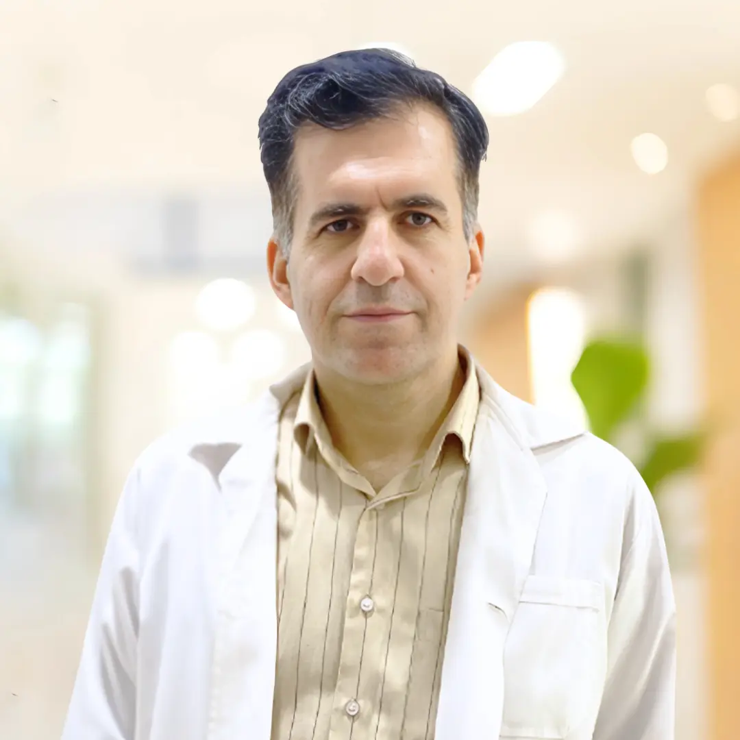
डॉ. शाहू मज़हरी
सलाहकार हृदय रोग विशेषज्ञ
एमडी, कार्डियोलॉजी में बोर्ड प्रमाणित
डॉ. शाहू मज़हरी DCDC दुबई हेल्थकेयर सिटी में गैर-आक्रामक कार्डियक इमेजिंग, कोरोनरी सीटी एंजियोग्राफी और निवारक कार्डियोलॉजी में विशेषज्ञता रखने वाले सलाहकार हृदय रोग विशेषज्ञ हैं।
संबंधित लेख
सीटी एंजियोग्राम क्या है?
सीटी एंजियोग्राम प्रक्रिया: स्कैन के दौरान क्या होता है
सीटी एंजियोग्राम सुरक्षा और जोखिम
दुबई में कार्डियोलॉजी: विशेषज्ञ हृदय देखभाल
More in Cardiology

ECG Cost in Dubai: From AED 200 (2026)
और पढ़ें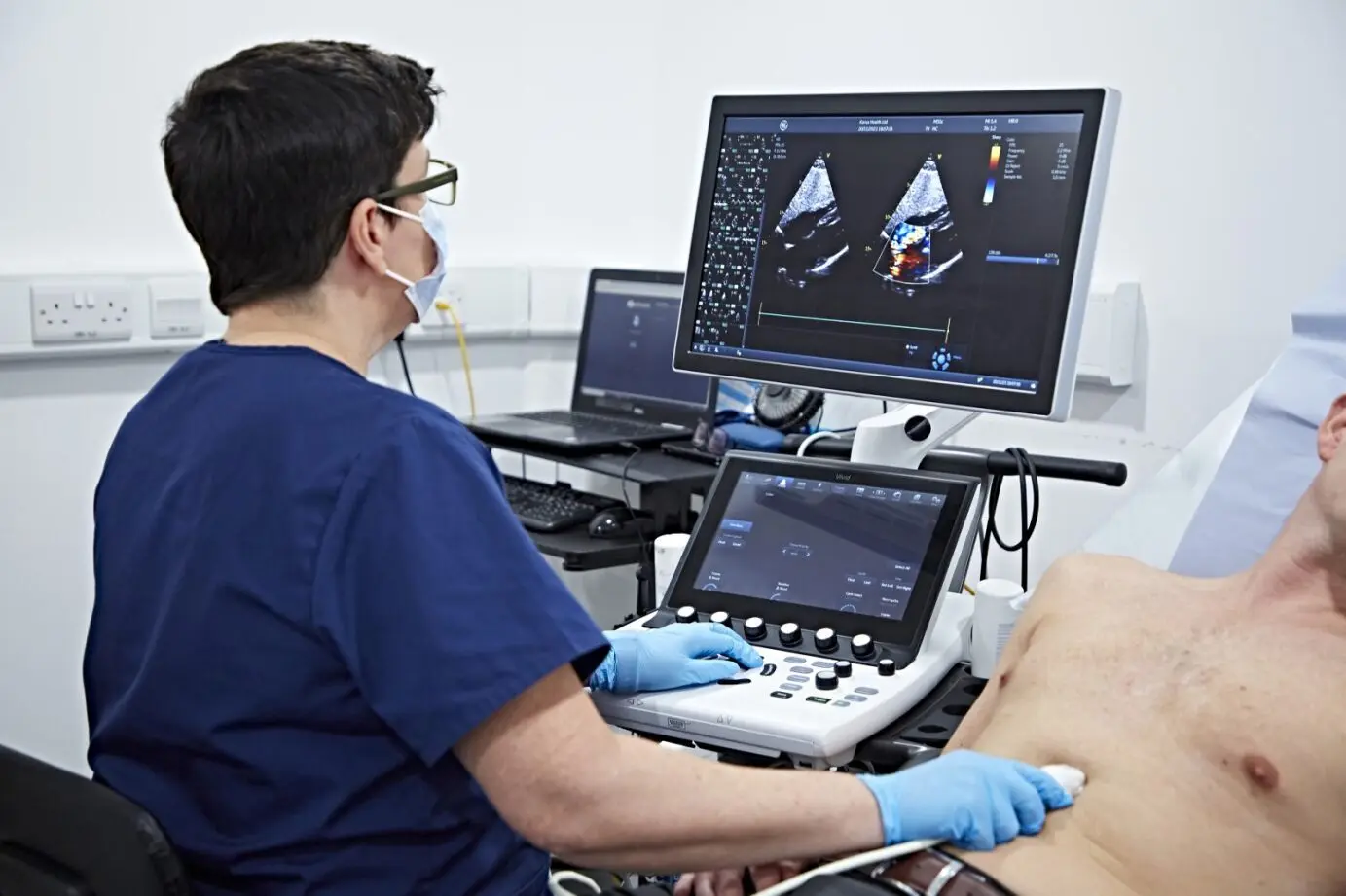
Echo at Home Dubai: From AED 799 (2026)
और पढ़ेंECG at Home Dubai: Cost & What to Expect (2026)
और पढ़ें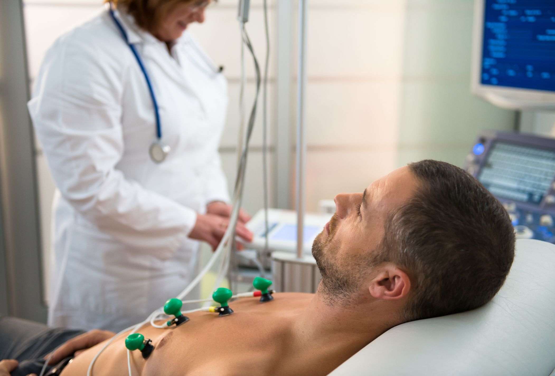
Holter Monitor vs ECG Dubai: Which Test? (2026)
और पढ़ें
Cardiac Rehabilitation Dubai: Recovery (2026)
और पढ़ें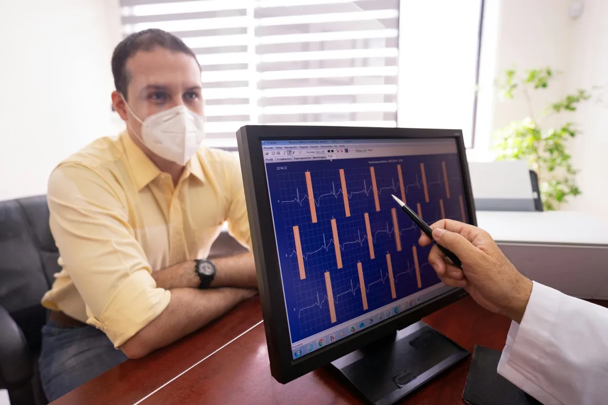
Troponin Test Dubai: Heart Attack Marker (2026)
और पढ़ें© 2026 Doctors Clinic Diagnostic Center (DCDC), Dubai Healthcare City. Originally published at https://doctorsclinicdubai.ae/blog/how-to-prepare-ct-angiogram. All rights reserved. Unauthorized reproduction is prohibited.