Wichtigste Erkenntnisse
- Verzichten Sie mindestens 24-48 Stunden vor Ihrer CT-Angiographie auf Koffein, um eine niedrige Herzfrequenz aufrechtzuerhalten
- Eine Ruheherzfrequenz unter 65 Schlägen pro Minute ist ideal für hochwertige Bilder; Betablocker können bei Bedarf gegeben werden
- Fasten Sie 4-6 Stunden vor dem Scan, nehmen Sie aber verschriebene Medikamente weiterhin mit Wasser ein
- Die Nierenfunktion (Kreatinin) sollte vor der Gabe von Kontrastmittel überprüft werden
- Tragen Sie bequeme, weite Kleidung und entfernen Sie allen Metallschmuck vor dem Scan
Eine sorgfältige Vorbereitung ist entscheidend für eine erfolgreiche CT-Angiographie. Anders als viele andere bildgebende Untersuchungen erfordert die koronare CT-Angiographie spezifische Schritte in den Tagen und Stunden vor Ihrem Termin, um die klarstmöglichen Bilder Ihrer Herzarterien zu gewährleisten. Die genaue Befolgung dieser Richtlinien kann den Unterschied zwischen einer diagnostischen Untersuchung und einer ausmachen, die wiederholt werden muss.
Diese Checkliste deckt alles ab, was Sie vor Ihrer CT-Angiographie wissen müssen, von diätetischen Einschränkungen und Medikamentenüberlegungen bis hin zu dem, was passiert, wenn Sie in der Klinik ankommen.

Warum die Vorbereitung für eine CT-Angiographie wichtig ist
Eine koronare CT-Angiographie erfasst Bilder Ihrer Herzarterien während eines einzelnen Herzschlags. Da das Herz ständig in Bewegung ist, muss der Scanner eine langsame und gleichmäßige Herzfrequenz haben, um scharfe, bewegungsfreie Bilder zu erzeugen. Faktoren wie Koffeinkonsum, Stress, Dehydrierung und bestimmte Medikamente können Ihre Herzfrequenz erhöhen und die Bildqualität verringern.
Zusätzlich verwendet die Untersuchung intravenöses Kontrastmittel, das Ihre Arterien auf den Bildern hervorhebt. Dieses Kontrastmittel erfordert eine ausreichende Nierenfunktion und Hydratation, um von Ihrem Körper sicher verarbeitet zu werden. Die richtige Vorbereitung berücksichtigt all diese Faktoren.
48 Stunden vor Ihrer CT-Angiographie
Koffein vollständig eliminieren
Koffein ist ein Stimulans, das Ihre Herzfrequenz erhöht und sie unregelmäßig machen kann. Sie sollten alle Koffeinquellen für mindestens 24 bis 48 Stunden vor Ihrem Scan meiden. Dazu gehören Kaffee, Tee, Energy-Drinks, Schokolade, koffeinhaltige Erfrischungsgetränke und bestimmte Medikamente oder Nahrungsergänzungsmittel, die Koffein enthalten.
Viele Patienten unterschätzen, wie stark Koffein ihre Herzfrequenz beeinflusst. Selbst entkoffeinierter Kaffee enthält kleine Mengen. Wenn Sie ein regelmäßiger Koffeinkonsument sind, gibt das Beginnen der Einschränkung zwei volle Tage vor dem Scan Ihrem Körper Zeit zur Anpassung und hilft, entzugsbedingte Herzfrequenzschwankungen zu vermeiden.
Kürzlich hatten wir einen Patienten, der am Morgen seiner geplanten CT-Angiographie eine Tasse Kaffee trank, ohne zu wissen, wie stark dies seine Untersuchung beeinflussen würde. Als er bei DCDC ankam, lag seine Herzfrequenz bei 92 Schlägen pro Minute, deutlich über dem Zielwert von 65 Schlägen pro Minute, der für klare Bilder benötigt wird. Trotz der Gabe von Betablockern konnten wir seine Herzfrequenz nicht ausreichend senken und mussten den Scan auf die folgende Woche verschieben. Der Patient erzählte uns später, dass er sich wünschte, die Koffeineinschränkung ernster genommen zu haben. Seine Erfahrung ist eine Erinnerung daran, dass dieser einzelne Vorbereitungsschritt bestimmen kann, ob Ihr Scan planmäßig stattfindet oder einen erneuten Besuch erfordert.
„Koffein ist der Grund Nummer eins, warum wir CT-Angiographien verschieben müssen," sagt Dr. Shahoo Mazhari, Facharzt für Kardiologie bei DCDC. „Patienten denken oft, eine Tasse Kaffee wird nichts ausmachen, aber selbst eine kleine Menge kann die Herzfrequenz genug erhöhen, um die Bildqualität zu beeinträchtigen. Das 48-Stunden-Fenster ohne Koffein ist nicht optional — es ist wesentlich."
Überprüfen Sie Ihre Medikamente mit Ihrem Arzt
Bestimmte Medikamente müssen möglicherweise vor Ihrer CT-Angiographie angepasst werden. Besprechen Sie alle aktuellen Medikamente mit Ihrem Kardiologen oder dem Bildgebungszentrum, einschließlich:
- Betablocker: Wenn Sie bereits Betablocker einnehmen, kann Ihr Arzt die Dosis anpassen, um eine Zielherzfrequenz unter 65 Schlägen pro Minute zu erreichen.
- Metformin: Patienten mit Diabetes, die Metformin einnehmen, können angewiesen werden, es für 48 Stunden nach dem Scan aufgrund einer möglichen Wechselwirkung mit Kontrastmittel und Nierenfunktion zu pausieren.
- Erektionsstörungsmedikamente: Medikamente wie Sildenafil (Viagra) sollten typischerweise 48 Stunden vorher abgesetzt werden, da sie mit Nitroglycerin interagieren können, das während des Scans verwendet werden kann.
- Blutverdünner: Im Allgemeinen wie gewohnt fortsetzen, aber das Team über alle Antikoagulanzien oder Thrombozytenaggregationshemmer informieren, die Sie einnehmen.
Bleiben Sie gut hydriert
Gute Hydratation unterstützt die Nierenfunktion, die für die Verarbeitung des während des Scans verwendeten Kontrastmittels wichtig ist. Trinken Sie in den zwei Tagen vor Ihrem Termin reichlich Wasser. Vermeiden Sie Alkohol, da er Dehydrierung verursachen und den Herzrhythmus beeinflussen kann.
Am Tag vor Ihrer CT-Angiographie
- Weiterhin Koffein und Alkohol meiden
- Trinken Sie über den Tag verteilt mindestens 1,5 bis 2 Liter Wasser
- Bereiten Sie bequeme, weite Kleidung ohne Metallreißverschlüsse, Druckknöpfe oder Bügel-BH vor
- Legen Sie allen Schmuck, Uhren und Piercings beiseite, um sie zu Hause zu lassen
- Bestätigen Sie Ihre Terminzeit und alle spezifischen Anweisungen der Klinik
- Schlafen Sie gut; Müdigkeit und schlechter Schlaf können Ihre Ruheherzfrequenz erhöhen
Am Tag Ihrer CT-Angiographie
Nüchternheitsanforderungen
Sie sollten 4 bis 6 Stunden vor Ihrem geplanten Scantermin nüchtern bleiben. Das bedeutet keine Nahrung oder Getränke außer stillem Wasser. Nüchternheit reduziert das Risiko von Übelkeit durch das Kontrastmittel und hilft, stabile physiologische Bedingungen während des Scans aufrechtzuerhalten.
Sie sollten jedoch weiterhin Ihre verschriebenen Medikamente mit kleinen Schlucken Wasser einnehmen, es sei denn, Ihr Arzt hat ausdrücklich etwas anderes angewiesen. Dies ist besonders wichtig für Blutdruckmedikamente und Betablocker, die helfen, Ihre Herzfrequenz kontrolliert zu halten.
Was Sie anziehen und mitbringen sollten
- Tragen Sie bequeme, weite Kleidung (Sie erhalten möglicherweise ein Krankenhaushemd)
- Entfernen Sie alle Metallgegenstände: Schmuck, Uhren, Gürtel, Haarspangen und Piercings
- Bringen Sie eine Liste Ihrer aktuellen Medikamente und Dosierungen mit
- Bringen Sie aktuelle Bluttestergebnisse mit, insbesondere Nierenfunktion (Kreatinin/eGFR), falls verfügbar
- Bringen Sie Ihre Emirates ID oder Ihren Reisepass und Ihre Versicherungskarte mit
- Bringen Sie alle früheren kardialen Bildgebungsberichte oder Überweisungsschreiben mit
Vorbereitung auf Ihre CT-Angiographie bei DCDC
Im Doctors Clinic Diagnostic Center in Dubai Healthcare City begleiten wir jeden Patienten durch die richtige CT-Angiographie-Vorbereitung, um die bestmöglichen Ergebnisse zu gewährleisten.
Herzfrequenzziel: Warum unter 65 BPM wichtig ist
Die optimale Herzfrequenz für eine koronare CT-Angiographie liegt unter 65 Schlägen pro Minute (bpm), wobei einige Zentren unter 60 bpm bevorzugen. Bei diesen Herzfrequenzen ist das Herz zwischen den Schlägen lang genug entspannt, damit der Scanner klare, bewegungsfreie Bilder der Koronararterien aufnehmen kann.
Wenn Sie in der Klinik ankommen, wird Ihre Herzfrequenz gemessen. Wenn sie über dem Zielbereich liegt, kann das medizinische Team einen kurzwirksamen Betablocker (typischerweise Metoprolol) oral oder intravenös verabreichen, um sie zu senken. Dies ist ein routinemäßiger und sicherer Teil der CT-Angiographie-Vorbereitung.
Faktoren, die Ihre Herzfrequenz erhöhen können und am Scantag vermieden werden sollten, sind Koffein, Rauchen, anstrengendes Training, Stress und das Hetzen zu Ihrem Termin. Kommen Sie 15 bis 20 Minuten früher, damit Sie Zeit haben, sich vor dem Scan zu entspannen.
„Wir führen bei DCDC jeden Monat über 1.000 diagnostische Scans durch, und die Patienten, die die besten Ergebnisse erzielen, sind diejenigen, die die Vorbereitungsschritte sorgfältig befolgen," sagt Dr. Shahoo Mazhari, Facharzt für Kardiologie bei DCDC. „Entspannt und gut hydriert mit einer niedrigen Herzfrequenz anzukommen, macht den entscheidenden Unterschied bei der Bildklarheit."
Nierenfunktionsprüfung: Warum Kreatinin wichtig ist
Das während einer CT-Angiographie verwendete Kontrastmittel (jodbasiert) wird von den Nieren verarbeitet und ausgeschieden. Vor dem Scan ist ein Bluttest erforderlich, der Kreatinin und die geschätzte glomeruläre Filtrationsrate (eGFR) misst, um zu bestätigen, dass Ihre Nieren gut genug funktionieren, um das Kontrastmittel sicher zu verarbeiten.
Dieser Bluttest sollte idealerweise innerhalb von 1 bis 3 Monaten vor Ihrem Scan durchgeführt werden. Patienten mit Diabetes, Bluthochdruck, bekannter Nierenerkrankung oder über 60 Jahren haben ein höheres Risiko für kontrastmittelbedingte Nierenprobleme und werden genauer überwacht.
Wenn Ihre Nierenfunktion grenzwertig ist, kann Ihr Arzt eine Vorhydratation mit intravenösen Flüssigkeiten vor und nach dem Scan empfehlen, um Ihre Nieren zu schützen. In seltenen Fällen kann der Scan verschoben werden, wenn die Nierenfunktion erheblich beeinträchtigt ist.
Allergieüberlegungen
Wenn Sie eine bekannte Allergie gegen jodbasiertes Kontrastmittel, Schalentiere haben oder in der Vergangenheit eine allergische Reaktion auf ein Kontrastmittel hatten, informieren Sie das medizinische Team rechtzeitig im Voraus. Eine Vormedikation mit Antihistaminika und/oder Steroiden kann arrangiert werden, um das Reaktionsrisiko zu reduzieren.
Leichte Reaktionen wie ein Wärmegefühl, metallischer Geschmack oder kurze Übelkeit während der Kontrastmittelinjektion sind häufig und normal. Echte allergische Reaktionen sind selten, aber beherrschbar, wenn das Team vorbereitet ist.
Checkliste zur CT-Angiographie-Vorbereitung
| Zeitpunkt | Maßnahme |
|---|---|
| 48 Stunden vorher | Alles Koffein stoppen (Kaffee, Tee, Energy-Drinks, Schokolade) |
| 48 Stunden vorher | Medikamente mit Ihrem Arzt besprechen (Metformin, ED-Medikamente) |
| 48 Stunden vorher | Alkohol vermeiden |
| 2 Tage vorher | Wasseraufnahme auf 1,5-2 Liter pro Tag erhöhen |
| 1 Tag vorher | Termin und Vorbereitungsanweisungen bestätigen |
| 1 Tag vorher | Bequeme Kleidung ohne Metall vorbereiten |
| Abend vorher | Ausreichend schlafen; späte Mahlzeiten vermeiden |
| 4-6 Stunden vorher | Mit dem Fasten beginnen (Wasser ist erlaubt) |
| Morgen des Scans | Verschriebene Medikamente mit kleinen Schlucken Wasser einnehmen |
| In der Klinik | 15-20 Minuten früher ankommen; Ausweis, Versicherung und Medikamentenliste mitbringen |
Befolgen Sie diesen Zeitplan, um eine optimale Vorbereitung auf Ihre CT-Angiographie sicherzustellen.
Was während der CT-Angiographie passiert
Das Verständnis des Verfahrens hilft, Ängste zu reduzieren. Bei Ihrer Ankunft wird eine IV-Leitung in Ihrem Arm für die Kontrastmittelinjektion gelegt. EKG-Elektroden werden an Ihrer Brust befestigt, um Ihren Herzrhythmus zu überwachen. Sie liegen auf einem schmalen Tisch, der in den CT-Scanner gleitet, der eine offene Ringform hat (kein geschlossener Tunnel wie ein MRT).
Möglicherweise erhalten Sie Nitroglycerin-Spray unter die Zunge, um Ihre Koronararterien zur besseren Darstellung zu erweitern. Während des Scans werden Sie gebeten, etwa 10 bis 15 Sekunden die Luft anzuhalten, während der Scanner Bilder aufnimmt. Die eigentliche Scanzeit beträgt typischerweise unter einer Minute, obwohl der gesamte Termin meist 30 bis 60 Minuten dauert.
Nach dem Scan: Was Sie erwarten können
- Trinken Sie nach dem Scan zusätzlich Wasser, um das Kontrastmittel aus Ihrem System zu spülen
- Nehmen Sie normales Essen und Trinken sofort nach dem Scan wieder auf
- Wenn Sie Betablocker erhalten haben, fühlen Sie sich möglicherweise leicht schläfrig; vermeiden Sie das Autofahren für 1-2 Stunden, wenn Sie betroffen sind
- Die Ergebnisse sind typischerweise innerhalb von 24-48 Stunden verfügbar
- Kontaktieren Sie die Klinik, wenn Sie ungewöhnliche Symptome wie Hautausschlag, Schwellung oder Atemnot bemerken
Bereiten Sie sich auf Ihre CT-Angiographie bei DCDC vor?
Im Doctors Clinic Diagnostic Center in Dubai Healthcare City begleiten wir jeden Patienten durch die richtige CT-Angiographie-Vorbereitung, um die bestmöglichen Ergebnisse zu gewährleisten. Mit über 1.000 diagnostischen Scans pro Monat und über 13 Jahren Erfahrung in Dubai Healthcare City wird unser erfahrenes Team Sie vor Ihrem Termin mit personalisierten Anweisungen kontaktieren. Wir begrüßen Patienten aus den gesamten VAE und international.
Verwandte Leistungen im DCDC
Fachkundige Betreuung und moderne Diagnostik in Dubai Healthcare City
Frequently Asked Questions
Fazit
Eine koronare CT-Angiographie ist eines der fortschrittlichsten nicht-invasiven Werkzeuge zur Bewertung Ihrer Herzarterien, aber ihre Genauigkeit hängt stark von der richtigen Vorbereitung ab. Indem Sie diese Richtlinien befolgen, insbesondere Koffein meiden, Hydratation aufrechterhalten und entspannt ankommen, geben Sie dem Bildgebungsteam die besten Bedingungen, um klare, diagnostische Bilder aufzunehmen.
Wenn Sie Fragen zu Ihrer spezifischen Vorbereitung haben, zögern Sie nicht, das Bildgebungszentrum oder Ihren Kardiologen vor Ihrem Termin zu kontaktieren. Die Situation jedes Patienten ist leicht unterschiedlich, und eine personalisierte Beratung gewährleistet die sicherste und effektivste Scanerfahrung. Informationen zu Preisen und Versicherungsdeckung finden Sie in unserem Leitfaden zu CT-Angiographie-Kosten in Dubai.
Quellen und Referenzen
Dieser Artikel wurde von unserem medizinischen Team überprüft und bezieht sich auf folgende Quellen:
- Society of Cardiovascular Computed Tomography - Leitlinien zur Patientenvorbereitung
- American College of Radiology - CT-Angiographie-Standards
- European Society of Cardiology - Protokolle für kardiale CT-Bildgebung
Medizinische Inhalte auf dieser Website werden von DHA-lizenzierten Ärzten überprüft. Siehe unsere redaktionelle Richtlinien für weitere Informationen.

Verfasst von
Dr. Shahoo Mazhari
Facharzt für Kardiologie
Dr. med., Facharzt für Kardiologie
Dr. Shahoo Mazhari ist Facharzt für Kardiologie mit Spezialisierung auf nicht-invasive kardiale Bildgebung, Koronar-CT-Angiographie und präventive Kardiologie bei DCDC Dubai Healthcare City.
Verwandte Artikel
Was ist eine CT-Angiographie?
CT-Angiographie-Verfahren: Was während des Scans passiert
CT-Angiographie Sicherheit und Risiken
Kardiologie in Dubai: Fachärztliche Herzversorgung
More in Cardiology

ECG Cost in Dubai: From AED 200 (2026)
Weiterlesen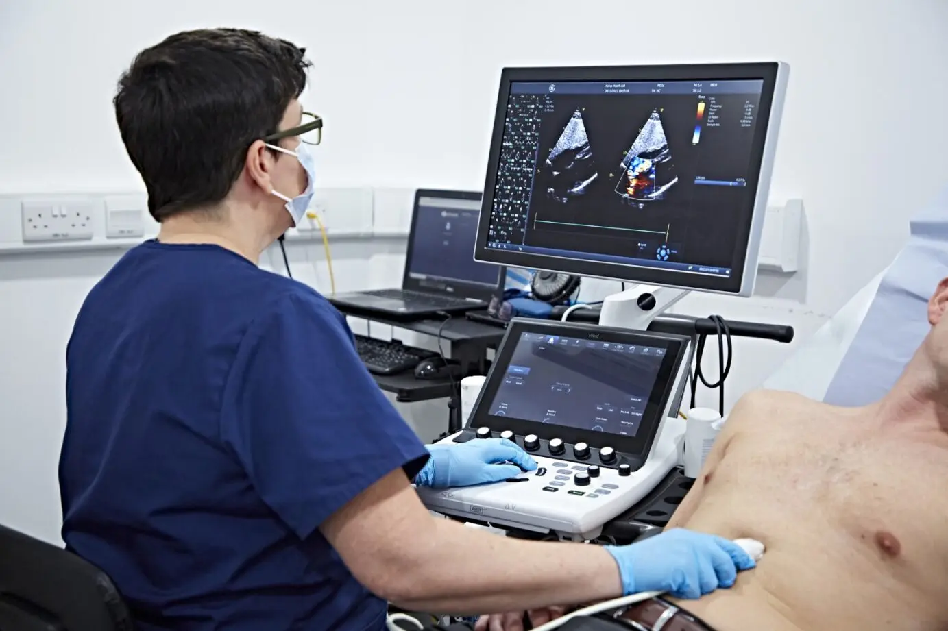
Echo at Home Dubai: From AED 799 (2026)
WeiterlesenECG at Home Dubai: Cost & What to Expect (2026)
Weiterlesen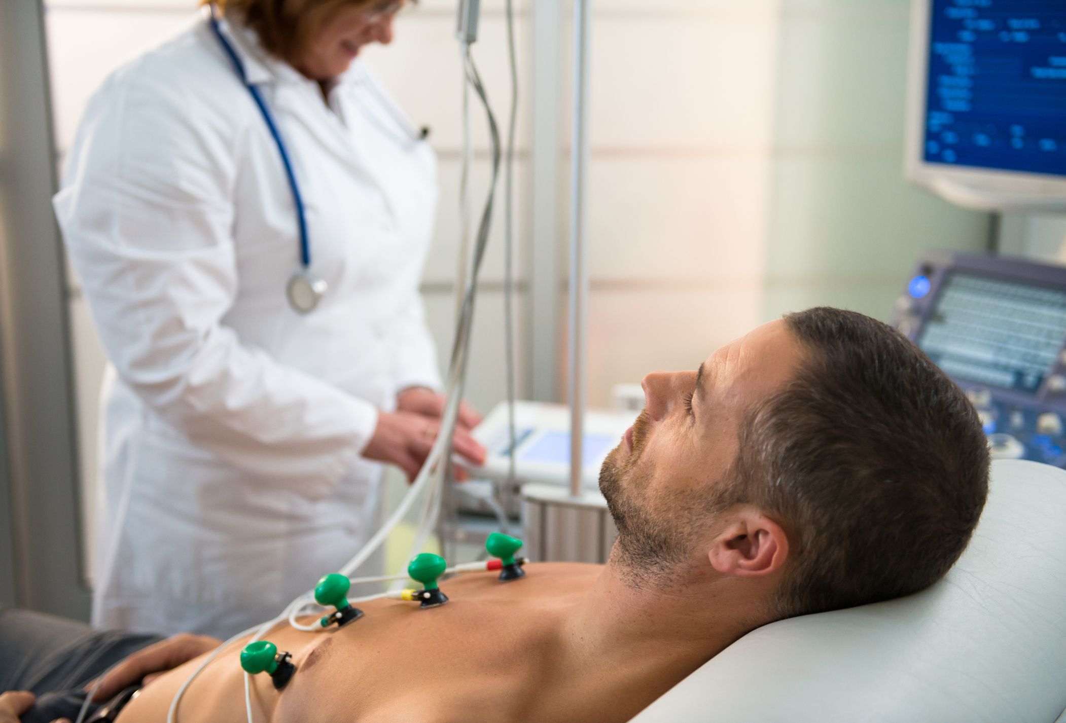
Holter Monitor vs ECG Dubai: Which Test? (2026)
Weiterlesen
Cardiac Rehabilitation Dubai: Recovery (2026)
Weiterlesen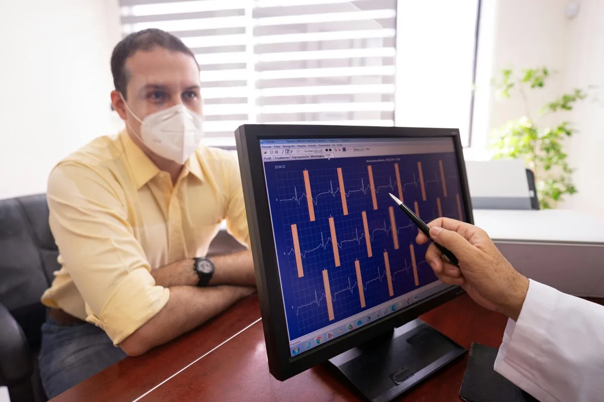
Troponin Test Dubai: Heart Attack Marker (2026)
Weiterlesen© 2026 Doctors Clinic Diagnostic Center (DCDC), Dubai Healthcare City. Originally published at https://doctorsclinicdubai.ae/blog/how-to-prepare-ct-angiogram. All rights reserved. Unauthorized reproduction is prohibited.