اہم نکات
- Most headaches and migraines do not require an MRI — the scan is reserved for headaches with red flag symptoms such as thunderclap onset, neurological deficits, or progressive worsening over weeks
- Brain MRI can detect serious causes of headaches including tumors, aneurysms, Chiari malformation, sinusitis, hydrocephalus, and cerebral venous thrombosis
- A neurologist or physician typically orders an MRI when headache pattern changes suddenly, does not respond to treatment, or is accompanied by vision changes, weakness, seizures, or fever
- MRI is preferred over CT for chronic and recurrent headache evaluation because it provides superior soft tissue detail with zero radiation exposure
- A normal brain MRI in a patient with severe headaches is actually reassuring — it confirms that the headaches are a primary disorder like migraine or tension-type headache, not caused by a dangerous structural problem
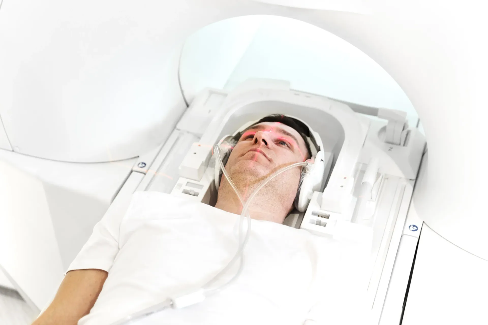
Headaches are one of the most common reasons patients visit a physician, and the question "Do I need an MRI for my headaches?" is one of the most frequent concerns raised in clinic. The vast majority of headaches — including migraines, tension-type headaches, and cluster headaches — are primary headache disorders with no underlying structural abnormality. However, certain warning signs indicate that a brain MRI is warranted to rule out potentially serious causes. Understanding when an MRI is necessary and what it can reveal empowers patients to make informed decisions about their care.


Red Flag Headache Symptoms That Require MRI
Neurologists and headache specialists use the term "red flags" to describe headache features that raise concern for a secondary cause — meaning the headache is a symptom of an underlying condition rather than a primary headache disorder itself. When any of the following red flags are present, brain imaging with MRI is strongly recommended:
- Thunderclap headache: A headache that reaches maximum intensity within seconds to one minute is a medical emergency. This "worst headache of my life" presentation can indicate a subarachnoid hemorrhage from a ruptured aneurysm, cerebral venous sinus thrombosis, or pituitary apoplexy. These patients typically receive an urgent CT scan first, followed by MRI and MRA for definitive evaluation.
- Progressive worsening over weeks: A headache that steadily increases in frequency, severity, or duration over days to weeks may indicate a growing intracranial mass (tumor), expanding aneurysm, hydrocephalus (fluid buildup in the brain), or chronic subdural hematoma. This pattern is fundamentally different from the fluctuating course of migraines.
- Headache with neurological symptoms: New weakness, numbness, tingling, vision changes, speech difficulty, coordination problems, personality changes, or cognitive decline accompanying headaches are red flags for structural brain pathology. MRI is essential in this scenario.
- New headache after age 50: The onset of a new headache type in a patient over 50 years old raises concern for temporal arteritis (giant cell arteritis), brain tumor, or cerebrovascular disease. These conditions become more common with age, and MRI is typically performed alongside blood tests.
- Headache with fever and stiff neck: While meningitis is diagnosed through lumbar puncture and blood work, MRI may be ordered if there is concern for brain abscess, encephalitis, or empyema. MRI with contrast is highly sensitive for these infectious complications.
- Headache that wakes you from sleep: Primary headaches can occur during sleep (hypnic headache, cluster headache), but new headaches that consistently wake a patient from sleep — especially with nausea, vomiting, or visual symptoms — require imaging to exclude raised intracranial pressure from a tumor or other mass.
- Headache triggered by coughing, straining, or exertion: Headaches provoked by Valsalva maneuvers (coughing, sneezing, bearing down) can indicate a Chiari malformation, in which the cerebellar tonsils protrude through the base of the skull. MRI is the definitive test for diagnosing Chiari malformations.
- Headache in immunocompromised patients: Patients with HIV, cancer, organ transplants, or those on immunosuppressive medications are at higher risk for opportunistic brain infections and lymphoma. A new headache in these patients almost always warrants MRI with contrast.
"The red flag approach is essential because it allows us to identify the small subset of headache patients who truly need imaging," explains Dr. Osama Elzamzami, Consultant Radiologist at DCDC. "Most patients presenting with typical migraine characteristics and a normal neurological examination do not require an MRI. But when red flags are present, brain MRI can be life-saving by detecting conditions that require urgent treatment."
What Can a Brain MRI Reveal in Headache Patients?
When a brain MRI is ordered for headache evaluation, the radiologist examines the brain parenchyma, blood vessels, meninges, sinuses, and surrounding structures in meticulous detail. The following conditions can be identified on brain MRI:
- Brain tumors: Both primary brain tumors (gliomas, meningiomas, acoustic neuromas) and metastatic tumors from cancers elsewhere in the body can cause headaches — typically progressive, worse in the morning, and associated with nausea or neurological deficits. Contrast-enhanced MRI is the gold standard for detecting and characterizing brain tumors.
- Intracranial aneurysms: Unruptured aneurysms may cause headaches, particularly when they enlarge and compress nearby structures. MRI angiography (MRA) can detect aneurysms as small as 3 millimeters. A ruptured aneurysm causing subarachnoid hemorrhage presents as a sudden thunderclap headache and is a life-threatening emergency.
- Chiari malformation: This structural abnormality involves downward displacement of the cerebellar tonsils through the foramen magnum. It characteristically causes headaches at the back of the head that worsen with coughing, straining, or bending forward. MRI of the brain and cervical spine is the definitive diagnostic tool.
- Sinusitis: While sinusitis is typically diagnosed clinically, MRI often incidentally reveals sinus inflammation in patients being scanned for headaches. Chronic sinusitis can mimic tension-type headaches or migraines, and MRI provides detailed visualization of sinus mucosal thickening, fluid levels, and polyps.
- Hydrocephalus: Abnormal accumulation of cerebrospinal fluid (CSF) in the ventricles causes headaches from raised intracranial pressure. MRI demonstrates enlarged ventricles and can identify the cause of obstruction. Normal-pressure hydrocephalus (NPH) in older adults presents with a triad of gait disturbance, cognitive decline, and urinary incontinence alongside headaches.
- Cerebral venous sinus thrombosis (CVST): Blood clots in the venous sinuses of the brain cause headaches that can mimic migraines or tension headaches. CVST is more common in young women, especially those on oral contraceptives or in the postpartum period. MRI with MR venography is the preferred diagnostic modality.
- Arteriovenous malformations (AVMs): These abnormal tangles of blood vessels can cause headaches, seizures, and neurological deficits. MRI detects AVMs with high sensitivity and helps plan treatment approaches including surgery, embolization, or radiosurgery.
- White matter changes: MRI may reveal white matter lesions that can be associated with migraine (particularly migraine with aura), small vessel disease, or early multiple sclerosis. While small white matter changes in migraine patients are generally considered benign, their presence may influence treatment decisions.
When Does a Neurologist Order an MRI for Headaches?
A neurologist evaluates headache patients through a careful history, physical examination, and neurological assessment before deciding whether imaging is necessary. The decision to order MRI is based on clinical guidelines, particularly those from the American Academy of Neurology (AAN) and the American College of Radiology (ACR). In general, MRI is ordered in the following scenarios:
- Atypical headache pattern: Headaches that do not fit the established criteria for any primary headache disorder (migraine, tension-type, cluster, or other trigeminal autonomic cephalalgias) warrant imaging to investigate secondary causes.
- Change in established headache pattern: A migraine patient whose headaches suddenly change in character, frequency, severity, or associated symptoms needs re-evaluation. A previously episodic migraine becoming daily, a new aura in a patient who never had aura, or a shift from bilateral to strictly unilateral headache are all reasons for imaging.
- Abnormal neurological examination: Any focal neurological deficit found on examination — such as papilledema (swelling of the optic disc), cranial nerve palsy, weakness, sensory loss, or abnormal reflexes — mandates urgent brain imaging.
- Treatment-resistant headaches: If headaches fail to respond to appropriate first-line and second-line preventive treatments, imaging may be ordered to ensure there is no underlying structural cause contributing to treatment failure.
- Patient reassurance with appropriate indications: In some cases, a neurologist may order an MRI to provide reassurance to a patient with significant anxiety about a serious brain condition, provided the clinical situation has some features that make imaging reasonable. A normal MRI can be therapeutically valuable by reducing catastrophizing and health anxiety.
It is important to note that routine screening MRI is not recommended for patients with typical migraine and a normal neurological examination. Multiple clinical guidelines and systematic reviews have confirmed that the yield of MRI in this population is extremely low — the vast majority of scans will be normal or show only incidental findings of no clinical significance. Ordering unnecessary imaging can lead to incidental findings that cause anxiety and trigger further unnecessary investigations.
Need a Brain MRI for Headache Evaluation?
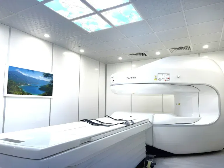
At Doctors Clinic Diagnostic Center in Dubai Healthcare City, we provide comprehensive brain MRI with expert consultant radiologist interpretation. Our structured reports give your neurologist the detailed findings needed for accurate diagnosis.
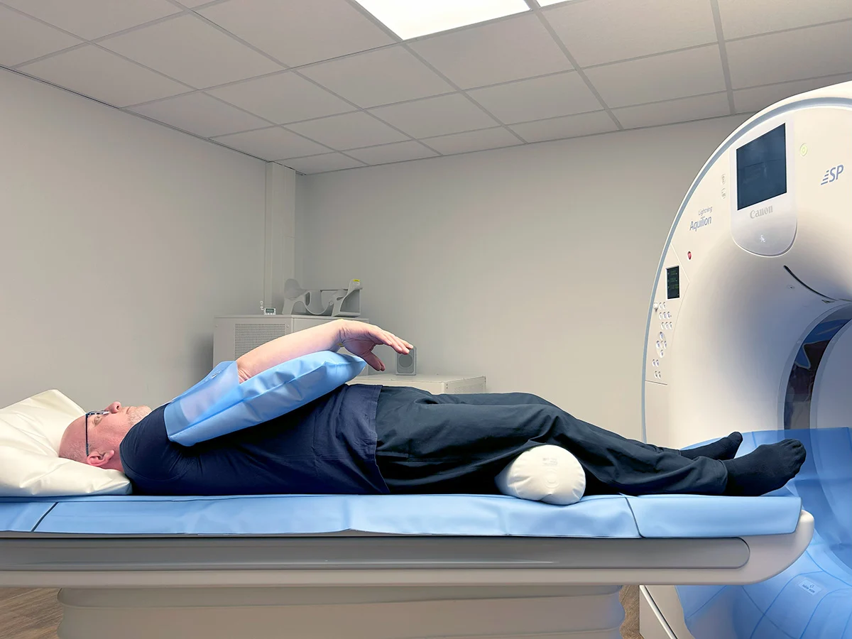
MRI vs CT Scan for Headaches: Which Is Better?
Both MRI and CT scan can image the brain, but they have very different roles in headache evaluation. Understanding when each is appropriate helps patients and physicians make the right choice.
| Factor | Brain MRI | Brain CT |
|---|---|---|
| Best for | Chronic/recurrent headaches, migraines, non-emergency evaluation | Acute emergencies, thunderclap headache, suspected hemorrhage |
| Soft tissue detail | Excellent — gold standard for brain | Limited |
| Radiation | None (zero radiation) | Yes (approximately 2 mSv) |
| Scan time | 30 – 60 minutes | 5 – 10 minutes |
| Tumor detection | Superior sensitivity | May miss small tumors |
| Aneurysm detection | Excellent with MRA | Good with CT angiography |
| Acute hemorrhage | Good but not first-line | Excellent — first-line choice |
| Chiari malformation | Definitive diagnostic tool | May miss or poorly characterize |
| Cost (approx.) | AED 1,200 – 2,000 | AED 500 – 900 |
CT is the first choice in acute emergencies. MRI is preferred for non-urgent headache evaluation.
In emergency settings — such as a patient presenting with a sudden thunderclap headache, the worst headache of their life, or a headache with rapid neurological deterioration — a CT scan is performed first because it is fast (5 minutes), widely available, and exceptionally sensitive for acute blood in or around the brain. If the CT is negative or inconclusive, MRI and sometimes lumbar puncture follow.
For non-emergency headache evaluation — investigating chronic migraines, progressive headaches, headaches with subtle neurological symptoms, or screening for structural abnormalities — MRI is clearly superior. It provides far better contrast resolution, can detect tumors, white matter changes, Chiari malformations, and vascular abnormalities that CT may miss entirely. Additionally, MRI uses no ionizing radiation, making it the safer option for young patients and those requiring serial imaging.
What Does a Normal MRI Mean When You Have Bad Headaches?
One of the most common questions patients ask after receiving their brain MRI results is: "If my MRI is normal, why do I still have such terrible headaches?" This is a critically important point to understand.
A normal brain MRI is actually good news. It means that your headaches are not caused by a brain tumor, aneurysm, hydrocephalus, structural malformation, or other dangerous intracranial pathology. This is the primary reason the MRI was ordered — to rule out these serious conditions. A normal scan confirms that your headaches are a primary headache disorder, which is what the vast majority of headaches are.
Primary headache disorders — including migraine, tension-type headache, cluster headache, and others — are neurological conditions involving abnormal brain function (neurochemical and electrical changes) rather than structural abnormalities. Migraine, in particular, involves dysfunction in pain-processing networks, cortical spreading depression, and sensitization of the trigeminal nerve system. None of these functional abnormalities are visible on standard MRI because the brain structure itself is normal.
Understanding this distinction is therapeutically important. Patients who know their brain is structurally normal can focus on evidence-based migraine management strategies including preventive medications, lifestyle modification (sleep hygiene, stress management, regular exercise), trigger identification and avoidance, and acute treatment optimization. The reassurance from a normal MRI can itself reduce headache frequency by alleviating the anxiety and catastrophizing that often worsen chronic pain conditions.
Occasionally, MRI in migraine patients may reveal small, nonspecific white matter lesions (bright spots on T2-weighted images). These are found more frequently in migraine sufferers — particularly those with aura — than in the general population. Research suggests these lesions are generally benign and do not indicate that the patient is at elevated risk for stroke or other serious neurological disease. Your neurologist will interpret these findings in the context of your overall clinical picture.
Cost of MRI for Headaches in Dubai
The cost of a brain MRI for headache evaluation in Dubai depends on the specific protocol ordered and whether contrast dye is required. At Doctors Clinic Diagnostic Center, pricing includes the scan, comprehensive consultant radiologist report, and digital images.
| MRI Protocol | Approximate Cost (AED) | Typical Indication |
|---|---|---|
| Brain MRI without contrast | 1,200 – 1,500 | Standard headache evaluation, migraine workup |
| Brain MRI with contrast | 1,500 – 2,000 | Suspected tumor, infection, or inflammation |
| Brain MRI with MRA | 1,800 – 2,200 | Suspected aneurysm or vascular malformation |
| Brain + cervical spine MRI | 2,200 – 2,800 | Suspected Chiari malformation, cervicogenic headache |
Prices are approximate and include radiologist report at DCDC. Most insurance plans cover MRI with appropriate clinical justification.
For most headache evaluations, a brain MRI without contrast is sufficient. This is the standard protocol for investigating chronic migraines, tension-type headaches, and most new headache presentations without red flag features. Contrast is added when there is specific clinical concern for tumors, infections, or inflammatory conditions. Your referring physician or neurologist will specify the appropriate protocol.
Brain MRI for headache evaluation is covered by most health insurance plans in the UAE when ordered by a physician with documented clinical justification. Common accepted indications include new-onset headaches with red flag features, headaches with abnormal neurological findings, headache pattern changes in established migraine patients, and headaches not responding to appropriate treatment.
Book Your Brain MRI at DCDC
At Doctors Clinic Diagnostic Center in Dubai Healthcare City, every brain MRI is interpreted by a consultant radiologist with extensive neuroradiology experience. Reports are delivered within 24 to 48 hours. Learn more about our MRI services.
DCDC میں متعلقہ خدمات
دبئی ہیلتھ کیئر سٹی میں ماہرانہ دیکھ بھال اور جدید تشخیص
اکثر پوچھے گئے سوالات
آخری خیالات
Not every headache needs an MRI, but recognizing when brain imaging is warranted can be the difference between catching a serious condition early and missing a critical diagnosis. Red flag symptoms — thunderclap headache, progressive worsening, neurological deficits, new headaches after age 50, and headaches that fail to respond to treatment — should prompt discussion with your physician about brain MRI.
If your MRI is normal, take it as the good news it is. Your headaches are a primary neurological disorder that, while painful and disruptive, is treatable with proper medical management. At Doctors Clinic Diagnostic Center in Dubai Healthcare City, our consultant radiologists provide expert brain MRI interpretation to give you and your neurologist the clarity needed for confident diagnosis and treatment planning.
ذرائع اور حوالہ جات
یہ مضمون ہماری طبی ٹیم نے جائزہ لیا ہے اور درج ذیل ذرائع کا حوالہ دیتا ہے:
- American Academy of Neurology - Neuroimaging Guidelines for Headache
- American College of Radiology - ACR Appropriateness Criteria for Headache
- International Headache Society - ICHD-3 Classification
- The Lancet Neurology - Migraine Pathophysiology and Management
- Radiological Society of North America - Brain MRI
اس سائٹ پر طبی مواد کا جائزہ DHA لائسنس یافتہ ڈاکٹرز نے لیا ہے۔ ہماری دیکھیں تحریری پالیسی مزید معلومات کے لیے۔

تحریر
Dr. Osama Elzamzami
Diagnostic Radiology
MD, FRCR
Dr. Osama Elzamzami is a Consultant Radiologist specializing in diagnostic imaging including MRI, CT, and ultrasound at DCDC Dubai Healthcare City.
متعلقہ مضامین
Brain MRI in Dubai: What It Detects, Cost & When You Need One
MRI Cost in Dubai: Complete Guide to Types & Pricing (2026)
MRI for Multiple Sclerosis: How MS Is Diagnosed & Monitored
MRI for Vertigo: When Dizziness Needs Brain & Spine Imaging
MRI with Contrast: What to Expect, Safety & Cost
Diagnostic Imaging میں مزید
CT Scan Cost Dubai: Prices & Types (2026)
مزید پڑھیں
DEXA Scan Dubai: Cost, T-Score & Who Needs It (2026)
مزید پڑھیں![Full Body MRI Cost Dubai: AED 5,000-15,000 [2026]](/wp-media/blog/full-body-mri-scan.webp)
Full Body MRI Cost Dubai: AED 5,000-15,000 [2026]
مزید پڑھیں
MRI Preparation Dubai: Complete Guide (2026)
مزید پڑھیں
CBCT Scan: 3D Dental Imaging Guide Dubai (2026)
مزید پڑھیں
Ultrasound Preparation Dubai: What to Expect (2026)
مزید پڑھیں© 2026 Doctors Clinic Diagnostic Center (DCDC), Dubai Healthcare City. Originally published at https://doctorsclinicdubai.ae/blog/mri-for-headaches-migraines. All rights reserved. Unauthorized reproduction is prohibited.