اہم نکات
- MRI detects ACL tears with greater than 95 percent sensitivity and specificity, making it the gold standard for diagnosing anterior cruciate ligament injuries without surgery
- MRI distinguishes between partial ACL tears (which may heal with rehabilitation) and complete ACL ruptures (which typically require surgical reconstruction in active individuals)
- ACL tears rarely occur in isolation — MRI simultaneously detects associated injuries including meniscus tears (present in 50 to 70 percent of ACL injuries), MCL sprains, bone bruises, and cartilage damage
- Characteristic bone bruise patterns on MRI (lateral femoral condyle and posterolateral tibial plateau) are highly specific for ACL injury and can confirm the diagnosis even when the ligament is difficult to visualize
- A knee MRI for ACL evaluation in Dubai costs between AED 900 and AED 1,400, and the scan should ideally be performed 2 to 7 days after injury for optimal diagnostic accuracy
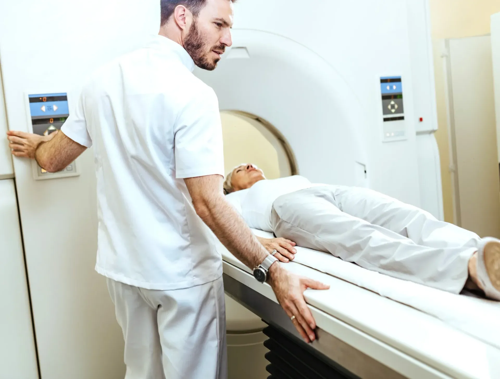
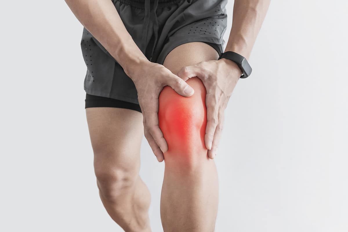
The anterior cruciate ligament (ACL) is one of the most commonly injured knee ligaments, particularly in athletes involved in pivoting sports such as football, padel, basketball, and skiing. An ACL tear can be a career-altering injury that requires accurate diagnosis to guide treatment decisions. While clinical examination by an experienced orthopedic surgeon can suggest an ACL tear with reasonable accuracy, MRI is the definitive diagnostic tool — it confirms the diagnosis, grades the severity, and reveals associated injuries that clinical examination alone cannot detect. This comprehensive guide explains how MRI diagnoses ACL tears, what the MRI findings look like, how accurate the test is, and when to get scanned after a knee injury in Dubai.


How Does MRI Detect an ACL Tear?
The ACL is a band of dense connective tissue that runs diagonally through the center of the knee, connecting the femur (thighbone) to the tibia (shinbone). Its primary function is to prevent the tibia from sliding forward relative to the femur and to provide rotational stability. On MRI, the normal ACL appears as a well-defined, low-signal (dark) band that runs from the posterior aspect of the lateral femoral condyle to the anterior intercondylar area of the tibia.
When the ACL is torn, MRI reveals specific changes that allow the radiologist to diagnose and grade the injury:
- Direct signs of ACL tear: The most reliable direct sign is discontinuity (a gap) in the ACL fibers. A complete tear shows a disrupted ligament with increased signal (brightness) replacing the normal dark appearance, often with lax or irregular fibers. In acute tears, the torn ligament ends may be edematous (swollen) and displaced. In chronic tears, the ACL may appear thin, absent, or scarred.
- Abnormal ligament signal and morphology: Even without a complete gap, abnormal increased signal within the ACL substance indicates fiber damage. The ligament may appear thickened, wavy, or horizontally oriented (loss of its normal oblique course). These findings suggest a partial tear or an acute injury with intact but damaged fibers.
- Bone bruise pattern (indirect sign): One of the most distinctive MRI findings in ACL injury is a characteristic bone bruise pattern involving the lateral femoral condyle and the posterolateral tibial plateau. These bone bruises result from the impaction of these bones against each other at the moment the ACL tears. This bone bruise pattern is so specific to ACL injury that it can confirm the diagnosis even when visualization of the ACL itself is suboptimal due to swelling or hemorrhage.
- Anterior tibial translation: MRI can demonstrate forward displacement of the tibia relative to the femur (anterior tibial subluxation), which indicates ACL insufficiency. When the posterior margin of the tibia extends more than 7 millimeters beyond the posterior margin of the femur on sagittal images, ACL injury is strongly suspected.
- Associated findings: MRI simultaneously evaluates the menisci, other knee ligaments (MCL, LCL, PCL), cartilage surfaces, and surrounding structures. The presence of a joint effusion (fluid in the knee) and a Segond fracture (avulsion fracture of the lateral tibial plateau) are additional indirect signs that support the ACL tear diagnosis.
"The beauty of MRI in ACL diagnosis is not just that it sees the torn ligament itself," explains Dr. Osama Elzamzami, Consultant Radiologist at DCDC. "It is that MRI reveals the full picture — the bone bruise pattern that confirms the mechanism of injury, the meniscus tears that occur in more than half of ACL injuries, the cartilage damage, and the condition of every other ligament. This complete assessment is essential for surgical planning and for setting realistic recovery expectations."
ACL Tear Grading on MRI: Partial vs Complete
MRI allows the radiologist to grade ACL injuries based on the degree of fiber disruption. This grading directly influences treatment decisions — particularly whether surgical reconstruction is recommended or whether rehabilitation alone may be sufficient.
- Grade I (sprain): The ACL fibers are intact but stretched. MRI shows edema (increased signal) around the ligament, but the fibers remain continuous and the ligament course is normal. Grade I sprains typically heal with rest, physiotherapy, and activity modification. The ligament is structurally intact but bruised.
- Grade II (partial tear): Some ACL fibers are torn while others remain intact. MRI shows partial discontinuity with increased signal within the ligament substance, and the remaining fibers may appear thin or irregular. Partial tears represent a spectrum — some are nearly complete and functionally unstable, while others are minor and heal well with rehabilitation. The percentage of fibers torn and the patient's functional demands determine whether surgery is needed.
- Grade III (complete tear): All ACL fibers are torn. MRI shows complete discontinuity of the ligament, often with the torn ends retracted and surrounded by hemorrhage or edema. In chronic cases, the ACL may be absent or appear as a thin, poorly defined remnant. Complete ACL tears in young, active individuals who wish to return to pivoting sports typically require surgical reconstruction (ACL reconstruction using a graft).
| ACL Injury Grade | MRI Findings | Stability | Typical Treatment |
|---|---|---|---|
| Grade I (sprain) | Edema around intact fibers, normal course | Stable | Rehabilitation, activity modification |
| Grade II (partial tear) | Partial discontinuity, thinned or irregular fibers | Variable | Rehabilitation or surgery (depends on instability and demands) |
| Grade III (complete tear) | Complete discontinuity, retracted ends, possible absent ligament | Unstable | ACL reconstruction recommended for active patients |
Treatment decisions depend on the grade of injury, associated injuries, the patient's age, activity level, and functional demands.
Associated Injuries: What MRI Finds Beyond the ACL
ACL tears rarely occur in isolation. The force that ruptures the ACL frequently damages other knee structures simultaneously. MRI is uniquely capable of detecting all of these associated injuries in a single examination, which is critical because unrecognized associated injuries significantly affect treatment outcomes. For related information, see our guide on Knee MRI Dubai: What It Shows & Cost.
- Meniscus tears (50 to 70 percent): Meniscal injuries are the most common associated finding in ACL tears. The lateral meniscus is more frequently torn in acute ACL injuries, while the medial meniscus is more commonly damaged in chronic ACL deficiency (due to repeated episodes of instability). MRI detects the type, location, and size of meniscal tears, determining whether they can be repaired (sutured) or require partial removal (meniscectomy) during ACL reconstruction surgery.
- MCL sprains (15 to 25 percent): The medial collateral ligament is frequently injured alongside the ACL, particularly in contact sports where a blow to the outside of the knee forces it inward. MRI grades MCL injuries from Grade I (mild sprain) to Grade III (complete tear). Most MCL injuries heal without surgery, but the surgeon needs to know the MCL status before planning ACL reconstruction.
- Bone bruises (80 percent or more): Bone marrow edema (bone bruising) is present in the vast majority of acute ACL tears, typically involving the lateral femoral condyle and posterolateral tibial plateau. While bone bruises are not fractures and typically resolve over 3 to 6 months, they indicate significant cartilage impact and may predispose to future cartilage degeneration.
- Cartilage damage (20 to 50 percent): The articular cartilage of the femoral condyles and tibial plateaus can be damaged during the injury or may develop progressive damage if the knee remains unstable. MRI detects cartilage fissuring, thinning, and full-thickness defects that influence the surgical approach and long-term prognosis.
- Posterolateral corner injuries: In severe knee injuries, the posterolateral structures (popliteus tendon, lateral collateral ligament, arcuate ligament complex) may also be damaged. These injuries are easy to miss on clinical examination but are clearly visible on MRI and must be addressed surgically to prevent ACL reconstruction failure.
How Accurate Is MRI for ACL Tears?
MRI is the most accurate non-invasive diagnostic tool for ACL injuries. Multiple large studies have established the following performance metrics:
- Sensitivity: 94 to 100 percent — MRI correctly identifies the vast majority of ACL tears. False negatives (missed tears) are rare and tend to occur with partial tears or chronic injuries where the ligament has scarred down.
- Specificity: 95 to 100 percent — MRI rarely diagnoses an ACL tear when the ligament is actually intact. False positives are uncommon.
- Accuracy: 95 percent or higher — When combining sensitivity and specificity, MRI achieves an overall diagnostic accuracy exceeding 95 percent for ACL tears, making it the gold standard imaging test.
- Meniscal tear detection: 90 to 95 percent — In the setting of ACL injury, MRI simultaneously detects meniscal tears with high accuracy, providing the surgeon with a complete pre-operative assessment.
The accuracy of MRI depends on image quality (field strength, appropriate sequences, patient cooperation), timing relative to injury, and radiologist experience. Having the scan interpreted by a radiologist experienced in musculoskeletal imaging ensures the highest diagnostic accuracy.
When Should You Get an MRI After a Knee Injury?
The optimal timing for MRI after a suspected ACL injury balances the need for timely diagnosis with the quality of the images obtained:
- Ideal window: 2 to 7 days after injury. By this time, the initial hemorrhage and swelling have partially subsided, improving image clarity. The ACL damage is still clearly visible, and associated injuries are well demonstrated.
- Too early (first 24 to 48 hours): Scanning immediately after injury is possible and can be diagnostic, but extensive hemorrhage and edema in the acute phase can sometimes make interpretation more challenging. In an emergency setting, such as a suspected fracture-dislocation, immediate MRI is appropriate.
- Delayed scanning (weeks to months): MRI can still diagnose ACL tears weeks or months after the injury. However, the appearance changes — acute edema resolves and the ligament may scar down, become resorbed, or develop a pseudo-continuous appearance (mucoid degeneration) that can make diagnosis slightly more challenging in some cases. Chronic ACL deficiency has its own characteristic MRI features.
In practice, if you suspect you have torn your ACL (a popping sensation, immediate swelling, difficulty walking, knee instability), see an orthopedic specialist within the first few days and arrange for an MRI within the first week. An X-ray should be obtained first to rule out fractures.
ACL MRI Cost in Dubai
An ACL evaluation is performed as part of a standard knee MRI. The cost in Dubai ranges from AED 900 to AED 1,400 without contrast. ACL evaluation does not typically require contrast dye. At Doctors Clinic Diagnostic Center in Dubai Healthcare City, pricing is all-inclusive — covering the scan, comprehensive radiologist report, and digital image copies. You may also find our Shoulder MRI Dubai: Rotator Cuff & Sports Injuries helpful.
| Knee MRI for ACL | Approximate Cost (AED) |
|---|---|
| Knee MRI (includes full ACL evaluation) | 900 – 1,400 |
| Both knees (bilateral) | 1,600 – 2,200 |
| Follow-up MRI after ACL reconstruction | 900 – 1,400 |
A standard knee MRI includes a complete evaluation of the ACL, PCL, menisci, collateral ligaments, cartilage, and bone marrow.
Most health insurance plans in the UAE cover knee MRI for suspected ACL injury when ordered by a licensed physician. DCDC assists with insurance pre-authorization.
Book Your ACL MRI at DCDC
At Doctors Clinic Diagnostic Center in Dubai Healthcare City, we offer same-week appointments for knee MRI scans with comprehensive evaluation of the ACL and all knee structures. Reports are delivered within 24 to 48 hours by consultant radiologists with musculoskeletal imaging expertise.
DCDC میں متعلقہ خدمات
دبئی ہیلتھ کیئر سٹی میں ماہرانہ دیکھ بھال اور جدید تشخیص
Frequently Asked Questions
Final Thoughts
MRI is the gold standard for diagnosing ACL tears, offering greater than 95 percent accuracy in detecting both partial and complete ruptures. Beyond confirming the ACL injury itself, MRI reveals the full scope of damage including meniscus tears, bone bruises, cartilage damage, and injuries to other ligaments — all of which are essential for surgical planning and recovery expectations. If you have experienced a knee injury with a popping sensation, swelling, or instability, an MRI provides the definitive diagnosis your treating physician needs.
At Doctors Clinic Diagnostic Center in Dubai Healthcare City, our consultant radiologists have extensive experience in musculoskeletal imaging and provide detailed, structured knee MRI reports that give orthopedic surgeons the precise information needed for treatment planning. Contact us to book your knee MRI.
ذرائع اور حوالہ جات
یہ مضمون ہماری طبی ٹیم نے جائزہ لیا ہے اور درج ذیل ذرائع کا حوالہ دیتا ہے:
- American College of Radiology - Appropriateness Criteria for Acute Knee Trauma
- Radiological Society of North America - MRI of ACL Injuries
- American Journal of Sports Medicine - MRI Accuracy for ACL Tears
- European Radiology - MRI Findings in ACL Injuries
- Dubai Health Authority - Diagnostic Imaging Standards
اس سائٹ پر طبی مواد کا جائزہ DHA لائسنس یافتہ ڈاکٹرز نے لیا ہے۔ ہماری دیکھیں تحریری پالیسی مزید معلومات کے لیے۔

تحریر
Dr. Osama Elzamzami
Diagnostic Radiology
MD, FRCR
Dr. Osama Elzamzami is a Consultant Radiologist specializing in diagnostic imaging including MRI, CT, and ultrasound at DCDC Dubai Healthcare City.
Related Articles
Knee MRI in Dubai: What It Shows, Cost & When You Need One
MRI Scan Dubai: Complete Guide to Types, Cost & Full Body MRI
ACL Injury Recovery: Complete Rehabilitation Guide
Shoulder MRI Dubai: Rotator Cuff, Labrum & Sports Injuries

Sports Injury Recovery in Dubai: Complete Treatment Guide
More in Diagnostic Imaging

X-Ray at Home Dubai: Cost & Guide (2026)
مزید پڑھیں
Ultrasound at Home Dubai: From AED 599 (2026)
مزید پڑھیں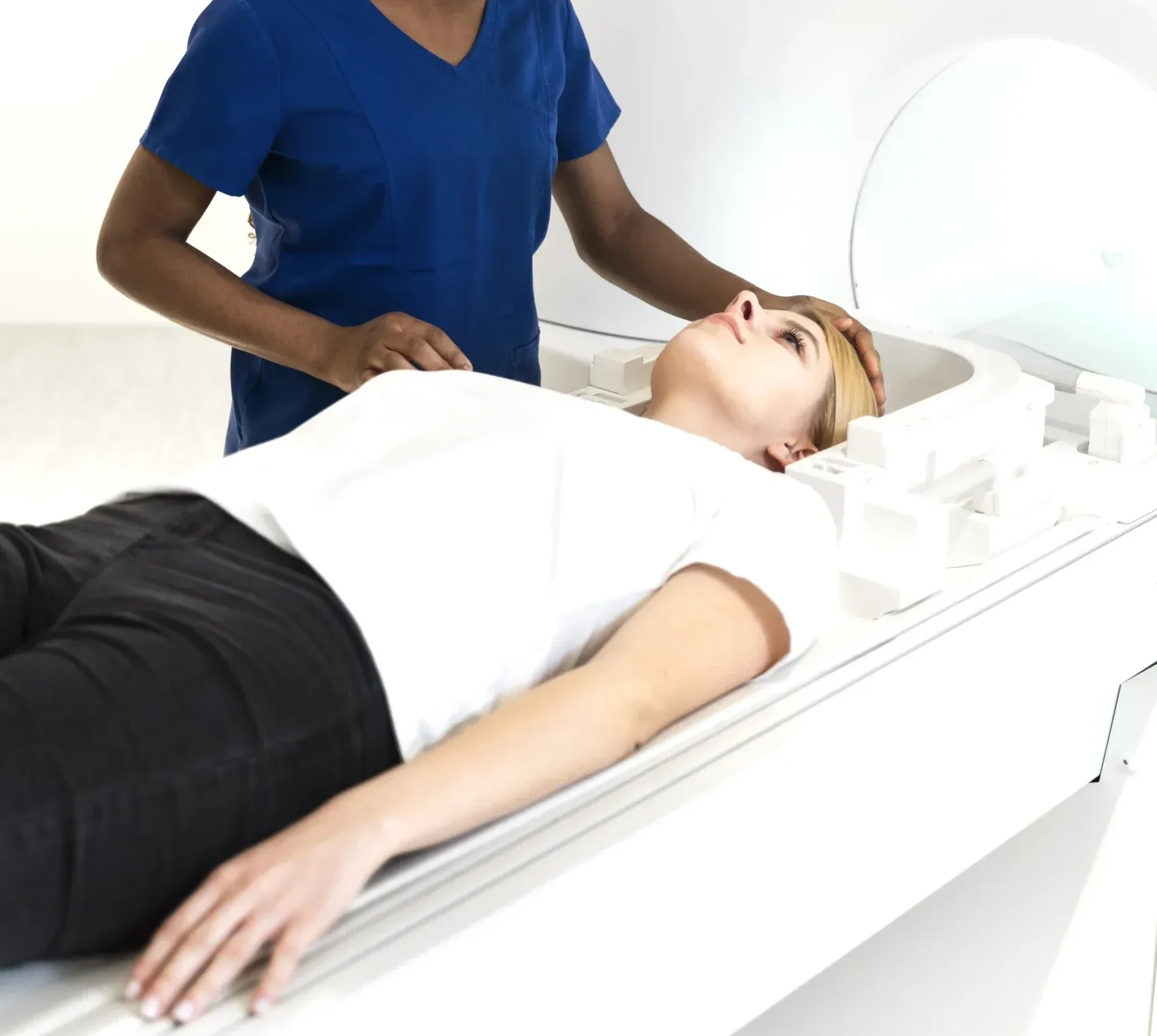
Neck MRI Dubai: What It Shows & Cost (2026)
مزید پڑھیں
X-Ray Scan Dubai: Complete Guide (2026)
مزید پڑھیں
MRI vs Ultrasound Dubai: Which Scan? (2026)
مزید پڑھیں
MRI vs X-Ray Dubai: Which Scan Do You Need? (2026)
مزید پڑھیں© 2026 Doctors Clinic Diagnostic Center (DCDC), Dubai Healthcare City. Originally published at https://doctorsclinicdubai.ae/blog/acl-mri-scan-dubai. All rights reserved. Unauthorized reproduction is prohibited.