Ключевые выводы
- ECG records the heart's electrical activity (rhythm, rate, conduction); echocardiogram images the heart's structure and pumping function
- ECG is essential for detecting arrhythmias, heart attacks, and conduction problems; echo detects valve disease, heart failure, and structural abnormalities
- One cannot replace the other - they provide fundamentally different information that cardiologists need together for complete cardiac assessment
- ECG takes 5-10 minutes and costs AED 200-500; echo takes 20-40 minutes and costs AED 850-2,000
- Most cardiac evaluations include both tests because electrical and structural problems often coexist
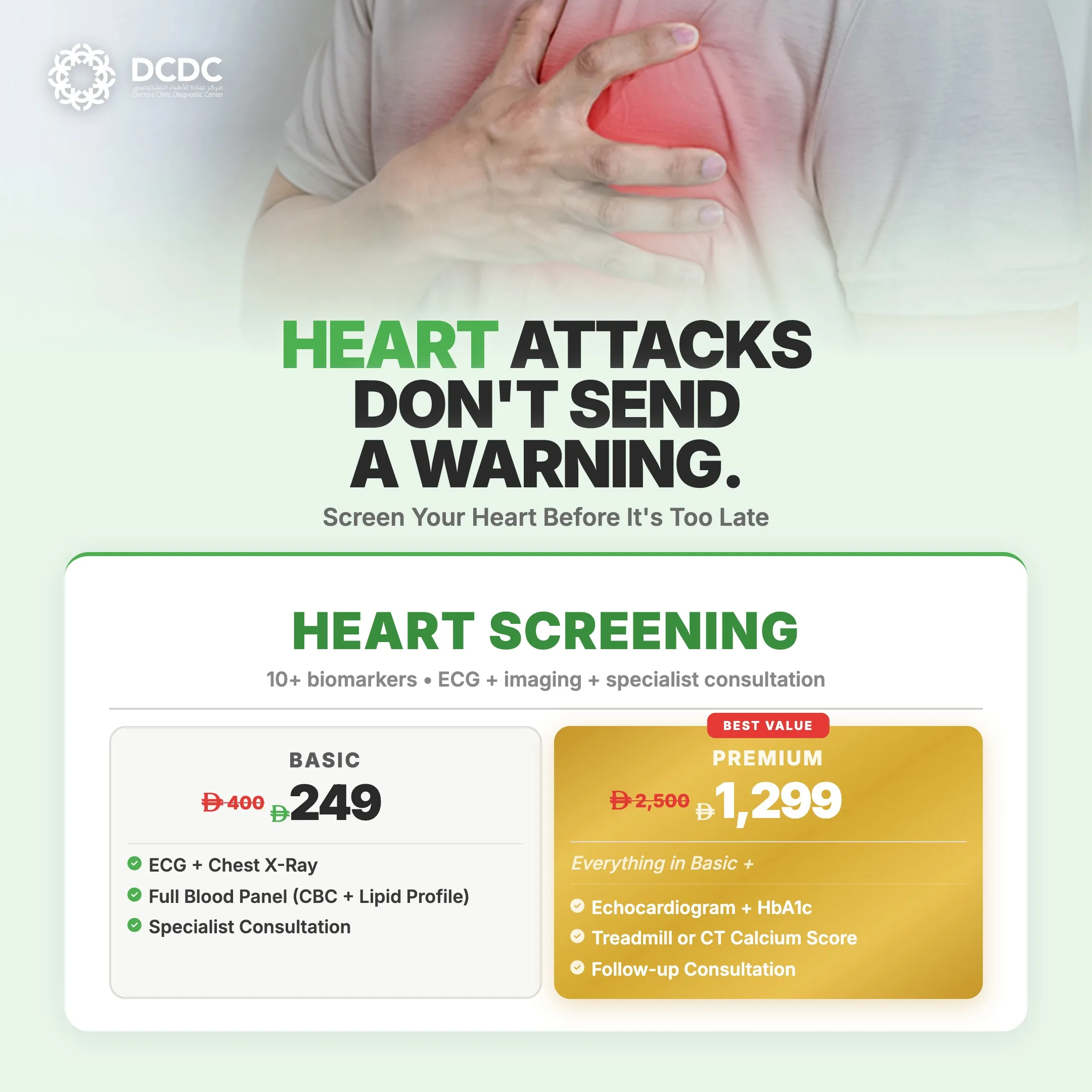
The ECG (electrocardiogram) and echocardiogram (echo) are the two most commonly ordered heart tests, but they measure entirely different aspects of cardiac function. An ECG records the electrical signals that control your heartbeat, while an echocardiogram uses ultrasound to create moving images of the heart's structure and pumping action. Understanding what each test tells you, and why one cannot replace the other, helps you appreciate their complementary roles in cardiac care.
Patients frequently ask whether they need both tests or if one is sufficient. The answer depends on your symptoms and clinical situation, but in most comprehensive cardiac evaluations, both are ordered because they provide fundamentally different information. This guide explains exactly what each test measures, compares them side by side, and clarifies when you need one, the other, or both.

What an ECG Measures
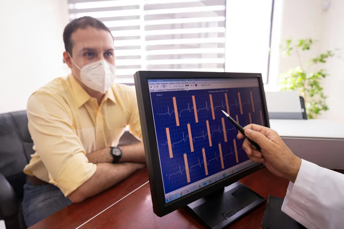
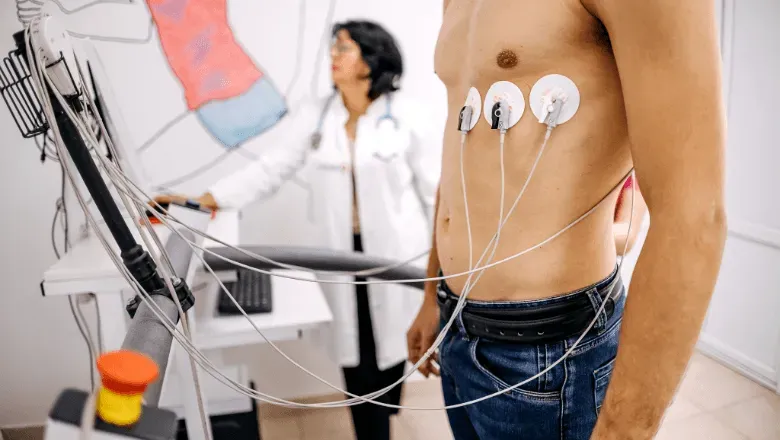
An electrocardiogram (ECG or EKG) records the electrical impulses that travel through your heart muscle to coordinate each heartbeat. Small adhesive electrodes (typically 10) are placed on your chest, arms, and legs. These electrodes detect the tiny electrical signals generated by the heart and display them as a characteristic waveform on paper or a screen.
The ECG waveform has distinct components (P wave, QRS complex, T wave) that correspond to specific electrical events in the cardiac cycle. A trained reader can identify abnormalities in these components to diagnose conditions including:
- Arrhythmias (abnormal heart rhythms): Atrial fibrillation, supraventricular tachycardia, ventricular tachycardia, heart block, and other rhythm disturbances
- Heart attack (myocardial infarction): ST-segment elevation or depression indicates acute ischemia or infarction. This is the first test performed in suspected heart attack and determines whether emergency intervention is needed
- Conduction abnormalities: Bundle branch blocks, Wolff-Parkinson-White syndrome, and prolonged QT interval, which can predispose to dangerous arrhythmias
- Heart rate: Bradycardia (slow rate), tachycardia (fast rate), and whether the rate is regular or irregular
- Chamber enlargement clues: ECG patterns can suggest left atrial enlargement, right atrial enlargement, or ventricular hypertrophy, though echo confirms these more accurately
- Electrolyte imbalances: Abnormal potassium, calcium, or magnesium levels can produce characteristic ECG changes
- Medication effects: Certain drugs (digoxin, beta-blockers, antiarrhythmics) produce identifiable ECG patterns
The ECG is performed in 5-10 minutes, is completely painless (electrodes are simply stuck to the skin), and provides immediate results. It is the most basic and universally available cardiac test, found in every hospital, clinic, and ambulance. For more detail on ECG testing, see our ECG test guide.
What an Echocardiogram Measures
An echocardiogram uses ultrasound waves (the same technology used in pregnancy scans) to create real-time moving images of the heart. A probe placed on the chest sends sound waves that bounce off heart structures and return to the probe, where they are converted into detailed images.
The echocardiogram reveals:
- Heart valve function: Whether valves open properly (stenosis assessment) and close completely (regurgitation assessment). This is the primary test for valve disease diagnosis and monitoring
- Ejection fraction: The percentage of blood pumped from the left ventricle with each beat (normal 55-70%). This is the most important measurement for heart failure diagnosis and monitoring
- Wall motion abnormalities: Areas of the heart that do not contract properly, indicating previous heart attack damage, ischemia, or cardiomyopathy
- Chamber dimensions: Precise measurement of all four heart chambers. Enlarged chambers suggest chronic valve disease, heart failure, or uncontrolled hypertension
- Wall thickness: Thickened walls (hypertrophy) from high blood pressure, hypertrophic cardiomyopathy, or other infiltrative diseases
- Blood flow patterns: Doppler technology measures the speed and direction of blood flow, quantifying the severity of valve problems and detecting abnormal communications between chambers
- Pericardial effusion: Fluid around the heart that may cause compression
- Congenital defects: Structural abnormalities present from birth such as septal defects (holes between chambers)
- Pulmonary artery pressure: Estimated from the velocity of tricuspid regurgitation, important for diagnosing pulmonary hypertension
The standard echocardiogram takes 20-40 minutes, is painless, uses no radiation, and requires no preparation. For a comprehensive guide including types of echo and cost, see our echocardiogram guide.
Side-by-Side Comparison
| Feature | ECG | Echocardiogram |
|---|---|---|
| What it measures | Electrical activity of the heart | Physical structure and pumping function |
| Technology | Electrodes on skin detecting electrical signals | Ultrasound waves creating real-time images |
| Duration | 5-10 minutes | 20-40 minutes |
| Preparation | None | None (for standard TTE) |
| Pain or discomfort | None | None (mild pressure from probe) |
| Radiation | None | None |
| Detects heart rhythm problems | Yes - primary test | No |
| Detects heart attack | Yes - ST changes (acute phase) | Yes - wall motion abnormalities (damage phase) |
| Detects valve disease | Indirect clues only | Yes - primary test with full quantification |
| Measures ejection fraction | No | Yes |
| Detects heart failure | Indirect clues | Yes - directly measures function and structure |
| Detects pericardial effusion | Indirect clues (low voltages) | Yes - directly visualized |
| Cost in Dubai (AED) | 200 - 500 | 850 - 2,000 |
| Results available | Immediately | Same day (after interpretation) |
| Portable / bedside | Yes - very portable | Yes - portable machines available |
| Frequency of use | Most common cardiac test worldwide | Most common cardiac imaging test |
ECG and echo test different aspects of cardiac function. Neither can replace the other.
Can an Echo Replace an ECG (or Vice Versa)?
No. This is one of the most important points to understand about cardiac testing. An echocardiogram cannot detect arrhythmias, heart blocks, or the acute electrical changes of a heart attack. An ECG cannot visualize valve disease, measure ejection fraction, or detect pericardial effusion. They test fundamentally different aspects of cardiac function.
Consider this analogy: an ECG is like listening to the electrical wiring of your house to check if the circuits are working properly. An echocardiogram is like using a camera to inspect the physical structure of the house, including the walls, doors, plumbing, and foundations. Both inspections are necessary for a complete picture of the house's condition, and finding a wiring problem does not mean the structure is fine, or vice versa.
In clinical practice, electrical and structural heart problems frequently coexist. Atrial fibrillation (detected on ECG) often causes or results from an enlarged left atrium (detected on echo). A previous heart attack (detected on ECG as Q waves) causes wall motion abnormalities and reduced ejection fraction (detected on echo). This is why cardiologists routinely order both tests together.
When You Need an ECG
- Palpitations (awareness of heartbeat, skipped beats, racing heart)
- Chest pain, especially acute onset
- Fainting (syncope) or near-fainting episodes
- Dizziness or lightheadedness
- Known or suspected arrhythmia monitoring
- Before starting certain medications (QT-prolonging drugs)
- Pre-operative cardiac assessment
- Sports participation clearance
- Follow-up after pacemaker or defibrillator implantation
- Routine health screening (especially men over 40 and women over 50)
When You Need an Echocardiogram
- Heart murmur detected on physical examination
- Unexplained shortness of breath, especially with exertion or lying flat
- Known heart failure requiring ejection fraction monitoring
- Assessment after heart attack to evaluate damage
- Before and after valve surgery
- Atrial fibrillation evaluation (checking for clots and structural cause)
- Suspected endocarditis (infection on heart valves)
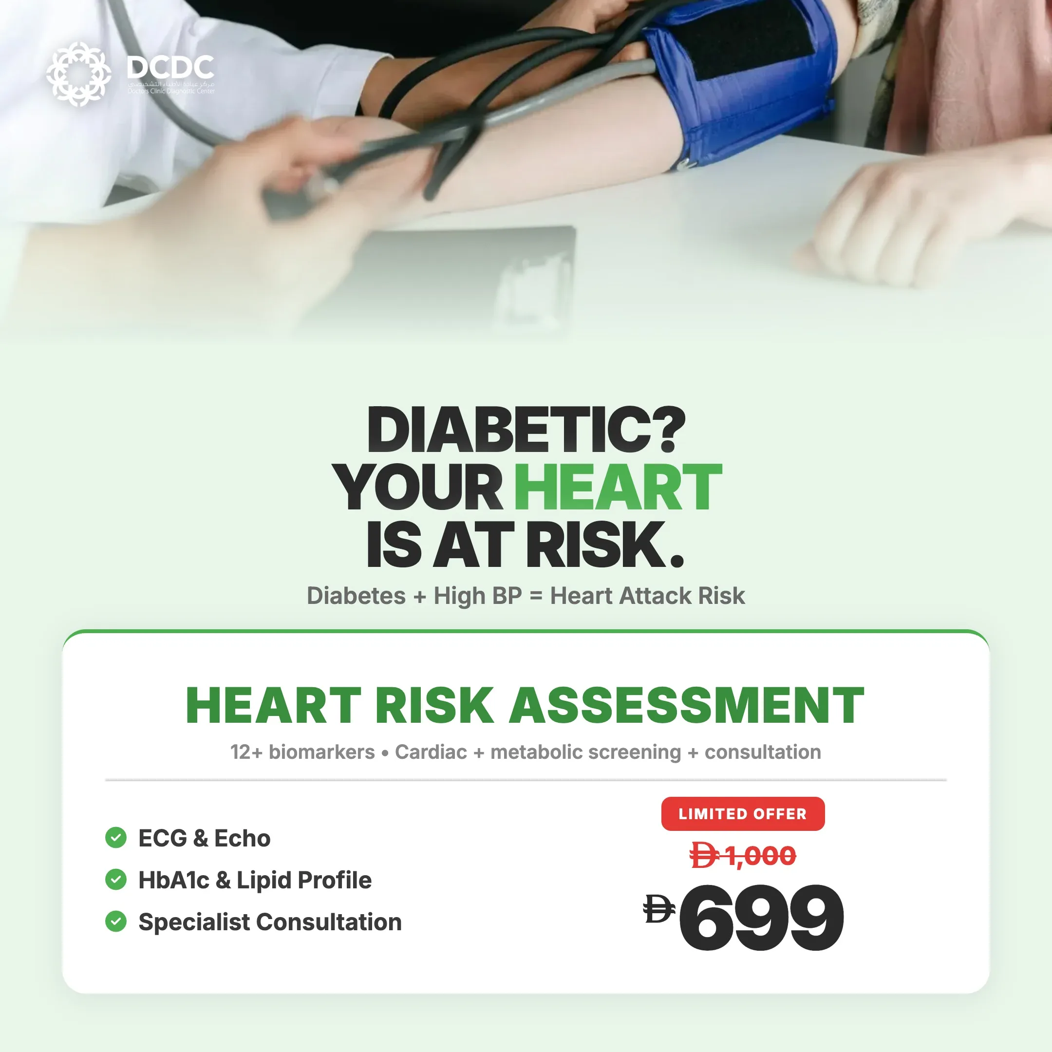
- Hypertension assessment for left ventricular hypertrophy
- Suspected pericardial effusion
- Congenital heart disease evaluation
When You Need Both Together
Most comprehensive cardiac evaluations include both ECG and echo. Specific scenarios where both are routinely ordered together include:
- New-onset heart failure: ECG to check for arrhythmias and evidence of old heart attack, echo to measure ejection fraction and assess valves
- Chest pain evaluation: ECG to rule out acute ischemia, echo to assess heart function and wall motion
- Atrial fibrillation workup: ECG confirms the rhythm, echo assesses atrial size, valve function, and checks for clots before cardioversion
- Pre-surgical cardiac clearance: ECG for rhythm, echo for function, especially before major surgery
- After heart attack: ECG to monitor for arrhythmias and conduction issues, echo to measure damage and ejection fraction
- Syncope (fainting) workup: ECG to look for conduction disease or arrhythmia, echo to rule out structural causes like aortic stenosis or hypertrophic cardiomyopathy
Cost Comparison in Dubai
| Test | Cost Range (AED) | What You Get |
|---|---|---|
| ECG (resting) | 200 - 500 | Heart rhythm, rate, and electrical pattern assessment |
| Standard echocardiogram (TTE) | 850 - 2,000 | Heart structure, valve function, ejection fraction, Doppler |
| ECG + echo combined with consultation | 1,500 - 3,500 | Both tests plus cardiologist interpretation and recommendations |
| 24-hour Holter ECG monitor | 800 - 1,500 | Continuous ECG recording for intermittent rhythm problems |
| Stress echo | 2,000 - 4,000 | Echo at rest and with exercise for coronary artery disease assessment |
ECG is a quick, affordable screening test. Echo provides more detailed information at higher cost. Both are typically covered by insurance.
Both tests are covered by most insurance plans in Dubai when ordered by a physician for medical indications. An ECG is often included in routine health checkup packages. A cardiology consultation ensures the right combination of tests is ordered for your specific situation.
"I never consider a cardiac evaluation complete with only one of these tests. An ECG tells me about the heart's electrical wiring. An echo tells me about its plumbing and construction. You need both perspectives to understand what is happening. I have seen patients with perfectly normal ECGs who have severe valve disease on echo, and patients with normal echo findings who have dangerous arrhythmias on ECG," explains Dr. Osama Elzamzami.
Complete Cardiac Assessment at DCDC
Our cardiology team provides both ECG and echocardiogram testing with expert interpretation for a comprehensive view of your heart health.
Heart Health Services at DCDC Dubai Healthcare City
At Doctors Clinic Diagnostic Center, we offer complete cardiac diagnostics including ECG, echocardiography, and cardiology consultations. Located in Dubai Healthcare City.
Связанные услуги в DCDC
Квалифицированная помощь и современная диагностика в Dubai Healthcare City
Frequently Asked Questions
Final Thoughts
The ECG and echocardiogram are the two foundational tests in cardiology, each providing essential but entirely different information about your heart. The ECG evaluates the electrical wiring; the echo evaluates the physical structure and pumping performance. Neither can replace the other, and most comprehensive cardiac assessments require both.
If your doctor has ordered one or both of these tests, understanding what each reveals helps you engage more meaningfully with your results and treatment plan. Both tests are painless, radiation-free, and widely available in Dubai with broad insurance coverage.
For complete cardiac diagnostics in Dubai, Doctors Clinic Diagnostic Center in Dubai Healthcare City offers ECG, echocardiography, and cardiology consultation services with experienced practitioners.
Источники и ссылки
Эта статья проверена нашей медицинской командой и ссылается на следующие источники:
- American Heart Association - ECG and Echo Testing
- American College of Cardiology - Appropriate Use Criteria
- European Society of Cardiology - Diagnostic Testing Guidelines
- American Society of Echocardiography
- British Heart Foundation - Heart Tests Explained
Медицинский контент на этом сайте проверяется врачами, лицензированными DHA. См. нашу редакционную политику для получения дополнительной информации.

Автор
Dr. Osama Elzamzami
Consultant Radiologist
MD, Radiology
Dr. Osama Elzamzami is a Consultant Radiologist at DCDC Dubai Healthcare City with expertise in diagnostic imaging and cardiac assessment.
Related Articles
Echocardiogram Dubai: Types, Cost & What It Shows

ECG Test Guide Dubai
Stress Test Dubai
Heart Health Prevention Dubai
More in Cardiology

Cardiac Rehabilitation Dubai: Recovery (2026)
Читать далееTroponin Test Dubai: Heart Attack Marker (2026)
Читать далее
Best Cardiologist in Dubai: Dr. Shahoo Mazhari at DCDC (2026)
Читать далееCardiologist in Dubai Healthcare City: Dr. Shahoo Mazhari at DCDC (2026)
Читать далее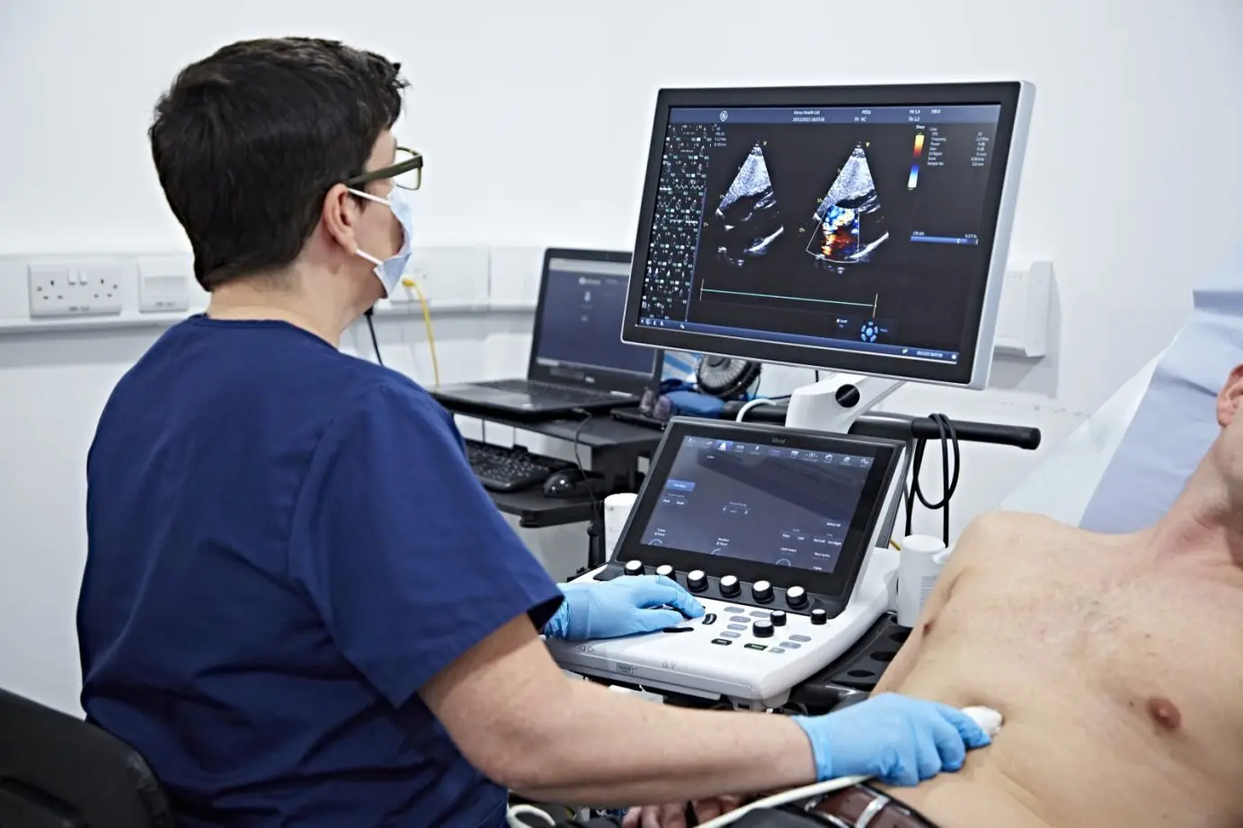
Echocardiogram (Echo) Test in Dubai: Cost, Procedure & What to Expect (2026)
Читать далееIranian Cardiologist in Dubai: Dr. Shahoo Mazhari at DCDC (2026)
Читать далее© 2026 Doctors Clinic Diagnostic Center (DCDC), Dubai Healthcare City. Originally published at https://doctorsclinicdubai.ae/blog/ecg-vs-echo-difference. All rights reserved. Unauthorized reproduction is prohibited.