
Points cles
- Uterine fibroids affect up to 70 percent of women by age 50, making them the most common benign tumors of the female reproductive tract, yet many women remain undiagnosed because fibroids are frequently asymptomatic
- Fibroids are classified by location — intramural (within the uterine wall), subserosal (on the outer surface), submucosal (projecting into the uterine cavity), and pedunculated (attached by a stalk) — and their position directly determines which symptoms they cause and which treatments are most appropriate
- Submucosal fibroids, even when small, are the type most likely to cause heavy menstrual bleeding and fertility problems, while large subserosal fibroids tend to cause pressure symptoms such as urinary frequency and pelvic pain
- Medical management — including hormonal contraceptives, GnRH agonists, tranexamic acid, and the newer oral GnRH antagonists — can effectively control symptoms and shrink fibroids without surgery in many women
- Myomectomy (surgical fibroid removal while preserving the uterus) is the preferred approach for women who wish to maintain fertility, with laparoscopic and hysteroscopic techniques offering shorter recovery times than open surgery
- At DCDC in Dubai Healthcare City, gynecology consultation with ultrasound assessment starts from AED 500, with on-site pelvic imaging and coordinated MRI scanning from AED 900 for surgical planning
- Fibroids are hormonally driven and tend to shrink after menopause when estrogen and progesterone levels decline — watchful waiting is a valid strategy for women with small, asymptomatic fibroids approaching menopause
Uterine fibroids (leiomyomas) are the most common benign tumors of the female reproductive system, affecting an estimated 20 to 40 percent of women of reproductive age and up to 70 percent of women by age 50. Despite their prevalence, many women in Dubai and the UAE remain undiagnosed because fibroids frequently cause no symptoms. When symptoms do occur — heavy menstrual bleeding, pelvic pain, urinary frequency, or difficulty conceiving — they can significantly impact quality of life. At Doctors Clinic Diagnostic Center (DCDC) in Dubai Healthcare City, our OB-GYN specialists provide comprehensive fibroid evaluation and treatment using evidence-based protocols tailored to each patient's symptoms, reproductive goals, and clinical findings. This guide covers everything you need to know about uterine fibroids — from types and symptoms to medical and surgical treatment options available in Dubai.
Fibroids are not cancerous and very rarely become malignant (the risk of a fibroid being a leiomyosarcoma is estimated at less than 1 in 1,000). However, the symptoms they produce — particularly heavy or prolonged menstrual bleeding — can lead to iron deficiency anemia, fatigue, and diminished quality of life. Additionally, depending on their size and location, fibroids can affect fertility and pregnancy outcomes. Understanding your fibroid type, available treatment options, and when intervention is necessary empowers you to make informed decisions alongside your gynecologist.
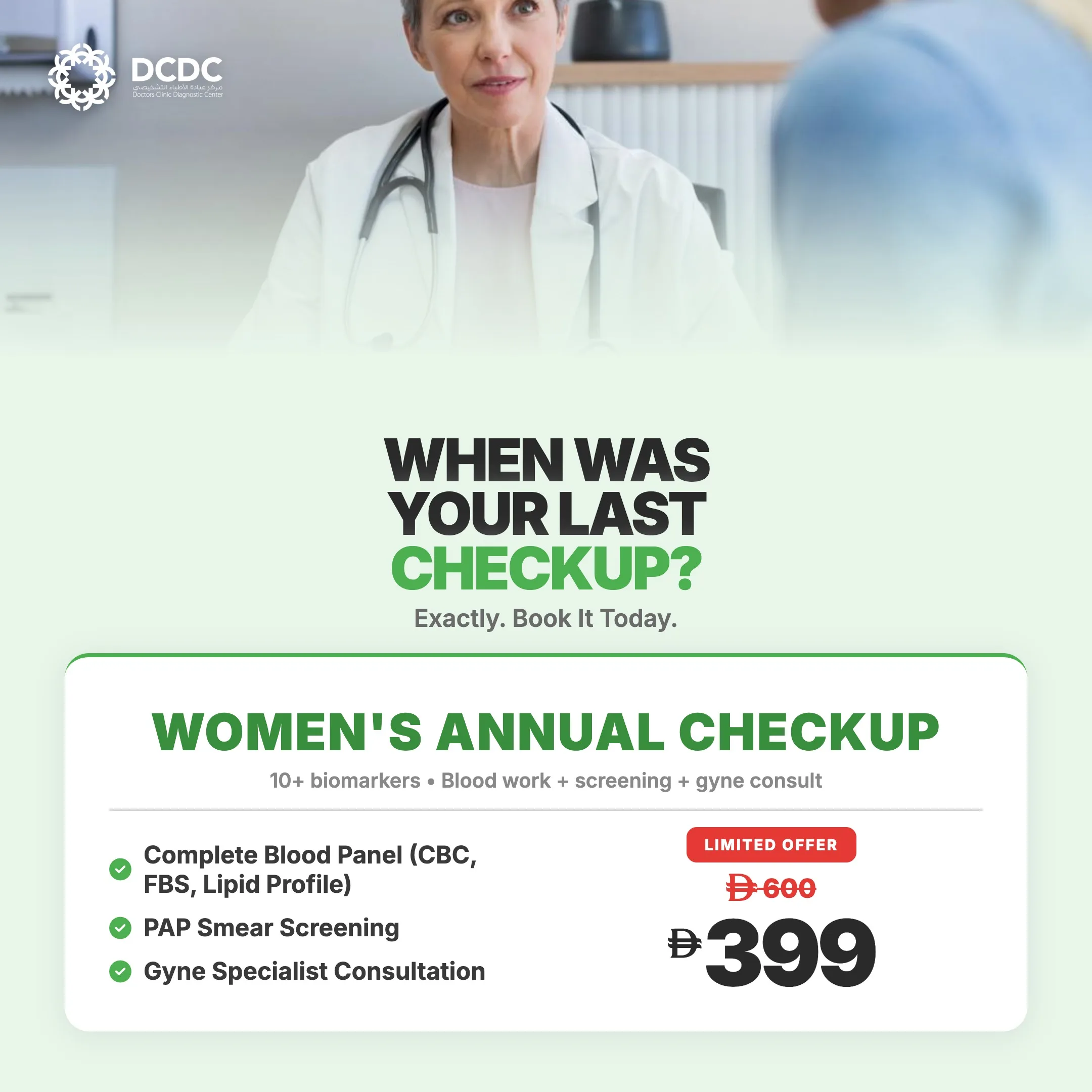
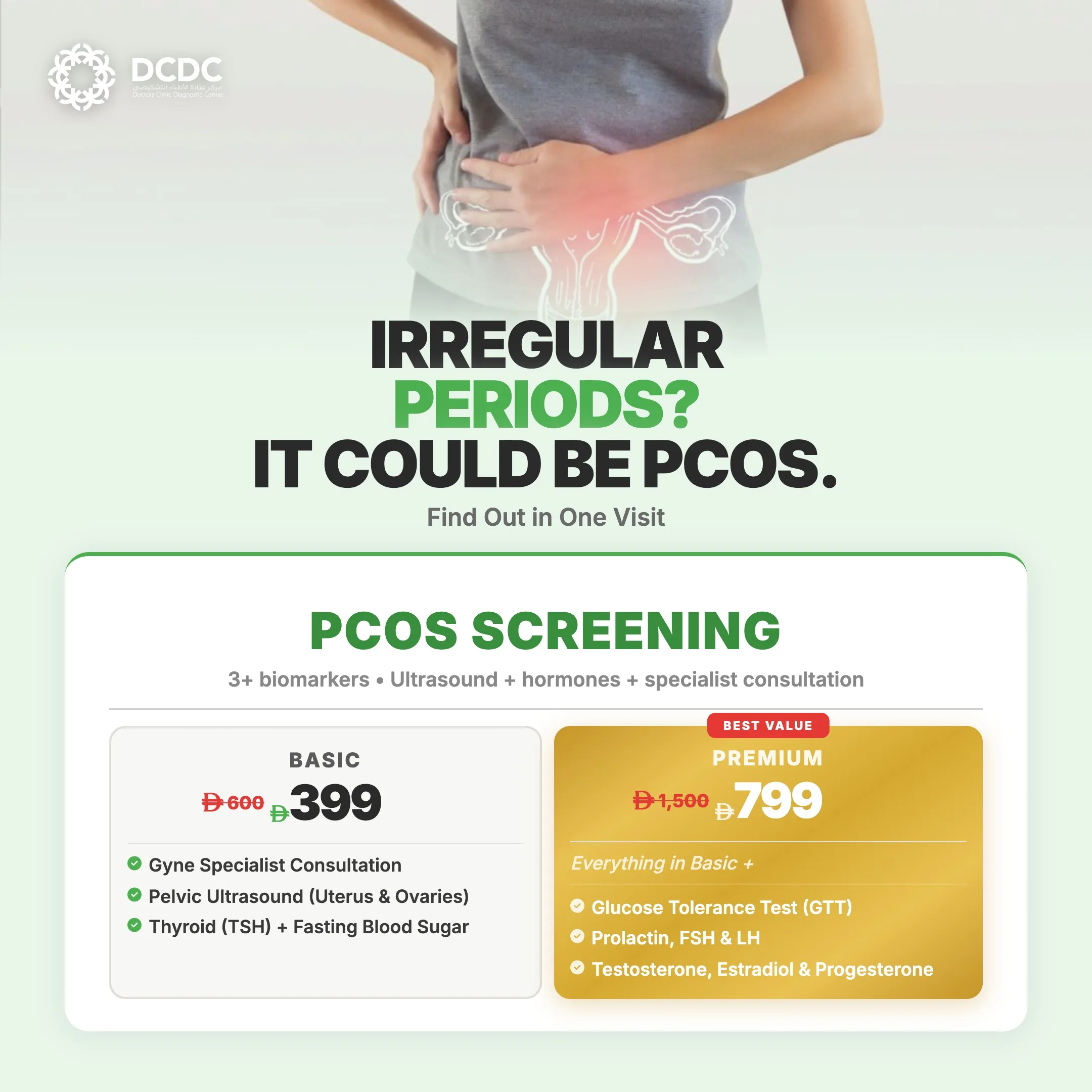
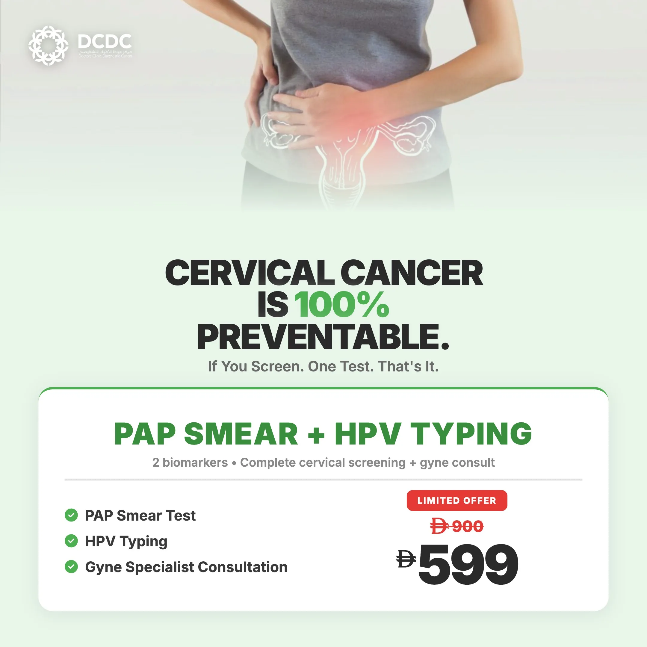
Health Screening Packages
Save with our bundled screening packages — specialist consultation included


What Are Uterine Fibroids?
Uterine fibroids are benign (non-cancerous) growths that develop from the smooth muscle tissue of the uterus (myometrium). They can range in size from tiny seedlings undetectable by the human eye to large masses that distort the shape and size of the uterus. Some women develop a single fibroid, while others have multiple fibroids of varying sizes throughout the uterus. The medical term for fibroids is leiomyomas or myomas.
Fibroids are hormonally responsive — their growth is driven primarily by estrogen and progesterone, the two main female reproductive hormones. This is why fibroids typically develop during the reproductive years (when these hormones are at their highest), tend to grow during pregnancy (when hormone levels surge), and generally shrink after menopause (when hormone levels decline). Each fibroid develops from a single smooth muscle cell that undergoes a genetic mutation, causing it to proliferate and form a distinct, firm, rubbery mass separate from the surrounding uterine tissue.
According to the American College of Obstetricians and Gynecologists (ACOG), fibroids are clinically apparent in approximately 25 percent of women, but histological studies suggest the true prevalence may be as high as 70 to 80 percent when accounting for small, asymptomatic fibroids. Risk factors include age (most common in women aged 30 to 50), family history, ethnicity (women of African descent have a 2 to 3 times higher risk), obesity, and early onset of menstruation. While fibroids share some symptom overlap with other gynecological conditions, they are distinct from ovarian cysts, which arise from the ovaries rather than the uterine muscle.
Types of Uterine Fibroids and Their Locations
The classification of fibroids is based on their location within the uterus. This distinction is clinically important because the position of a fibroid directly determines the symptoms it causes and the treatment options available. The International Federation of Gynecology and Obstetrics (FIGO) classification system uses a numbering system (0 through 8) to precisely describe fibroid location, but the four broad categories below cover the essential types.
| Fibroid Type | Location | Common Symptoms | Treatment Approach |
|---|---|---|---|
| Intramural | Within the muscular wall of the uterus | Heavy menstrual bleeding, pelvic pressure, uterine enlargement | Medication; myomectomy (laparoscopic or open) if large or symptomatic |
| Subserosal | On the outer surface of the uterus | Pelvic pressure, back pain, bladder or bowel compression | Watchful waiting if asymptomatic; laparoscopic myomectomy if symptomatic |
| Submucosal | Projecting into the uterine cavity | Heavy or prolonged periods, intermenstrual bleeding, infertility | Hysteroscopic myomectomy (gold standard); often requires removal even when small |
| Pedunculated | Attached to the uterus by a stalk (can be subserosal or submucosal) | Varies by location; stalk torsion can cause acute pelvic pain | Surgical removal if symptomatic or at risk of torsion |
Fibroid classification by location, associated symptoms, and typical treatment approach. The FIGO subclassification provides more granular detail for surgical planning.
Intramural fibroids are the most common type, accounting for approximately 70 percent of all fibroids. They grow within the muscular wall of the uterus and can cause the uterus to enlarge as they increase in size. Subserosal fibroids grow outward from the outer wall of the uterus and tend to cause pressure symptoms rather than menstrual irregularity. Submucosal fibroids are the least common (approximately 5 to 10 percent of fibroids) but are clinically the most significant because they distort the uterine cavity — even small submucosal fibroids (1 to 2 cm) can cause disproportionately heavy bleeding and interfere with embryo implantation.
Uterine Fibroid Symptoms: When to Seek Help
Approximately 20 to 50 percent of women with fibroids experience symptoms severe enough to seek medical attention. The remainder have asymptomatic fibroids that are discovered incidentally during routine pelvic examinations or imaging studies performed for other reasons. Symptoms depend on the number, size, and location of fibroids. You should consult a gynecologist if you experience any of the following:
- Heavy menstrual bleeding (menorrhagia): This is the most common symptom of fibroids, particularly intramural and submucosal types. Bleeding may be heavy enough to soak through a pad or tampon every hour for several consecutive hours, pass large blood clots, or last longer than 7 days. Chronic heavy bleeding often leads to iron deficiency anemia, causing fatigue, weakness, dizziness, and shortness of breath.
- Pelvic pressure and pain: Large fibroids can create a sensation of fullness, pressure, or heaviness in the lower abdomen and pelvis. Some women describe it as similar to early pregnancy pressure. Fibroids pressing on adjacent structures can cause specific symptoms — bladder pressure leading to urinary frequency or urgency, rectal pressure leading to constipation, and lower back pain from compression of spinal nerves.
- Prolonged or irregular periods: Fibroids can cause periods to last longer than 7 days, or cause bleeding or spotting between periods (intermenstrual bleeding). Submucosal fibroids are particularly associated with prolonged bleeding episodes.
- Abdominal enlargement: Large or multiple fibroids can cause the uterus to grow to the size of a 4 to 5 month pregnancy or larger. Women may notice their abdomen growing larger, clothes fitting differently, or palpable firmness in the lower abdomen.
- Painful intercourse (dyspareunia): Fibroids located on the anterior (front) wall of the uterus or near the cervix can cause pain during sexual intercourse, particularly with deep penetration.
- Difficulty conceiving: Submucosal fibroids and large intramural fibroids that distort the uterine cavity can impair fertility by preventing embryo implantation or blocking the fallopian tubes. Fibroids are identified as a contributing factor in approximately 5 to 10 percent of infertility cases.
- Pregnancy complications: Women with fibroids face higher risks of miscarriage, preterm labor, placental abruption, malpresentation (breech position), and cesarean delivery. Fibroid degeneration during pregnancy — when a growing fibroid outgrows its blood supply — can cause severe acute pain.
If you are experiencing heavy periods, chronic pelvic pain, or unexplained abdominal growth, do not assume it is normal. These symptoms warrant evaluation to determine whether fibroids — or another gynecological condition such as endometriosis or adenomyosis — are the underlying cause.
What Causes Fibroids to Grow?
The exact cause of uterine fibroids remains incompletely understood, but research has identified several interconnected factors that contribute to their development and growth. Fibroids are considered a multifactorial condition involving genetic predisposition, hormonal influences, and environmental factors.
- Hormonal factors — estrogen and progesterone: Fibroids contain more estrogen and progesterone receptors than normal uterine muscle tissue. Both hormones stimulate fibroid growth, which is why fibroids develop during the reproductive years, enlarge during pregnancy, and typically shrink after menopause. Conditions that increase estrogen exposure — such as early menarche, obesity (adipose tissue converts androgens to estrogen), and never having been pregnant — are associated with higher fibroid risk.
- Genetic mutations: Research has identified specific genetic mutations associated with fibroid development, particularly in the MED12 gene (found in up to 70 percent of fibroids) and the HMGA2 gene. These mutations cause individual smooth muscle cells to proliferate abnormally. Family history is a strong risk factor — women with a first-degree relative with fibroids have a 2 to 3 times higher risk of developing them.
- Growth factors: Substances that promote cell growth, such as insulin-like growth factor (IGF), transforming growth factor-beta (TGF-beta), and basic fibroblast growth factor (bFGF), are produced in excess within fibroid tissue. These growth factors create a local environment that sustains fibroid growth independently of systemic hormone levels.
- Extracellular matrix (ECM) overproduction: Fibroids produce excessive extracellular matrix — the structural material between cells — which makes them firm and fibrous. The ECM also stores growth factors and acts as a reservoir that sustains fibroid growth. This is why fibroids feel characteristically hard and rubbery on examination.
- Ethnicity: Women of African descent develop fibroids at 2 to 3 times the rate of Caucasian women, tend to develop them at younger ages, and are more likely to have larger, more numerous, and more symptomatic fibroids. This disparity appears to have both genetic and environmental components.
- Lifestyle and environmental factors: Obesity (BMI greater than 25) increases fibroid risk, with each 10 kg of weight gain associated with a 21 percent increase in risk. Vitamin D deficiency — common in the UAE despite abundant sunshine due to indoor lifestyles and sun-protective clothing — has been linked to increased fibroid risk in several studies. High red meat consumption and alcohol intake have also been associated with higher prevalence. Women dealing with hormonal imbalances may be at greater risk of conditions like fibroids that respond to hormone levels.
How Are Uterine Fibroids Diagnosed?
Fibroid diagnosis typically begins with a clinical history and pelvic examination, followed by imaging studies to confirm the presence, number, size, and location of fibroids. Accurate diagnosis is essential because treatment decisions depend heavily on fibroid characteristics. The diagnostic workup at DCDC includes the following components:
- Pelvic examination: During a bimanual pelvic examination, your gynecologist can detect an enlarged, irregularly shaped uterus suggestive of fibroids. Large fibroids may be palpable through the abdominal wall. However, a pelvic exam alone cannot determine the exact number, size, or location of fibroids — imaging is required.
- Transvaginal ultrasound (TVUS): This is the first-line imaging modality for fibroid evaluation. A small ultrasound probe is inserted into the vagina to produce detailed images of the uterus and surrounding structures. TVUS can identify fibroids as small as 1 cm, determine their location (intramural, subserosal, submucosal), measure their dimensions, and assess the endometrial lining. For a comprehensive explanation of the procedure, see our pelvic ultrasound guide. At DCDC, on-site ultrasound with targeted assessment is available during your consultation visit.
- Transabdominal ultrasound: Used in conjunction with TVUS, particularly when the uterus is significantly enlarged. The transabdominal approach provides a wider field of view to assess very large fibroids or multiple fibroids that extend beyond the pelvis.
- Saline infusion sonohysterography (SIS): Sterile saline is infused into the uterine cavity during ultrasound to better delineate submucosal fibroids and their relationship to the endometrial cavity. This technique is particularly useful for surgical planning when hysteroscopic myomectomy is being considered.
- Pelvic MRI: MRI is the most accurate imaging modality for fibroid mapping. It provides superior soft-tissue contrast, can precisely determine the number, size, and location of every fibroid, and can distinguish fibroids from adenomyosis (a condition that can mimic fibroids on ultrasound). MRI is recommended before surgical planning, particularly for myomectomy. For more details, see our pelvic MRI imaging guide. At DCDC, coordinated MRI scanning for surgical planning starts from AED 900.
- Blood tests: A complete blood count (CBC) is essential to check for iron deficiency anemia caused by heavy menstrual bleeding. Thyroid function tests (TSH) and hormone levels may be ordered to exclude other causes of abnormal uterine bleeding. In selected cases, additional blood work including a metabolic panel and coagulation studies may be warranted.
- Hysteroscopy: A thin, lighted telescope is inserted through the cervix into the uterine cavity, allowing direct visualization of the endometrial surface and any submucosal fibroids. Hysteroscopy is both diagnostic and therapeutic — small submucosal fibroids can be removed during the same procedure.
Fibroid Treatment Options: Medical vs Surgical
The management of uterine fibroids ranges from watchful waiting (for small, asymptomatic fibroids) to major surgery (for large, symptomatic fibroids or those affecting fertility). Treatment decisions are individualized based on symptom severity, fibroid characteristics (number, size, location), your age, reproductive goals, and personal preferences. Dr. Parisa Dini and the OB-GYN team at DCDC prioritize a stepwise approach — beginning with the least invasive option and escalating only when necessary. In every case, the goal is symptom relief while preserving the uterus whenever possible, particularly for women who wish to have children.
The two broad categories of treatment are medical management (medications to control symptoms and shrink fibroids) and surgical intervention (procedures to remove fibroids or, in some cases, the uterus itself). Many women can be managed effectively with medication alone, and surgery is reserved for cases where medical therapy fails, fibroids are too large or numerous for medical management, or fertility-preserving removal is needed.
Medications for Fibroid Management
Medical therapy for fibroids focuses on two goals: controlling symptoms (primarily heavy bleeding and pain) and reducing fibroid size. While medications do not permanently eliminate fibroids — they typically regrow when treatment is stopped — they can effectively manage symptoms, bridge the gap to menopause (when fibroids shrink naturally), and reduce fibroid size before surgery to make procedures safer and less invasive.
- Hormonal contraceptives (combined pills, patch, ring): Combined oral contraceptive pills do not shrink fibroids but can significantly reduce menstrual bleeding and regulate cycle length. They are often the first-line approach for women with mild to moderate bleeding symptoms who do not have large fibroids. The levonorgestrel-releasing intrauterine device (Mirena IUD) is particularly effective — it reduces menstrual blood loss by 70 to 90 percent and can be used in women with fibroids, provided the uterine cavity is not significantly distorted.
- Tranexamic acid: An antifibrinolytic medication taken only during menstruation (typically 3 to 5 days) that reduces bleeding by 30 to 55 percent by stabilizing blood clots within the uterine lining. Tranexamic acid is non-hormonal, making it suitable for women who cannot or prefer not to use hormonal therapy. It does not affect fibroid size.
- GnRH agonists (leuprolide, goserelin): These injectable medications suppress estrogen production to menopausal levels, causing fibroids to shrink by 30 to 50 percent over 3 to 6 months. GnRH agonists are highly effective but cause menopausal side effects (hot flushes, vaginal dryness, bone density loss) that limit their use to 6 months. They are primarily used as preoperative treatment to shrink large fibroids before surgery, reduce intraoperative bleeding, and potentially allow a less invasive surgical approach.
- GnRH antagonists (relugolix, linzagolix, elagolix): These newer oral medications represent a significant advance in fibroid pharmacotherapy. Unlike GnRH agonists, they work immediately (no initial hormonal flare), can be taken as a daily tablet, and when combined with low-dose add-back hormonal therapy, allow long-term use with fewer menopausal side effects and reduced bone density loss. Relugolix combination therapy has been shown to reduce heavy menstrual bleeding in over 70 percent of women. These medications are increasingly available in the UAE.
- Nonsteroidal anti-inflammatory drugs (NSAIDs): Ibuprofen, mefenamic acid, and naproxen can reduce menstrual pain (dysmenorrhea) and modestly decrease bleeding by inhibiting prostaglandin production. NSAIDs are often used in combination with other therapies rather than as standalone treatment.
- Iron supplementation: While not a fibroid treatment per se, iron supplementation is an essential component of care for women with heavy menstrual bleeding and resulting iron deficiency anemia. Oral iron (ferrous sulfate, ferrous fumarate) or intravenous iron infusion (for moderate to severe anemia or oral intolerance) should be initiated based on ferritin and hemoglobin levels.
Get a Fibroid Assessment at DCDC Dubai
At Doctors Clinic Diagnostic Center in Dubai Healthcare City, our OB-GYN specialists provide comprehensive fibroid evaluation including pelvic ultrasound assessment, hormonal profiling, and individualized treatment planning. Book your consultation — initial gynecology assessment from AED 500.
DCDC is rated 4.8/5 on Google from 1,000+ reviews. MOHAP-licensed in DHCC with direct billing for 20+ insurance providers including Daman, AXA, and Bupa.
Surgical Options for Fibroids
Surgery is recommended when fibroids cause severe symptoms that do not respond to medical management, when fibroids are large (generally greater than 5 cm) or rapidly growing, when fertility is impaired, or when the diagnosis is uncertain and tissue examination is needed to exclude malignancy. The choice of surgical technique depends on fibroid number, size, location, and whether preservation of the uterus is desired.
- Hysteroscopic myomectomy: The gold standard for submucosal fibroids. A resectoscope is inserted through the cervix (no abdominal incision), and the fibroid is removed under direct visualization. This procedure is performed as day surgery, has a rapid recovery (most women return to normal activities within 2 to 3 days), and preserves the uterus and fertility. It is suitable for fibroids that are predominantly within the uterine cavity (FIGO types 0, 1, and some type 2).
- Laparoscopic myomectomy: Fibroids are removed through 3 to 4 small abdominal incisions (0.5 to 1.5 cm) using minimally invasive techniques. This approach offers shorter hospital stays (typically 1 to 2 days), less postoperative pain, smaller scars, and faster recovery (2 to 4 weeks) compared to open surgery. It is best suited for a limited number of subserosal or intramural fibroids up to approximately 10 to 12 cm. Robot-assisted laparoscopic myomectomy offers enhanced precision for complex cases.
- Open (abdominal) myomectomy: Performed through a larger abdominal incision (similar to a cesarean section), open myomectomy is reserved for very large fibroids (greater than 12 to 15 cm), numerous fibroids (typically more than 4 to 5), or deeply embedded intramural fibroids that cannot be safely removed laparoscopically. Recovery time is 4 to 6 weeks. Despite longer recovery, open myomectomy allows the surgeon to palpate the entire uterus and remove fibroids that might be missed with minimally invasive approaches.
- Uterine artery embolization (UAE): A minimally invasive radiology procedure in which tiny particles are injected into the uterine arteries to block blood supply to fibroids, causing them to shrink by 40 to 60 percent over 3 to 6 months. UAE is an option for women who want to avoid surgery, but it is generally not recommended for women planning future pregnancies because its effects on fertility and pregnancy outcomes are not fully established.
- MRI-guided focused ultrasound surgery (MRgFUS/HIFU): A non-invasive procedure that uses focused ultrasound waves guided by MRI to heat and destroy fibroid tissue without incisions. It is suitable for selected fibroids (limited number, accessible location) and offers the shortest recovery time, but retreatment rates are higher than surgical options and availability in the UAE is limited.
- Hysterectomy (uterus removal): The only definitive treatment that guarantees fibroids will not recur. Hysterectomy is reserved for women who have completed childbearing, have severe symptoms unresponsive to other treatments, or who prefer definitive management. It can be performed vaginally, laparoscopically, or abdominally depending on uterine size and surgical factors. Hysterectomy is a major decision and should be discussed thoroughly with your gynecologist, as it is irreversible.
At DCDC, Dr. Parisa Dini evaluates each patient individually and coordinates with specialized surgeons when surgical intervention is needed. Her approach prioritizes medication before surgery and fertility-preserving techniques whenever possible. For women requiring surgery, DCDC provides comprehensive preoperative assessment including MRI mapping, blood work, and anesthesia consultation to ensure optimal outcomes.
Fibroids and Fertility: What You Need to Know
The relationship between fibroids and fertility is one of the most common concerns among women of reproductive age diagnosed with fibroids in Dubai. While most women with fibroids conceive and carry pregnancies successfully, certain fibroid types can impair fertility and increase pregnancy risks. Understanding which fibroids affect fertility — and which do not — is critical for making informed treatment decisions.
- Submucosal fibroids and fertility: These have the strongest evidence for impairing fertility. By distorting the uterine cavity, submucosal fibroids can prevent embryo implantation, disrupt endometrial blood supply, and cause early miscarriage. Studies show that removing submucosal fibroids before conception improves pregnancy rates by approximately 70 percent. Hysteroscopic myomectomy is recommended for women with submucosal fibroids who are planning pregnancy.
- Intramural fibroids and fertility: The impact of intramural fibroids on fertility is more debated. Evidence suggests that large intramural fibroids (greater than 4 to 5 cm) that distort the uterine cavity may reduce implantation rates and increase miscarriage risk, while small intramural fibroids that do not distort the cavity likely have minimal impact. The decision to remove intramural fibroids before fertility treatment is made on a case-by-case basis.
- Subserosal fibroids and fertility: These fibroids grow on the outer surface of the uterus and generally do not affect fertility or pregnancy outcomes. Removal is usually not recommended solely for fertility reasons unless the fibroid is very large or pedunculated with torsion risk.
- Fibroids during pregnancy: Approximately 10 to 30 percent of women with fibroids experience complications during pregnancy. Fibroids tend to grow during the first trimester (stimulated by rising hormone levels) and may undergo red degeneration — a painful condition occurring when a fibroid outgrows its blood supply. Fibroids near the placental site increase the risk of placental abruption, while those near the cervix can obstruct vaginal delivery.
- Post-myomectomy pregnancy planning: After myomectomy, most gynecologists recommend waiting 3 to 6 months before attempting conception to allow the uterus to heal. Women who have had extensive myomectomy involving deep uterine wall incisions may be advised to deliver via cesarean section to reduce the risk of uterine rupture during labor.
If you have been diagnosed with fibroids and are planning pregnancy, a thorough evaluation of fibroid type, size, and location is essential to determine whether treatment is needed before conception. Women with PCOS and fibroids face compounded fertility challenges — for guidance on managing PCOS, read our PCOS treatment and diagnosis guide.
What to Expect at DCDC for Fibroid Evaluation
At Doctors Clinic Diagnostic Center, we provide a structured, patient-centered approach to fibroid evaluation. From your first visit to treatment planning, our goal is to give you clear answers and an individualized management strategy. Here is what to expect when you visit our clinic in Building 64, Dubai Healthcare City.
- Step 1 — Arrival and registration: DCDC is located in Building 64, Dubai Healthcare City (DHCC). Upon arrival, our reception team will assist with registration, insurance verification, and medical history forms. We work with 20+ insurance providers including Daman, AXA, and Bupa, and our team handles pre-authorization for you.
- Step 2 — Consultation with your gynecologist: You will meet with a female OB-GYN specialist (Dr. Parisa Dini or another member of our gynecology team) who will review your symptoms, menstrual history, reproductive goals, and relevant medical history in detail. Dr. Dini specializes in fertility-preserving approaches and prioritizes medication before surgery whenever clinically appropriate.
- Step 3 — Pelvic ultrasound assessment: On-site transvaginal and/or transabdominal ultrasound is performed during your consultation visit to identify and map fibroids. Our ultrasound assessment focuses on fibroid number, size, location (using FIGO classification), relationship to the endometrial cavity, and assessment of ovarian health. This eliminates the need for a separate imaging appointment.
- Step 4 — Blood work and hormonal profiling: If indicated, blood samples are collected on-site for complete blood count (to assess for anemia), thyroid function, and hormonal profiling. Results are typically available within 24 to 48 hours.
- Step 5 — Treatment plan discussion: Based on the clinical findings, your gynecologist will explain the diagnosis in clear, understandable terms and discuss all available treatment options — from watchful waiting and medical management to surgical referral. You will receive a written treatment plan with a clear timeline, follow-up schedule, and expected outcomes.
- Step 6 — Coordinated MRI and surgical referral: If surgical planning is needed, DCDC coordinates pelvic MRI scanning (from AED 900) for detailed fibroid mapping and arranges referral to a gynecological surgeon for procedures such as myomectomy or hysteroscopy. Our team manages the entire referral pathway to ensure seamless, coordinated care.

Obstetrics & Gynecology Specialist
English, Farsi, Arabic
Dr. Parisa Dini is an OB-GYN specialist at DCDC in Dubai Healthcare City. She provides comprehensive gynecological care with a focus on fertility-preserving approaches and evidence-based treatment for conditions including fibroids, ovarian cysts, endometriosis, and PCOS. Dr. Dini prioritizes conservative management and medication before recommending surgical intervention.
Fibroid Treatment Cost in Dubai: Pricing Guide
Understanding the cost of fibroid diagnosis and treatment in Dubai helps you plan your healthcare effectively. The total cost depends on the severity of your condition, the diagnostic tests required, and the chosen treatment approach. The following table outlines typical costs at DCDC in Dubai Healthcare City. Most health insurance plans in the UAE cover fibroid-related consultations and diagnostic imaging when medically indicated.
| Service | Price Range (AED) | Notes |
|---|---|---|
| Gynecology Consultation | From AED 500 | Initial OB-GYN assessment with clinical history and pelvic examination |
| Pelvic Ultrasound (TVUS) | Available on-site | Fibroid mapping, size measurement, and cavity assessment |
| Pelvic MRI | From AED 900 | Detailed fibroid mapping for surgical planning |
| Complete Blood Count (CBC) | AED 80–150 | Anemia screening for women with heavy bleeding |
| Hormonal Profile | AED 350–800 | Thyroid, estrogen, progesterone, and related hormones |
| Follow-Up Consultation | AED 300–500 | Treatment monitoring and plan adjustment |
Prices are approximate and reflect typical ranges at DCDC Dubai Healthcare City. Contact us for exact pricing and insurance coverage details.
At DCDC, gynecology consultation with ultrasound assessment starts from AED 500. The total cost of an initial fibroid diagnostic workup — including the consultation, pelvic ultrasound, blood tests, and hormonal profiling — typically ranges from AED 1,200 to AED 2,000. If pelvic MRI is required for surgical planning, an additional cost from AED 900 applies. DCDC offers direct billing with 20+ insurance providers, and our team assists with pre-authorization to maximize your coverage. Self-pay patients benefit from transparent pricing with no hidden fees.
Living with Fibroids: Lifestyle and Monitoring
Not all fibroids require immediate treatment. For women with small, asymptomatic fibroids — particularly those approaching menopause — watchful waiting with regular monitoring is a valid and evidence-based strategy. Active surveillance involves periodic ultrasound examinations (typically every 6 to 12 months) to track fibroid size and assess for new symptoms. Between monitoring visits, certain lifestyle strategies can support overall gynecological health and may help manage mild fibroid-related symptoms.
- Maintain a healthy weight: Obesity increases estrogen production from adipose tissue, which can stimulate fibroid growth. Maintaining a BMI within the healthy range (18.5 to 24.9) through balanced nutrition and regular exercise may slow fibroid progression. Even a modest weight loss of 5 to 10 percent of body weight can reduce circulating estrogen levels.
- Eat a balanced, anti-inflammatory diet: Studies suggest that diets rich in fruits, vegetables, and dairy products (particularly vitamin D-fortified dairy) may be associated with lower fibroid risk. Conversely, high consumption of red meat and alcohol has been linked to increased risk. The Mediterranean diet — rich in olive oil, fish, legumes, and whole grains — provides anti-inflammatory benefits that may help manage fibroid-related inflammation.
- Vitamin D optimization: Emerging research suggests that vitamin D deficiency may be associated with increased fibroid risk and growth. In the UAE, despite year-round sunshine, vitamin D deficiency is remarkably common due to indoor lifestyles and sun-protective clothing. Discuss vitamin D testing and supplementation with your physician.
- Exercise regularly: Regular physical activity — at least 150 minutes per week of moderate-intensity exercise — helps maintain a healthy weight, reduces estrogen levels, and improves overall well-being. Exercise can also help manage the fatigue, mood changes, and pain associated with fibroids.
- Manage stress: Chronic stress elevates cortisol levels, which can disrupt the hormonal balance and potentially contribute to fibroid growth. Yoga, meditation, and deep-breathing practices have been shown to reduce stress hormones and may provide symptomatic relief for women living with fibroids.
- Track your symptoms: Keep a menstrual diary recording the duration, heaviness, and pattern of your periods, along with any pain, pressure symptoms, or other concerns. This information is invaluable for your gynecologist when assessing fibroid progression and determining whether treatment should be escalated.
- Do not skip follow-up appointments: Even if you feel well, regular follow-up ultrasounds allow your gynecologist to detect changes in fibroid size or characteristics before symptoms develop. Rapid growth — particularly in postmenopausal women — warrants further investigation to exclude the very rare possibility of leiomyosarcoma.
Book Your Fibroid Evaluation at DCDC Dubai
Do not wait for symptoms to worsen. At Doctors Clinic Diagnostic Center in Dubai Healthcare City, our OB-GYN specialists provide thorough fibroid evaluation, on-site ultrasound imaging, and individualized treatment plans. Initial consultation from AED 500. 98% patient satisfaction. Call us or message on WhatsApp today.
DCDC is MOHAP-licensed in Dubai Healthcare City with a 4.8/5 Google rating from 1,000+ reviews. We offer direct billing with 20+ insurance providers including Daman, AXA, and Bupa.
Fibroids vs Other Gynecological Conditions
Fibroids share symptoms with several other gynecological conditions, making accurate diagnosis essential. Heavy menstrual bleeding, pelvic pain, and pressure symptoms can also be caused by adenomyosis, endometriosis, ovarian cysts, endometrial polyps, or (rarely) uterine malignancy. Understanding these distinctions helps you and your physician arrive at the correct diagnosis and treatment plan.
- Fibroids vs adenomyosis: Adenomyosis occurs when endometrial tissue grows into the muscular wall of the uterus, causing diffuse uterine enlargement, heavy bleeding, and severe menstrual cramps. Fibroids are discrete masses, while adenomyosis is a diffuse process. The two conditions frequently coexist — up to 20 to 30 percent of women with fibroids also have adenomyosis. MRI is the best imaging modality for distinguishing between them.
- Fibroids vs ovarian cysts: Ovarian cysts arise from the ovaries and are typically fluid-filled, whereas fibroids are solid masses arising from the uterine muscle. Transvaginal ultrasound can readily distinguish between the two. For a detailed overview of ovarian cysts, see our ovarian cyst symptoms and treatment guide.
- Fibroids vs endometriosis: Endometriosis involves endometrial-like tissue growing outside the uterus, causing pelvic pain, painful periods, and infertility. While both conditions cause pelvic pain and can affect fertility, endometriosis typically causes more pain relative to bleeding, whereas fibroids cause more bleeding relative to pain. Women can have both conditions simultaneously. For more information, read our endometriosis symptoms and diagnosis guide.
- Fibroids vs endometrial polyps: Endometrial polyps are localized overgrowths of the endometrial lining that project into the uterine cavity. Like submucosal fibroids, polyps can cause heavy or irregular bleeding and infertility. Saline infusion sonohysterography or hysteroscopy can differentiate polyps from submucosal fibroids.
Frequently Asked Questions
Services associés au DCDC
Soins spécialisés et diagnostics avancés à Dubai Healthcare City
Frequently Asked Questions
Final Thoughts
Uterine fibroids are extremely common — affecting the majority of women at some point in their lives — yet they remain widely misunderstood. Many women endure years of heavy menstrual bleeding, chronic fatigue from anemia, pelvic discomfort, and fertility struggles before seeking evaluation, often because they assume their symptoms are simply normal. They are not. If heavy periods, pelvic pressure, or difficulty conceiving are affecting your quality of life, a thorough gynecological assessment can identify the cause and open the door to effective treatment.
The landscape of fibroid treatment has advanced significantly. Today, most women with symptomatic fibroids can be managed with medical therapy, minimally invasive procedures, or fertility-preserving surgery — hysterectomy is no longer the default answer. From hormonal medications and the newer oral GnRH antagonists to hysteroscopic and laparoscopic myomectomy, the options are more effective and less invasive than ever before.
At Doctors Clinic Diagnostic Center in Dubai Healthcare City, we provide comprehensive fibroid care from initial diagnosis through treatment and long-term monitoring. Our approach — led by Dr. Parisa Dini and our OB-GYN team — emphasizes accurate diagnosis with on-site ultrasound, conservative management first, and fertility preservation whenever possible. Whether you need reassurance that your fibroids can be safely monitored, medical therapy to control your symptoms, or referral for surgical intervention, we are here to guide you through every step. Contact us to schedule your fibroid consultation today.
Sources et references
Cet article a ete revise par notre equipe medicale et fait reference aux sources suivantes :
- American College of Obstetricians and Gynecologists (ACOG) - Uterine Fibroids
- Mayo Clinic - Uterine Fibroids: Symptoms and Causes
- NHS - Fibroids: Overview, Symptoms, and Treatment
- Cleveland Clinic - Uterine Fibroids: Management and Treatment
- The New England Journal of Medicine - Uterine Fibroids (Stewart, 2015)
- World Health Organization - Reproductive Health: Uterine Fibroids Prevalence Data
Le contenu medical de ce site est revise par des medecins agrees DHA. Voir notre politique editoriale pour plus d'informations.
Redige par
Dr. Parisa Dini
Obstetrics & Gynecology
MD, OB-GYN
Dr. Parisa Dini is an Obstetrics & Gynecology specialist at Doctors Clinic Diagnostic Center (DCDC) in Dubai Healthcare City.
Related Articles

Ovarian Cyst Dubai: Symptoms & Treatment
Endometriosis Dubai: Symptoms & Diagnosis
PCOS Treatment & Diagnosis Dubai: Guide
Pelvic Ultrasound for Women Dubai: Types & Guide

Women's Health Screening Dubai: Tests by Age
More in Gynecology

Menstrual Cramps Dubai: Relief & When to Worry (2026)
Lire la suiteIrregular Periods Treatment Dubai (2026)
Lire la suite
Premature Ovarian Failure Dubai: Guide (2026)
Lire la suiteOvarian Cyst Dubai: Symptoms & Treatment (2026)
Lire la suiteBest Gynecologist in Dubai: Dr. Parisa Dini at DCDC (2026)
Lire la suiteFemale Gynecologist in Dubai: Dr. Parisa Dini at DCDC Healthcare City (2026)
Lire la suite© 2026 Doctors Clinic Diagnostic Center (DCDC), Dubai Healthcare City. Originally published at https://doctorsclinicdubai.ae/blog/uterine-fibroids-treatment-dubai. All rights reserved. Unauthorized reproduction is prohibited.