Key Takeaways
- Persistent headaches that progressively worsen, especially morning headaches with nausea or headaches that change in character, are the most common brain tumor symptom — but most headaches are NOT caused by tumors
- New-onset seizures in an adult with no prior history of epilepsy are one of the strongest indicators for brain imaging — up to 30% are caused by a structural brain lesion
- MRI is the gold standard for brain tumor detection, offering far superior soft tissue contrast compared to CT — MRI with gadolinium contrast is the definitive imaging study
- The three most common brain tumor types are gliomas (arising from brain tissue), meningiomas (arising from the brain covering, usually benign), and metastatic tumors (spreading from cancer elsewhere in the body)
- Early detection dramatically improves outcomes: meningioma 5-year survival exceeds 90%, and even aggressive glioblastoma survival improves with earlier diagnosis and treatment
The fear of a brain tumor is one of the most common reasons people seek medical imaging. A persistent headache, a visual disturbance, or an unexplained episode of confusion can trigger intense anxiety — and understandably so. Brain tumors, while relatively uncommon (affecting approximately 25 per 100,000 people per year), are among the most feared diagnoses in medicine. The good news is that most people experiencing these symptoms do not have a brain tumor. Tension headaches, migraines, stress, and benign conditions are overwhelmingly more common. But there are specific patterns and combinations of symptoms that should prompt urgent medical evaluation and brain imaging. This guide explains what those warning signs are, when an MRI scan is genuinely indicated, and what happens if something abnormal is found.
Understanding brain tumor symptoms requires knowing what is normal and what is not. A single headache after a stressful day is normal. A headache that has been worsening over weeks and is now accompanied by morning nausea is not. A moment of forgetfulness is normal. Progressive difficulty finding words over months is not. Context and progression matter enormously. This guide walks through the key warning signs that neurologists and radiologists use to determine when brain imaging is warranted, how MRI and CT compare for brain tumor detection, and what the different types of brain tumors look like on imaging.


The Warning Signs: When to Be Concerned
Brain tumors produce symptoms through two mechanisms: by directly pressing on or infiltrating brain tissue (causing neurological deficits specific to the area affected) and by increasing intracranial pressure (causing generalised symptoms like headache, nausea, and altered consciousness). The specific symptoms depend on the tumor location, size, growth rate, and whether there is surrounding brain swelling (oedema). However, certain symptom patterns are more strongly associated with brain tumors and should prompt medical evaluation.
Persistent, Progressive Headaches
Headache is the most common brain tumor symptom, present in about 50% of patients at the time of diagnosis. However, the critical distinction is between ordinary headaches and "red flag" headaches. Brain tumor headaches typically have specific characteristics: they are new in onset (particularly in someone over 50 who has not been a lifelong headache sufferer), they progressively worsen over weeks to months, they are often worse in the morning (due to increased intracranial pressure from lying flat overnight), they may be accompanied by nausea and vomiting (especially projectile vomiting without nausea), and they may worsen with coughing, straining, or bending forward. A headache that is "the worst headache of my life" or "completely different from any headache I have had before" also warrants urgent evaluation — though this pattern is more typical of subarachnoid haemorrhage than tumor.
New-Onset Seizures
A seizure in an adult who has no prior history of epilepsy is one of the strongest indicators for brain imaging. Seizures are the presenting symptom in approximately 20-40% of brain tumor patients and occur at some point during the disease course in up to 60% of patients with low-grade gliomas. The seizures can be generalised (tonic-clonic, affecting the whole body) or focal (affecting one limb, one side of the face, or causing a brief episode of unusual sensation, déjà vu, or involuntary movement). Any first-time seizure in an adult requires brain imaging — typically an urgent MRI — to rule out a structural cause.
Cognitive and Personality Changes
Tumors in the frontal lobe — the brain's executive centre — can cause subtle but progressive personality and cognitive changes that may initially be attributed to stress, depression, or ageing. These include difficulty concentrating, memory problems, impaired judgment and decision-making, apathy or loss of initiative, disinhibition (saying or doing things that are socially inappropriate), emotional flatness, and difficulty with planning and organisation. Family members often notice these changes before the patient does. Tumors in the temporal lobe can cause emotional changes, memory difficulties, and language problems.
Vision Changes
Visual symptoms depend on tumor location. Tumors near the optic chiasm (such as pituitary adenomas) classically cause bitemporal hemianopia — loss of peripheral vision in both eyes. Occipital lobe tumors can cause loss of vision on one side (homonymous hemianopia). Tumors affecting the cerebellum or brainstem can cause double vision (diplopia). Any progressive visual change that does not have an obvious ophthalmological explanation should prompt brain imaging. Papilloedema — swelling of the optic disc visible during an eye examination — is a sign of raised intracranial pressure and warrants urgent brain imaging.
Other Warning Signs
- Balance and coordination problems: Tumors in the cerebellum cause progressive clumsiness, difficulty walking, and problems with fine motor tasks — often mistaken for inner ear problems or ageing
- Speech difficulties: Tumors in Broca's area (frontal lobe) cause difficulty producing speech; tumors in Wernicke's area (temporal lobe) cause difficulty understanding language
- Weakness or numbness: Progressive weakness or numbness on one side of the body (hemiparesis or hemianesthesia) suggests a lesion in the contralateral motor or sensory cortex
- Hearing changes: Unilateral hearing loss, especially with tinnitus, can indicate an acoustic neuroma (vestibular schwannoma) at the cerebellopontine angle
- Hormonal changes: Pituitary tumors can cause hormonal abnormalities — missed periods, galactorrhoea, growth hormone excess (acromegaly), or Cushing's syndrome
- Nausea without GI cause: Persistent nausea and vomiting without gastrointestinal explanation, especially if worse in the morning, may indicate raised intracranial pressure
Types of Brain Tumors: Primary vs Metastatic
Brain tumors are broadly classified into primary (originating in the brain) and metastatic (spreading to the brain from cancer elsewhere in the body). Metastatic brain tumors are actually more common than primary ones — approximately 150,000 people per year develop brain metastases compared to about 80,000 primary brain tumors. The most common cancers that spread to the brain are lung, breast, melanoma, renal, and colorectal cancers.
| Tumor Type | Origin | Behaviour | 5-Year Survival | Common Age Group |
|---|---|---|---|---|
| Meningioma | Meninges (brain covering) | Usually benign, slow-growing | > 90% | 40-70 years |
| Glioblastoma (GBM) | Glial cells (brain tissue) | Aggressive, fast-growing | 6-7% (median survival 15 months) | 55-65 years |
| Low-grade glioma | Glial cells | Slow-growing, may transform | 50-80% | 20-40 years |
| Pituitary adenoma | Pituitary gland | Usually benign | > 95% | 30-60 years |
| Acoustic neuroma | Vestibular nerve sheath | Benign, slow-growing | > 95% | 30-60 years |
| Metastatic tumors | Lung, breast, melanoma, etc. | Variable, often multiple | Depends on primary cancer | Any age |
Common brain tumor types, behaviour, and survival statistics
Meningiomas account for approximately 30-40% of all primary brain tumors. They arise from the meninges — the membranes covering the brain — and are usually benign (WHO grade I). Many meningiomas are discovered incidentally on imaging done for other reasons and never cause symptoms. Small, asymptomatic meningiomas are often monitored with serial MRI rather than treated immediately. When treatment is needed, surgical removal is often curative.
Gliomas are tumors arising from the glial cells that support neurons. They range from low-grade (WHO grade I-II) to high-grade (WHO grade III-IV). Glioblastoma (WHO grade IV) is the most aggressive and most common malignant primary brain tumor in adults. Despite advances in surgery, radiation, and chemotherapy (particularly temozolomide), median survival for glioblastoma remains approximately 15 months. Early detection, while not changing the fundamental biology of the tumor, can improve surgical outcomes and quality of life.
Metastatic brain tumors often present as multiple lesions on MRI and are typically found in patients with a known cancer diagnosis. However, in some cases, the brain metastasis is the first sign of an undiagnosed cancer elsewhere. When multiple brain lesions are found in a patient without a known cancer history, a comprehensive cancer workup is initiated, including CT of the chest, abdomen, and pelvis.
Concerned About Neurological Symptoms?
If you are experiencing persistent headaches, seizures, or other neurological changes, book a consultation at DCDC Dubai Healthcare City. Brain MRI with specialist reporting available.
MRI vs CT for Brain Tumors: Why MRI Is Superior
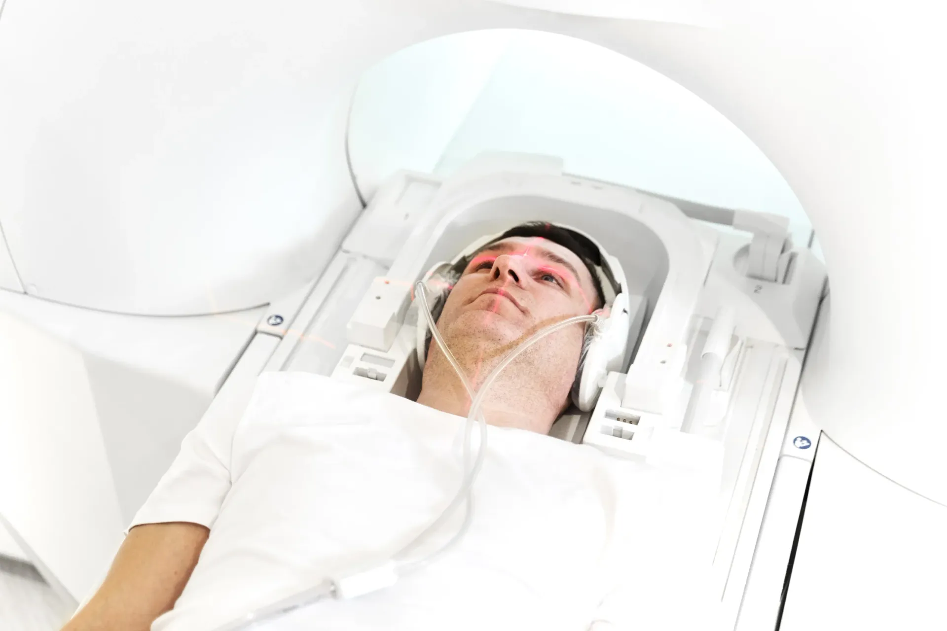
MRI (Magnetic Resonance Imaging) is the gold standard for brain tumor detection and characterisation. It provides dramatically better soft tissue contrast than CT, meaning it can distinguish between different types of brain tissue, oedema, tumor, and normal structures with far greater precision. MRI can detect tumors as small as a few millimetres, identify the precise location of a tumor relative to critical brain structures (eloquent cortex, motor strip, speech areas), and provide information about tumor biology through advanced sequences. For related information, see our guide on Brain MRI Dubai: What It Detects & Cost.
CT has a role in emergency settings — it is faster (5 minutes vs 30-45 minutes for MRI), widely available, and good at detecting acute haemorrhage, large masses with mass effect, and hydrocephalus. If you present to an emergency department with sudden severe headache or focal neurological deficits, a CT is often the first imaging study. However, CT has significant limitations for brain tumor detection: it misses many small tumors, poorly differentiates tumor from oedema, and provides limited information about tumor type and grade.
| Feature | Brain MRI | Brain CT |
|---|---|---|
| Soft tissue contrast | Excellent — distinguishes tumor, oedema, normal tissue | Limited — poor differentiation between tissue types |
| Small tumor detection | Detects lesions as small as 2-3mm | May miss lesions under 10mm |
| Posterior fossa imaging | Excellent — no bone artifact | Poor — bone artifact obscures cerebellum and brainstem |
| Radiation exposure | None (uses magnetic fields) | Yes (ionising radiation) |
| Scan time | 30-45 minutes | 5-10 minutes |
| With contrast | Gadolinium — shows blood-brain barrier disruption | Iodinated contrast — less informative for tumors |
| Cost (Dubai) | AED 1,500-3,500 | AED 600-1,500 |
Brain MRI vs CT comparison for tumor detection
MRI with Gadolinium Contrast
When a brain tumor is suspected, MRI is almost always performed with gadolinium contrast. Gadolinium is a paramagnetic contrast agent injected intravenously during the scan. In areas where the blood-brain barrier is disrupted — as occurs in most malignant tumors and some benign ones — gadolinium leaks into the tumor tissue and "lights up" on post-contrast images. This enhancement pattern provides critical diagnostic information: meningiomas show intense, homogeneous enhancement; glioblastomas show ring-like enhancement with a dark necrotic centre; low-grade gliomas may show little or no enhancement; and metastases typically show nodular enhancement. Gadolinium is generally very safe, with serious adverse reactions occurring in fewer than 1 in 100,000 administrations, though it is avoided in patients with severe kidney disease.
What Happens After an Abnormal Brain MRI Finding?
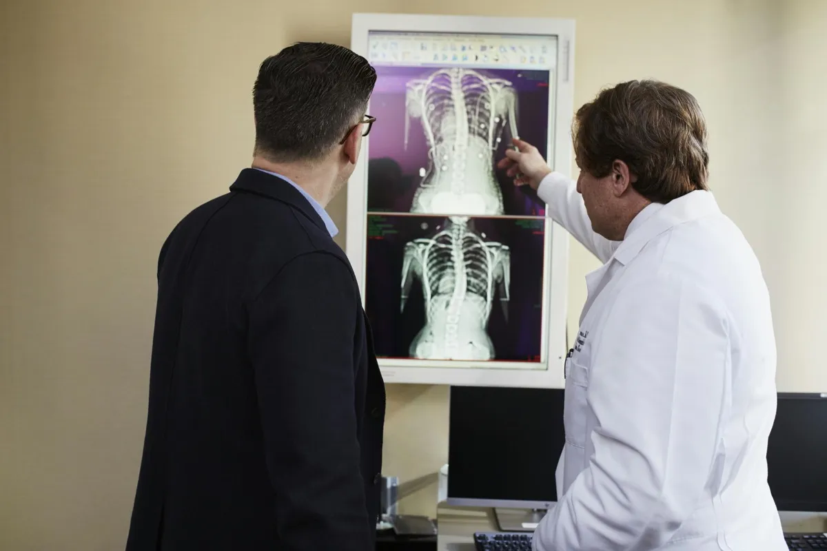
If your brain MRI shows an abnormality, the next steps depend entirely on what is found. Not every abnormal finding is a tumor, and not every tumor requires immediate treatment. The radiologist's report will describe the finding in detail — location, size, enhancement pattern, mass effect, surrounding oedema — and provide a differential diagnosis (a list of possible explanations ranked by probability). Here is what typically happens:
- Incidental meningioma (small, asymptomatic): Often monitored with serial MRI every 6-12 months initially, then annually — many never require treatment
- Suspected high-grade glioma: Urgent referral to neurosurgery for surgical planning — advanced MRI sequences (spectroscopy, perfusion, diffusion tensor imaging) may be added
- Multiple lesions suggesting metastases: Comprehensive cancer workup including CT chest/abdomen/pelvis, PET scan, and targeted biopsies to identify the primary cancer
- Pituitary mass: Referral to endocrinology for hormonal evaluation and to neurosurgery for surgical assessment if the mass is large or causing visual symptoms
- Non-neoplastic finding: Many MRI abnormalities are not tumors — demyelination (multiple sclerosis), abscess, vascular malformation, or post-inflammatory changes may mimic tumors on imaging
- Indeterminate lesion: Short-interval follow-up MRI (typically 3-6 months) to assess for growth — stability over time suggests a benign or low-grade process
It is important to understand that a brain MRI finding does not equal a brain tumor diagnosis. Many incidental findings on brain MRI are benign and require no treatment. Conversely, some findings require tissue diagnosis (biopsy or surgical resection) to determine the exact tumor type and guide treatment. The radiologist, referring physician, and neurosurgeon work together to determine the most appropriate next step based on the imaging characteristics and the patient's clinical presentation.
Survival Rates and the Impact of Early Detection
Survival rates for brain tumors vary enormously depending on the tumor type, grade, location, patient age, and how early the tumor is detected. For benign tumors like meningiomas and acoustic neuromas, five-year survival rates exceed 90%, and many patients are effectively cured with surgery. For low-grade gliomas, five-year survival ranges from 50-80%, and many patients live for decades with appropriate treatment. The challenge lies with high-grade gliomas — particularly glioblastoma, where median survival remains approximately 15 months despite aggressive treatment.
Early detection matters even for aggressive tumors. A smaller tumor is more likely to be completely resectable, which is the strongest prognostic factor in glioblastoma surgery. A tumor detected before it causes significant symptoms allows treatment to begin when the patient is in better neurological condition, tolerating surgery and subsequent chemo-radiation more effectively. For benign tumors, early detection often means the tumor can be monitored rather than treated — saving the patient from unnecessary surgery.
However, unlike breast, colon, or lung cancer, there is currently no recommended routine screening for brain tumors in the general population. The relatively low incidence of brain tumors (compared to other cancers) and the cost and time required for brain MRI mean that population-based screening is not cost-effective. Brain imaging is recommended when there are specific neurological symptoms or signs that suggest a possible intracranial lesion. The key is recognising when symptoms warrant investigation — which is the primary purpose of this guide.
When to Get a Brain MRI: A Practical Decision Guide
Knowing when to seek brain imaging can be difficult — you do not want to overreact to every headache, but you also do not want to dismiss symptoms that could indicate a serious problem. Here is a practical guide based on what neurologists and radiologists consider "red flag" symptoms: You may also find our MRI for Headaches: When You Need a Brain Scan helpful.
- Get urgent brain imaging (same day): First-time seizure, sudden severe "thunderclap" headache, sudden onset of weakness or numbness on one side, sudden vision loss, sudden difficulty speaking, rapidly deteriorating consciousness
- Get brain MRI within 1-2 weeks: Progressive headaches worsening over weeks, new persistent headache in a person over 50, headaches that wake you from sleep, progressive personality or cognitive changes noticed by others, new-onset balance or coordination problems, progressive visual changes not explained by eye examination
- Discuss with your doctor: Chronic headaches that have been stable for months or years (less likely to be tumor-related), intermittent dizziness without progression, occasional visual disturbances associated with migraine, family history of brain tumors (does not automatically warrant screening)
- Probably not needed for: Tension headaches that respond to over-the-counter medication and have been present for years without change, headaches clearly triggered by stress, dehydration, or alcohol, and anxiety-driven requests without neurological symptoms
At DCDC Dubai Healthcare City, brain MRI scans are available with short waiting times and reporting by specialist radiologists experienced in neuroradiology. If your doctor has recommended brain imaging, or if you are experiencing concerning neurological symptoms, our team can arrange an MRI with or without gadolinium contrast based on the clinical indication. We provide detailed radiology reports with imaging analysis and, when needed, can facilitate urgent referrals to neurology or neurosurgery.
Book a Brain MRI
Advanced brain MRI with specialist reporting at DCDC Dubai Healthcare City. Same-week appointments available.
MRI with and without gadolinium contrast available
Related Services at DCDC
Expert care and advanced diagnostics at Dubai Healthcare City
Frequently Asked Questions
Know the Signs, Act on the Right Ones
The purpose of this guide is not to create anxiety but to create informed awareness. The vast majority of headaches, vision changes, and memory lapses are not caused by brain tumors. But a small percentage are — and in those cases, recognising the pattern early and getting the right imaging can make an enormous difference in outcome. A meningioma found before it causes symptoms can be monitored safely. A glioma found while it is still small and resectable has better surgical outcomes. Even metastatic disease identified early allows for earlier initiation of targeted treatment.
The key patterns to watch for are progression (symptoms that steadily worsen over weeks to months), new neurological deficits (weakness, numbness, speech difficulty, vision changes that were not present before), and new-onset seizures in an adult. If you recognise these patterns in yourself or a family member, do not wait and hope they will resolve on their own. See a doctor, get a neurological examination, and if brain imaging is recommended, get an MRI rather than a CT for the most thorough evaluation.
At DCDC Dubai Healthcare City, our MRI department provides advanced brain imaging with specialist neuroradiology reporting. Whether you have been referred by your doctor or are concerned about neurological symptoms, we offer same-week appointments, MRI with and without gadolinium contrast, and comprehensive reporting that guides the next step in your care.
Sources & References
This article was reviewed by our medical team and references the following sources:
- American Brain Tumor Association — Brain Tumor Statistics
- National Cancer Institute — Adult Central Nervous System Tumors
- American College of Radiology — ACR Appropriateness Criteria for Headache
- Radiological Society of North America — Brain Tumor Imaging
- World Health Organization — Classification of Tumours of the Central Nervous System (5th Edition)
Medical content on this site is reviewed by DHA-licensed physicians. See our editorial policy for more information.

Written by
Dr. Osama Sanduka
Specialist Diagnostic Radiology
MD, Diagnostic Radiology Specialist
Dr. Osama Sanduka is a Specialist in Diagnostic Radiology with extensive experience in MRI, CT imaging, and neuroimaging. He practices at DCDC Dubai Healthcare City.
Related Articles
Brain MRI Scan Dubai: Complete Guide
MRI with Contrast: What You Need to Know
Complete Guide to MRI Scan in Dubai
Cancer Screening in Dubai: Tests, Costs & What You Need
More in Diagnostic Imaging
Ultrasound at Home Dubai: From AED 599 (2026)
Read More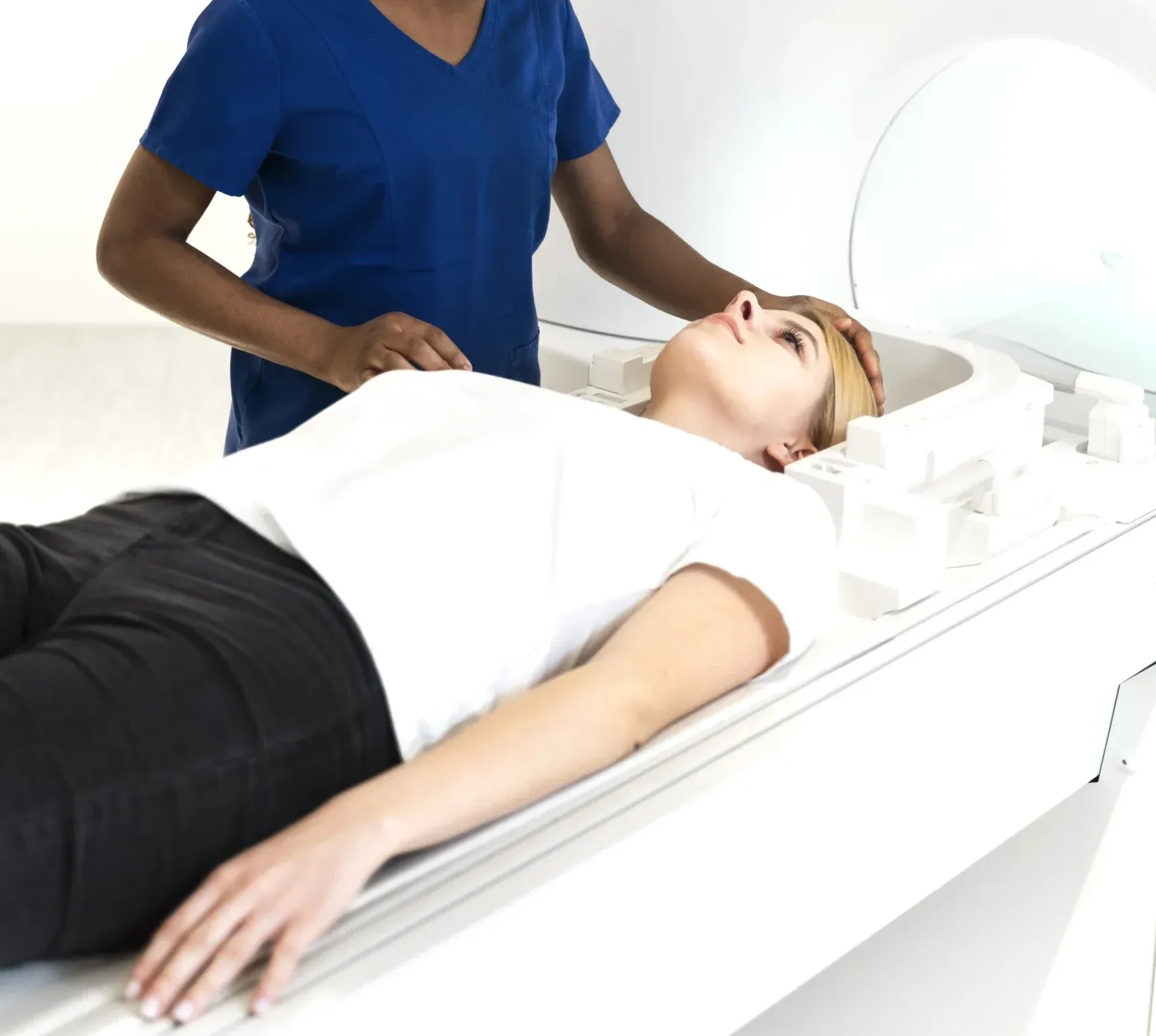
Neck MRI Dubai: What It Shows & Cost (2026)
Read More
X-Ray Scan Dubai: Complete Guide (2026)
Read More
MRI vs Ultrasound Dubai: Which Scan? (2026)
Read More
MRI vs X-Ray Dubai: Which Scan Do You Need? (2026)
Read More
Kidney Ultrasound vs CT Dubai: Which Test? (2026)
Read More© 2026 Doctors Clinic Diagnostic Center (DCDC), Dubai Healthcare City. Originally published at https://doctorsclinicdubai.ae/blog/brain-tumor-signs-imaging. All rights reserved. Unauthorized reproduction is prohibited.