النقاط الرئيسية
- Cardiac MRI is the gold standard for assessing heart muscle tissue, providing detailed images of myocarditis, cardiomyopathy, valve disease, congenital heart defects, and cardiac tumors without any radiation exposure
- Unlike echocardiography, cardiac MRI can distinguish between scar tissue, inflammation, edema, and healthy myocardium — critical for diagnosing conditions like myocarditis and determining whether damaged heart muscle can recover
- Cardiac MRI in Dubai costs approximately AED 3,000–6,000 depending on the protocol complexity and whether contrast (gadolinium) is used
- The scan takes 45–75 minutes and requires you to lie still inside the MRI bore while performing brief breath-holds. No radiation is involved — only magnetic fields and radio waves
- Cardiac MRI and CT angiogram are complementary, not interchangeable: CT angiogram excels at imaging coronary arteries, while cardiac MRI excels at evaluating the heart muscle itself
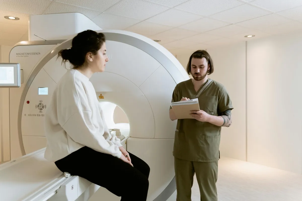
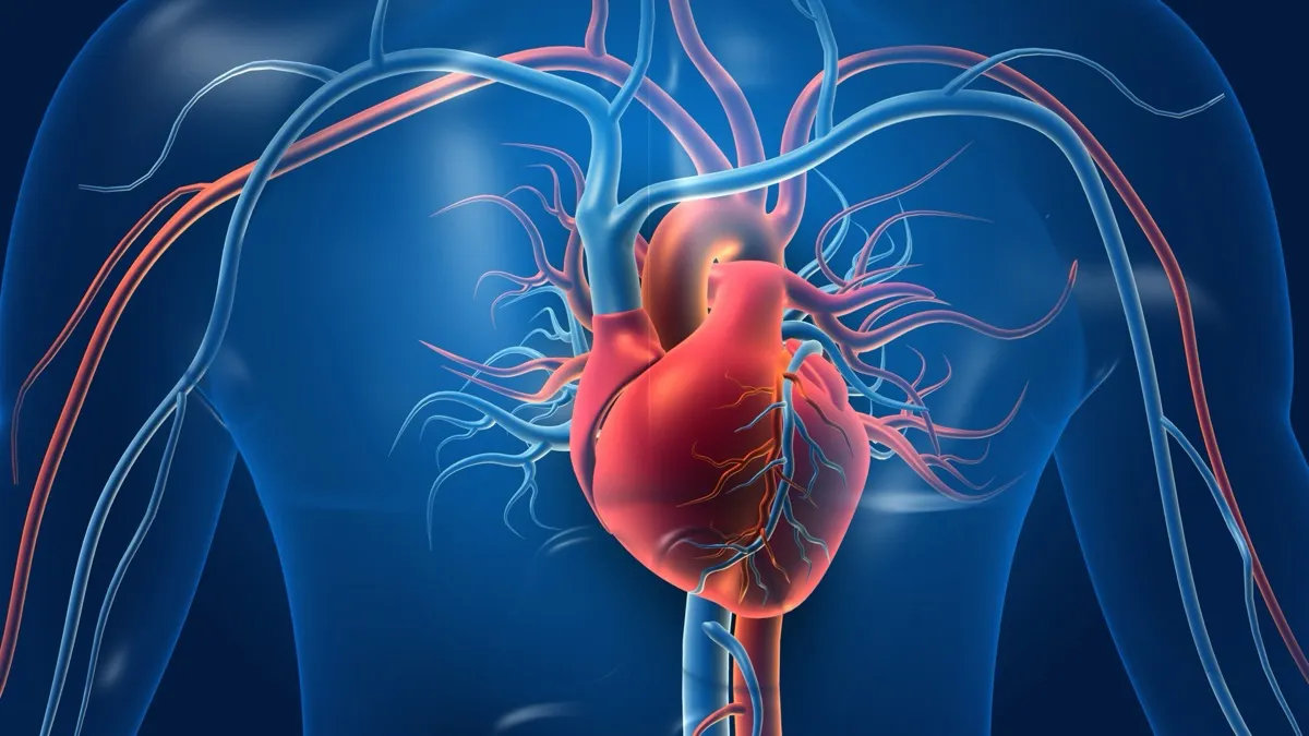
When your cardiologist needs to see beyond what an echocardiogram or ECG can show, cardiac MRI delivers the most detailed, comprehensive view of your heart available in modern medicine. Using powerful magnetic fields and radio waves — with zero radiation — a cardiac MRI scan produces high-resolution images of the heart muscle, chambers, valves, and surrounding structures that no other imaging modality can match.
Cardiac MRI is increasingly used to diagnose and monitor conditions such as myocarditis (heart muscle inflammation), cardiomyopathy (weakened heart muscle), valvular heart disease, congenital heart defects, and cardiac masses. For patients in Dubai seeking the most advanced cardiac imaging available, this guide covers what cardiac MRI shows, how it compares to echocardiography and CT angiogram, what to expect during the scan, the approximate cost, and how to prepare for your appointment at DCDC Dubai Healthcare City.

What Does a Cardiac MRI Show?
Cardiac MRI (also called CMR — Cardiac Magnetic Resonance) is uniquely capable of providing tissue characterization of the heart muscle. This means it can not only show the heart’s structure and function (like an echo), but also distinguish between healthy muscle, scar tissue, inflammation, edema, fat infiltration, and iron overload. No other imaging modality provides this level of tissue detail.
Myocarditis (Heart Inflammation)
Myocarditis is inflammation of the heart muscle, often caused by viral infections (including COVID-19), autoimmune conditions, or toxic exposures. It can cause chest pain, heart failure, and dangerous arrhythmias. Cardiac MRI is the non-invasive gold standard for diagnosing myocarditis. It detects myocardial edema (swelling) on T2-weighted images and identifies areas of inflammation and necrosis through late gadolinium enhancement (LGE). These findings help cardiologists distinguish myocarditis from a heart attack — both of which can present with similar symptoms and elevated cardiac enzymes.
Cardiomyopathy
Cardiomyopathy refers to diseases of the heart muscle that make it harder for the heart to pump blood. Cardiac MRI is essential for diagnosing and classifying the different types. Dilated cardiomyopathy shows an enlarged, weakened heart with reduced ejection fraction. Hypertrophic cardiomyopathy (HCM) shows abnormally thickened heart walls, and MRI can identify the pattern and extent of thickening as well as areas of fibrosis that predict arrhythmia risk. Arrhythmogenic right ventricular cardiomyopathy (ARVC) shows fat and fibrous replacement of the right ventricular wall. Cardiac amyloidosis and sarcoidosis show characteristic LGE patterns that help differentiate them from other causes of heart failure.
Valve Disease
While echocardiography is typically the first-line imaging tool for valvular heart disease, cardiac MRI provides superior quantification of valve regurgitation (leaking) and stenosis (narrowing). MRI can precisely measure regurgitant volumes and fractions, assess the impact of valve disease on heart chamber volumes and function, and help determine the optimal timing for valve surgery or repair. It is particularly valuable when echo images are technically limited or when precise quantification is needed for surgical planning.
Congenital Heart Disease
Cardiac MRI is invaluable for evaluating congenital heart defects in both children and adults. It provides detailed 3D anatomy of complex structural abnormalities including atrial and ventricular septal defects, tetralogy of Fallot, coarctation of the aorta, and transposition of the great arteries. For patients who have undergone corrective heart surgery, MRI monitors for complications and assesses the long-term function of surgical repairs. Because it uses no radiation, cardiac MRI is ideal for young patients who may need repeated imaging throughout their lives.
Cardiac Tumors and Masses
Cardiac MRI is the definitive imaging tool for evaluating masses found in or around the heart. Its tissue characterization capability can often distinguish between benign tumors (myxomas, fibromas, lipomas), malignant tumors (sarcomas, metastases), and thrombus (blood clots) without needing a biopsy. This differentiation guides treatment decisions and surgical planning.
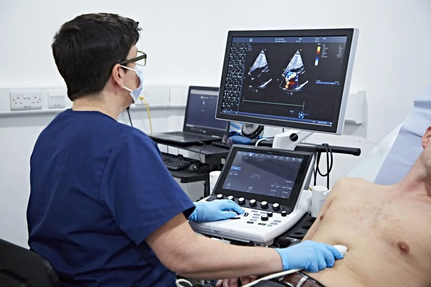
Cardiac MRI vs Echocardiogram vs CT Angiogram
Patients often ask: “Why do I need a cardiac MRI if I already had an echo or CT scan?” Each modality excels in different areas. Your cardiologist chooses the right test based on the specific clinical question.
| Factor | Cardiac MRI | Echocardiogram | CT Angiogram |
|---|---|---|---|
| Primary strength | Heart muscle tissue characterization | Real-time heart function and valve assessment | Coronary artery anatomy |
| Radiation | None | None | Yes (low-dose ionizing radiation) |
| Duration | 45–75 minutes | 20–40 minutes | 10–20 minutes |
| Contrast used | Gadolinium (when needed) | None (saline bubbles for specific tests) | Iodine-based |
| Myocarditis detection | Gold standard | Limited (reduced function may be seen) | Not used for this |
| Cardiomyopathy assessment | Gold standard | Good for initial assessment | Limited |
| Coronary artery imaging | Limited (emerging technique) | Cannot image coronary arteries | Gold standard |
| Valve disease | Excellent quantification | First-line screening tool | Limited |
| Approximate cost (Dubai) | AED 3,000–6,000 | AED 800–1,500 | AED 1,500–3,000 |
| Claustrophobia | Can be challenging (enclosed bore) | Not an issue | Rarely an issue (open ring) |
These are complementary tools. Your cardiologist will recommend the appropriate imaging based on your clinical question.
In many cases, a patient may need more than one imaging modality. For example, an initial echocardiogram may detect a dilated heart with reduced function, and a cardiac MRI is then ordered to determine the cause (ischemic vs non-ischemic cardiomyopathy) by looking for patterns of scar tissue. Similarly, a CT angiogram may confirm coronary artery disease, and a cardiac MRI may then be used to assess whether the affected heart muscle is still viable or permanently scarred.
Discuss Cardiac MRI with a Specialist
Book a cardiology consultation at DCDC to determine if cardiac MRI is the right test for your heart concern.
How to Prepare for a Cardiac MRI
Cardiac MRI preparation is straightforward, but there are important safety considerations related to the powerful magnetic field used by the scanner. For related information, see our guide on CT Angiogram Preparation: Step-by-Step Guide.
- Metal implant screening: The MRI magnetic field is extremely strong. You must inform the radiology team about any metallic implants, including pacemakers, defibrillators (ICDs), cochlear implants, metallic heart valves, joint replacements, metal clips, or retained shrapnel. Some modern pacemakers are MRI-conditional, but this must be verified and special protocols followed
- Fasting: A light fast of 2–4 hours before the scan is typically recommended, especially if gadolinium contrast will be used, to reduce the risk of nausea
- Kidney function: If contrast (gadolinium) will be administered, a recent blood creatinine test is required to confirm adequate kidney function. Gadolinium is very rarely associated with nephrogenic systemic fibrosis (NSF) in patients with severe kidney disease
- Clothing: Wear comfortable clothing without metal. All metal items including jewelry, watches, belts, hairpins, hearing aids, and removable dental work must be left outside the scan room
- Medications: Continue all regular medications unless your cardiologist instructs otherwise. Beta-blockers may be administered before the scan to slow the heart rate for better image quality if your resting rate is above 80 bpm
- Breath-hold practice: During the scan, you will be asked to hold your breath for 10–15 seconds at a time. The radiographer will coach you through an intercom. Practicing calm, steady breath-holds beforehand can help
- Allow enough time: Cardiac MRI takes 45–75 minutes. Including check-in and preparation, plan for a visit of approximately 90 minutes
What to Expect During the Cardiac MRI
Understanding the procedure helps reduce anxiety, especially since cardiac MRI is longer than most other imaging tests.
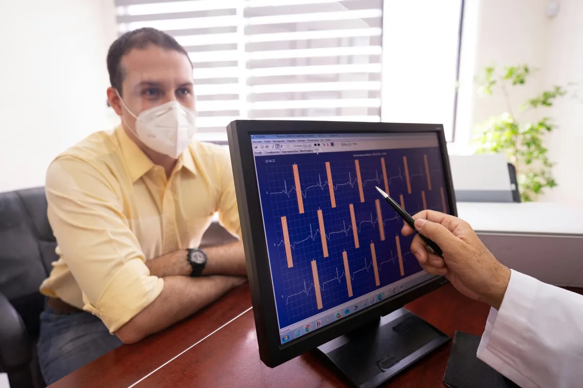
- ECG leads: Small electrode patches are placed on your chest to monitor your heart rhythm throughout the scan. The MRI system synchronizes image acquisition with your heartbeat (cardiac gating) to produce sharp, motion-free images
- IV placement: A small cannula is inserted in your arm if gadolinium contrast is planned. The contrast is injected partway through the scan to create late gadolinium enhancement (LGE) images that reveal scar tissue and inflammation
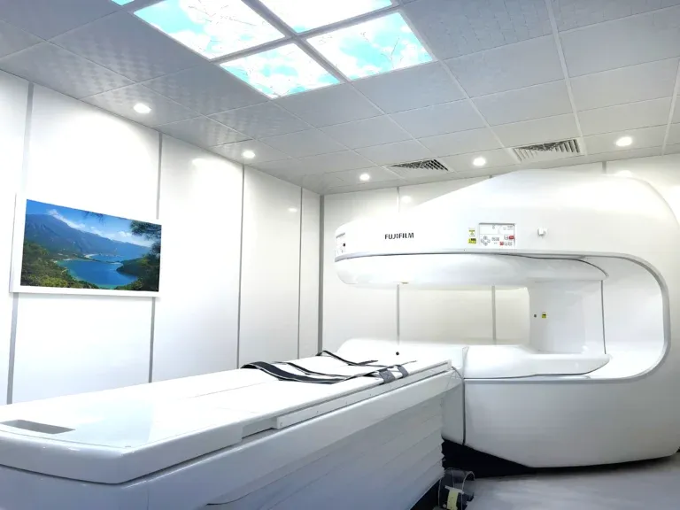
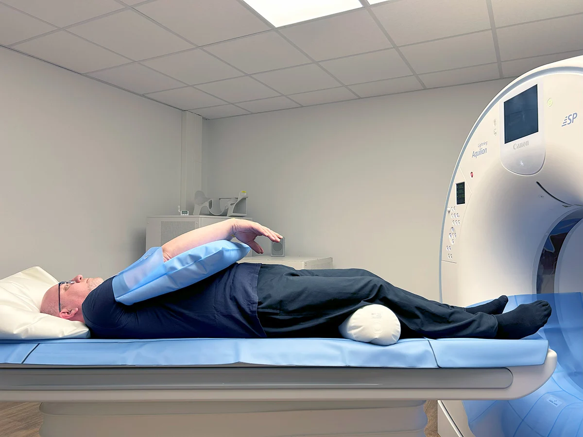
- Entering the scanner: You lie on a padded table that slides into the MRI bore (a cylindrical tunnel approximately 60 cm in diameter). The bore is well-lit and ventilated, and you can communicate with the radiographer at any time through an intercom. You will be given earplugs or headphones because the scanner produces loud knocking and buzzing sounds during image acquisition
- Breath-holds: The radiographer will instruct you through the intercom: “Breathe in… breathe out… breathe in… hold your breath.” Each hold lasts 10–15 seconds. Between breath-holds, you breathe normally. Most cardiac MRI protocols require 30–50 breath-holds throughout the exam
- Contrast injection: Approximately halfway through, gadolinium contrast is injected through the IV. You may feel a brief cool sensation in your arm. After a 10-minute wait (to allow contrast to distribute and wash out from normal tissue), the final set of images (LGE) is acquired
- Total time in scanner: 45–75 minutes depending on the clinical question and protocol complexity. The entire visit takes approximately 90 minutes
If you experience claustrophobia, inform the team before the scan. Strategies include listening to music through MRI-compatible headphones, keeping your eyes closed, using a mirror attachment that lets you see outside the bore, or taking a mild sedative (arranged in advance with your doctor). At DCDC, our radiographers are experienced in helping anxious patients through the scan comfortably.
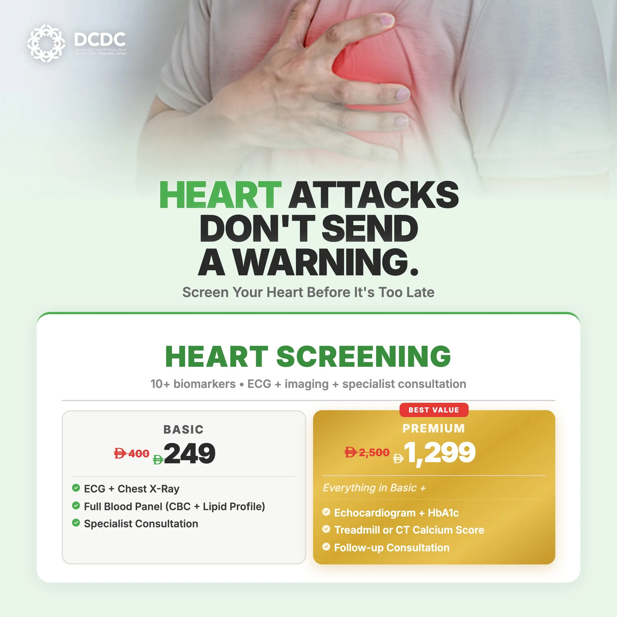
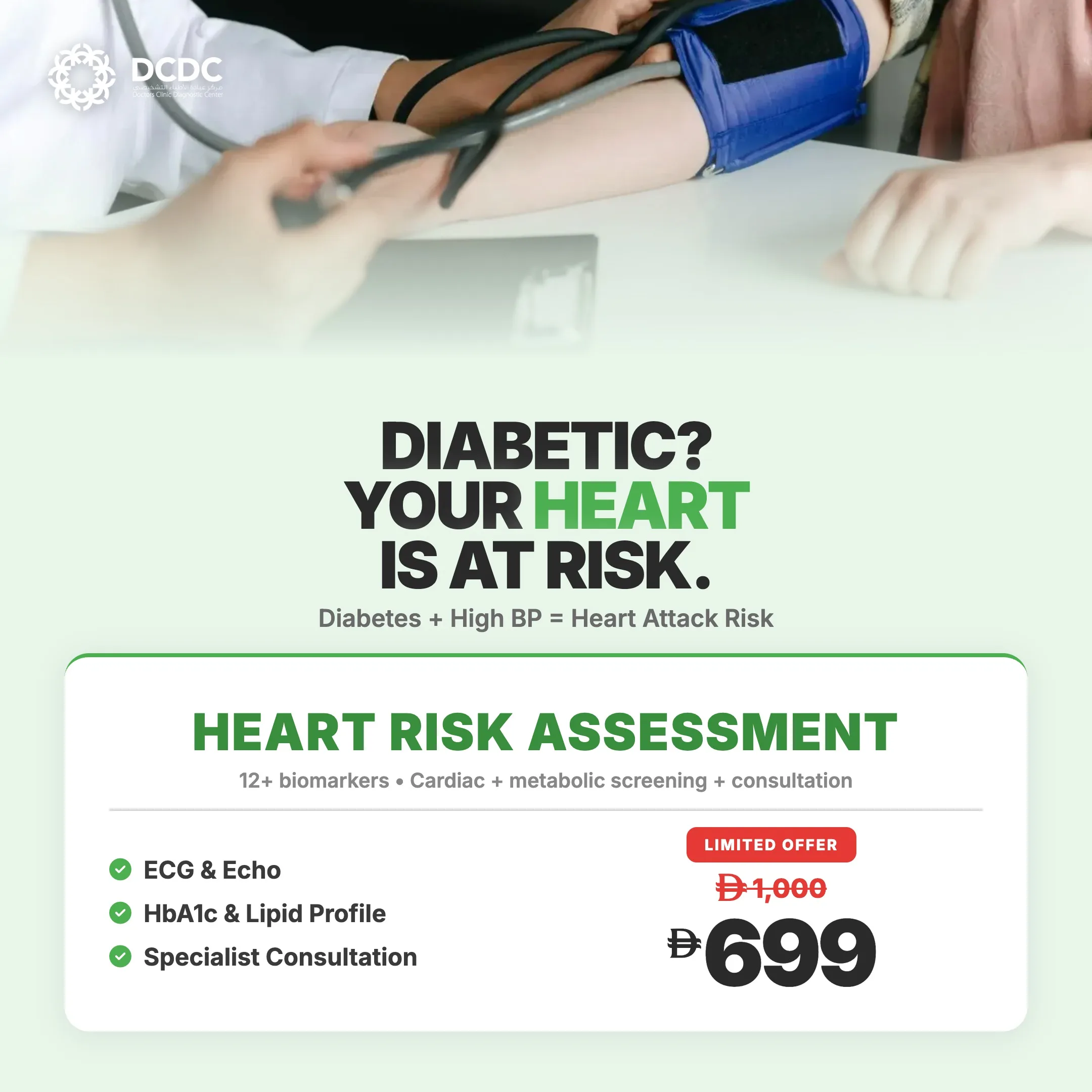
Cardiac MRI Cost in Dubai
Cardiac MRI is a specialized imaging study that requires advanced equipment, specialized protocols, and expert interpretation. The cost reflects this complexity.
| Cardiac MRI Protocol | Approximate Cost (AED) |
|---|---|
| Basic cardiac MRI (structure and function) | 3,000 – 4,000 |
| Cardiac MRI with gadolinium contrast (LGE) | 4,000 – 5,500 |
| Cardiac MRI stress test (adenosine/regadenoson stress perfusion) | 5,000 – 6,500 |
| Cardiac MRI for congenital heart disease | 4,000 – 6,000 |
Prices are approximate and vary by facility. Contact DCDC for current pricing and insurance verification.
Many Dubai insurance plans cover cardiac MRI when ordered by a cardiologist with a clinical indication. Pre-authorization is typically required. At DCDC, our team handles insurance verification and pre-authorization before your appointment so you know your out-of-pocket cost in advance.
While cardiac MRI costs more than an echocardiogram, it often prevents more invasive and expensive procedures. For example, a cardiac MRI that confirms myocarditis can prevent an unnecessary cardiac catheterization. An MRI that shows viable heart muscle can justify coronary bypass surgery that might otherwise be considered futile. The diagnostic precision of cardiac MRI frequently saves both money and unnecessary risk for the patient.
When Is Cardiac MRI the Right Choice?
Cardiac MRI is not a first-line screening tool — it is a specialized investigation ordered when your cardiologist needs information that echocardiography, ECG, or CT cannot provide. Common scenarios include: You may also find our Open MRI Dubai: Cost, Benefits & Who Needs It helpful.
- Suspected myocarditis: Chest pain and elevated troponin in a young patient with normal coronary arteries — MRI confirms or rules out inflammation
- Heart failure of unknown cause: Echo shows reduced function but the cause is unclear. MRI tissue characterization reveals whether the cause is ischemic (coronary artery disease) or non-ischemic (cardiomyopathy, myocarditis, infiltrative disease)
- Hypertrophic cardiomyopathy (HCM): Risk stratification — MRI quantifies the extent of myocardial fibrosis, which is the strongest predictor of sudden cardiac death risk in HCM patients
- Cardiac mass or tumor: A mass seen on echo needs further characterization. MRI determines whether it is a benign tumor, malignant tumor, thrombus, or normal anatomical variant
- Congenital heart disease follow-up: Adult patients with repaired congenital defects need serial monitoring without repeated radiation exposure
- Viability assessment before revascularization: Before coronary bypass surgery, MRI determines whether scarred heart muscle is still viable and would benefit from restored blood flow
- Post-COVID cardiac evaluation: Patients with persistent cardiac symptoms after COVID-19 infection — MRI can detect subclinical myocarditis or pericarditis
- Athlete screening: Evaluation of athletes with unexplained cardiac symptoms, arrhythmias, or family history of sudden cardiac death to rule out HCM, ARVC, or myocarditis
If your cardiologist has recommended a cardiac MRI, it is because this test provides information that is critical for your diagnosis and treatment plan and cannot be obtained through simpler imaging methods.
Book Your Cardiac MRI
Schedule a cardiac MRI at DCDC Dubai Healthcare City. Expert interpretation by experienced radiologists with results within 24–48 hours.
خدمات ذات صلة في DCDC
رعاية متخصصة وتشخيص متقدم في مدينة دبي الطبية
الأسئلة الشائعة
خلاصة القول
Cardiac MRI provides a level of detail about the heart muscle that no other imaging modality can match. When your cardiologist needs to know whether your heart muscle is inflamed, scarred, thickened, infiltrated, or structurally abnormal, cardiac MRI delivers answers that guide treatment decisions with precision. It accomplishes this without any radiation, making it safe for repeated monitoring over time.
In Dubai, cardiac MRI is increasingly recognized as an essential tool in the diagnosis and management of complex heart conditions. From post-COVID myocarditis evaluation to hypertrophic cardiomyopathy risk stratification to pre-surgical viability assessment, cardiac MRI fills diagnostic gaps that echocardiography and CT angiography cannot address.
At DCDC Dubai Healthcare City, cardiac MRI studies are performed on modern MRI equipment and interpreted by experienced consultant radiologists in collaboration with your referring cardiologist. If your doctor has recommended a cardiac MRI, or if you would like to discuss whether this test is appropriate for your heart symptoms, contact our team to schedule a cardiology consultation or book your imaging appointment.

كتبه
Dr. Osama Elzamzami
استشاري أشعة
دكتوراه في الطب، الأشعة
Dr. Osama Elzamzami is a Consultant Radiologist specializing in diagnostic imaging including X-ray, CT, MRI, and ultrasound at DCDC Dubai Healthcare City.
مقالات ذات صلة
MRI Cost in Dubai: Complete Guide to Types & Pricing (2026)
Full Body MRI Cost Dubai: Complete Price Guide (2026)
CT Angiogram Cost in Dubai: Complete Guide

Heart Health Prevention in Dubai: Your Guide
CTA vs Calcium Score: Which Heart Test Do You Need?
المزيد في Cardiology
Stress Test vs Echo Dubai: Which Heart Test? (2026)
اقرأ المزيدAngiogram Dubai: Types, Cost & Guide (2026)
اقرأ المزيدECG Test Dubai: Cost, Types & Results (2026)
اقرأ المزيدEchocardiogram (Echo) Test in Dubai: Cost, Procedure & What to Expect (2026)
اقرأ المزيدHolter Monitor Test Dubai: Cost & Guide (2026)
اقرأ المزيدECG Cost in Dubai: From AED 200 (2026)
اقرأ المزيد© 2026 Doctors Clinic Diagnostic Center (DCDC), Dubai Healthcare City. Originally published at https://doctorsclinicdubai.ae/blog/cardiac-mri-dubai. All rights reserved. Unauthorized reproduction is prohibited.