النقاط الرئيسية
- تتراوح تكلفة الرنين المغناطيسي للبطن في دبي عادةً بين 1,200 و2,000 درهم، مع بروتوكولات متخصصة كـ MRCP أو دراسات التباين الخاص بالكبد في الطرف الأعلى
- يُعدّ الرنين المغناطيسي للبطن المعيار الذهبي لتوصيف آفات الكبد والكشف عن سرطان الكبد (HCC) لدى مرضى التشمع وتقييم الكتل الكلوية وتصوير القنوات الصفراوية (MRCP) دون إجراءات تداخلية
- يُوفر الرنين المغناطيسي تباينًا أفضل للأنسجة الرخوة مقارنةً بالأشعة المقطعية لتقييم الكبد والبنكرياس، ولا يعرّض المريض للإشعاع المؤيِّن
- يشمل التحضير عادةً الصيام 4-6 ساعات قبل الفحص لتقليل نشاط الأمعاء وضمان امتلاء المرارة لأفضل تصوير
- يستغرق الفحص نحو 30-50 دقيقة وكثيرًا ما يتضمن حقن صبغة تباين لتوصيف آفات الأعضاء بدقة
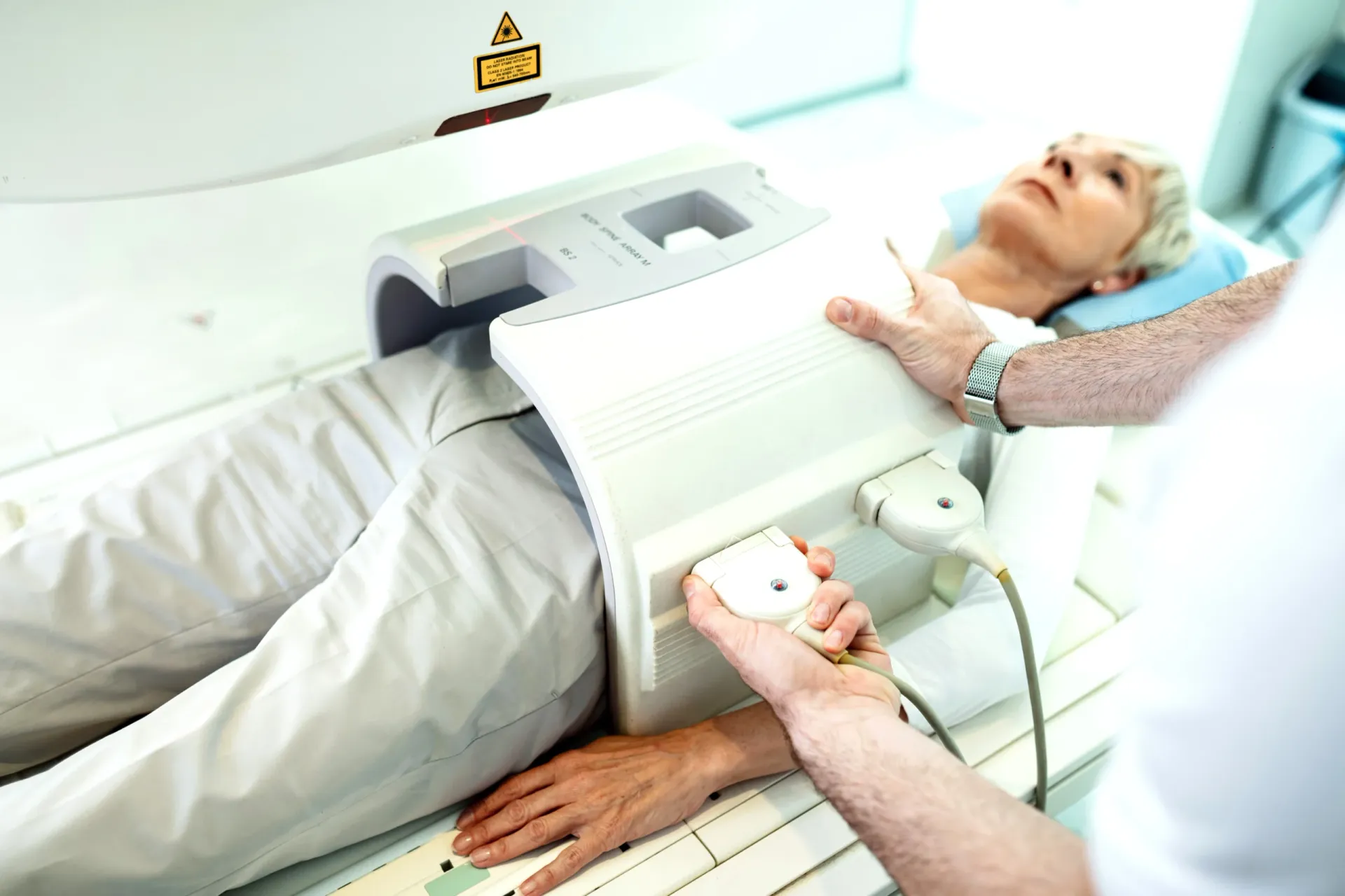
يُعدّ فحص الرنين المغناطيسي للبطن أدق أداة تصوير لتقييم الأعضاء الصلبة والمجوفة في البطن، بما فيها الكبد والكلى والبنكرياس والطحال والغدد الكظرية والقنوات الصفراوية. في حين تُعدّ الأشعة المقطعية في الغالب أول فحص مقطعي مقطعي يُؤمر لأعراض البطن، يُوفر الرنين المغناطيسي تباينًا أفضل للأنسجة الرخوة يُعدّ حاسمًا لتوصيف آفات الكبد والكشف المبكر عن سرطانه وتقييم الكتل الكلوية المعقدة وتصوير الجهاز الصفراوي.


ما الذي يُظهره الرنين المغناطيسي للبطن؟
يُنتج الرنين المغناطيسي للبطن صورًا تفصيلية لجميع أعضاء البطن باستخدام المجالات المغناطيسية وموجات الراديو بدلًا من الإشعاع. تتمثل أبرز مزاياه في تباين الأنسجة الرخوة الفائق — القدرة على التمييز بين أنواع مختلفة من الأنسجة داخل الأعضاء، وهو أمر ضروري لتوصيف الكتل والآفات. يُعدّ الرنين المغناطيسي الوسيلة التصويرية المفضلة حين تكون نتائج الأشعة المقطعية غير حاسمة أو حين يُفضَّل التصوير دون إشعاع.
تُقيَّم الأعضاء والحالات التالية شيوعًا بالرنين المغناطيسي للبطن:
تصوير الكبد
- توصيف آفات الكبد: هذا هو أكثر أسباب الرنين المغناطيسي للبطن شيوعًا. حين يكشف الموجات فوق الصوتية أو الأشعة المقطعية عن آفة كبدية، يُوفر الرنين المغناطيسي توصيفًا قاطعًا. باستخدام أنماط التعزيز التباين عبر مراحل متعددة، يُميّز الرنين المغناطيسي بين الآفات الحميدة والأورام الخبيثة. يمكن تشخيص كثير من آفات الكبد بثقة بالرنين المغناطيسي دون خزعة.
- فحص سرطان الخلايا الكبدية (HCC): لمرضى تشمع الكبد، يُعدّ الرنين المغناطيسي أحد أدوات المراقبة الرئيسية للكشف المبكر عن سرطان الخلايا الكبدية. يُصنّف نظام LI-RADS مشاهدات الكبد لدى المرضى المعرضين للخطر.
- تشمع الكبد والتهرب الكبدي: يمكن للرنين المغناطيسي اكتشاف وقياس محتوى الدهون الكبدية ودرجة التليف باستخدام تسلسلات متخصصة، وهو ذو أهمية خاصة في تصاعد وباء مرض الكبد الدهني غير الكحولي.
- فرط الحديد (داء الهيموكروماتوز): يُعدّ الرنين المغناطيسي الأداة غير التداخلية المحددة لاكتشاف وقياس فرط الحديد الكبدي، وهو ضروري لمرضى الهيموكروماتوز الوراثي والثلاسيميا.
تصوير الكلى
- توصيف الكتلة الكلوية: حين توجد كتلة كلوية غير محددة الطبيعة، يُوفر الرنين المغناطيسي توصيفًا نسيجيًا إضافيًا ويُفيد خاصةً في التمييز بين الأورام الصلبة والأكياس المعقدة وغيرها.
- تقييم الشرايين الكلوية: يمكن لتصوير الأوعية بالرنين المغناطيسي (MRA) تقييم الشرايين الكلوية للكشف عن التضيق دون إشعاع.
- عدوى الكلى وانسدادها: يُقيّم الرنين المغناطيسي عدوى الكلى المعقدة والانسداد البولي خاصةً حين تكون الأشعة المقطعية مُقيَّدة.
الجهاز الصفراوي (MRCP)
- MRCP (تصوير القنوات الصفراوية والبنكرياس بالرنين المغناطيسي): MRCP هي تقنية رنين مغناطيسي متخصصة تُنتج صورًا تفصيلية للقنوات الصفراوية والمرارة وقناة البنكرياس دون حقن تباين أو إجراءات تداخلية. وقد حلّت إلى حد بعيد محل ERCP التشخيصية لتقييم حصيات القنوات الصفراوية والتضيقات والشذوذات الخلقية.
البنكرياس والطحال والغدد الكظرية
- تقييم البنكرياس: يُوفر الرنين المغناطيسي تصويرًا ممتازًا للبنكرياس ويُفيد خاصةً في الكشف عن الآفات الكيسية البنكرياسية (IPMN وغيرها) وتوصيفها.
- آفات الغدد الكظرية: يمكن للرنين المغناطيسي باستخدام تصوير الإزاحة الكيميائية التمييز بين الأورام الحميدة للقشرة الكظرية والآفات المثيرة للقلق دون خزعة في معظم الحالات.
- تقييم الطحال: يُوصّف الرنين المغناطيسي آفات الطحال ويُقيّم حجمه وخصائص إشارته.
"الرنين المغناطيسي للبطن لا غنى عنه حين نحتاج إلى توصيف آفة لم تستطع الموجات فوق الصوتية أو الأشعة المقطعية تحديدها بالكامل،" يُوضّح Dr. Osama Elzamzami، استشاري الأشعة في DCDC.
كم تكلفة الرنين المغناطيسي للبطن في دبي؟
تتراوح تكلفة الرنين المغناطيسي للبطن في دبي عمومًا بين 1,200 و2,000 درهم. البروتوكولات المتخصصة كدراسات التباين الخاص بالكبد وـ MRCP تكون في الطرف الأعلى. في Doctors Clinic Diagnostic Center بـ Dubai Healthcare City، يشمل السعر الفحص والتباين (إن لزم) وتقرير الأشعة التفصيلي ونسخًا رقمية من الصور.
| نوع الرنين المغناطيسي للبطن | التكلفة التقريبية (درهم) |
|---|---|
| رنين مغناطيسي للبطن بدون تباين | 1,200 – 1,600 |
| رنين مغناطيسي للبطن مع تباين (غادولينيوم) | 1,500 – 2,000 |
| رنين مغناطيسي للكبد مع تباين خاص بالكبد | 1,800 – 2,500 |
| MRCP (تصوير القنوات الصفراوية) | 1,200 – 1,800 |
| رنين مغناطيسي مجمّع للبطن والحوض | 2,200 – 3,000 |
الأسعار تقريبية وتشمل تقرير الأشعة في DCDC. تواصل معنا للحصول على الأسعار الدقيقة.
يُستخدم الرنين المغناطيسي للبطن مع التباين في معظم الحالات لأن نمط تعزيز الآفات عبر مراحل مختلفة ضروري للتشخيص الدقيق.
تُغطي معظم خطط التأمين الصحي في الإمارات الرنين المغناطيسي للبطن حين تُؤمره طبيب مرخص بمؤشر سريري موثق. يُساعد DCDC المرضى في الحصول على الموافقة المسبقة من التأمين.
الرنين المغناطيسي للبطن مقابل الأشعة المقطعية مقابل الموجات فوق الصوتية
يؤدي كل نظام تصوير دورًا محددًا في تقييم البطن. للمزيد، راجع دليلنا حول تكلفة الموجات فوق الصوتية في دبي.
| العامل | الموجات فوق الصوتية | الأشعة المقطعية | الرنين المغناطيسي |
|---|---|---|---|
| التكلفة | 400 – 800 درهم | 800 – 1,500 درهم | 1,200 – 2,000 درهم |
| الإشعاع | لا يوجد | متوسط إلى مرتفع | لا يوجد |
| وقت الفحص | 15 – 30 دقيقة | 5 – 10 دقائق | 30 – 50 دقيقة |
| تباين الأنسجة الرخوة | متوسط | جيد | ممتاز (فائق) |
| توصيف آفات الكبد | جيد للاكتشاف الأولي | جيد | ممتاز (المعيار الذهبي) |
| تصوير القنوات الصفراوية (MRCP) | محدود | متوسط | ممتاز (يُحلّ محل ERCP التشخيصية) |
| توصيف الكتلة الكلوية | جيد (خط أول) | جيد | ممتاز (حل المشكلات) |
| تقييم كيسات البنكرياس | محدود | جيد | ممتاز |
| قياس دهون/حديد الكبد | نوعي فقط | محدود | ممتاز (كمي) |
| الأفضل لـ | الفحص الأولي، الإجراءات الموجّهة | الطوارئ، أمراض الأمعاء، الحصيات | التوصيف القاطع للآفات، تقييم القنوات الصفراوية |
الموجات فوق الصوتية هي فحص التصوير الأولي للبطن. الأشعة المقطعية سريعة ومتاحة على نطاق واسع. الرنين المغناطيسي يُوفر التوصيف القاطع حين تكون الفحوصات الأخرى غير حاسمة.
الموجات فوق الصوتية هي الفحص الأول المناسب لمعظم شكاوى البطن. الأشعة المقطعية هي الأداة الرئيسية للتقييم الطارئ. الرنين المغناطيسي هو أداة حل المشكلات القاطعة حين تكون نتائج الأشعة المقطعية أو الموجات فوق الصوتية غير حاسمة.
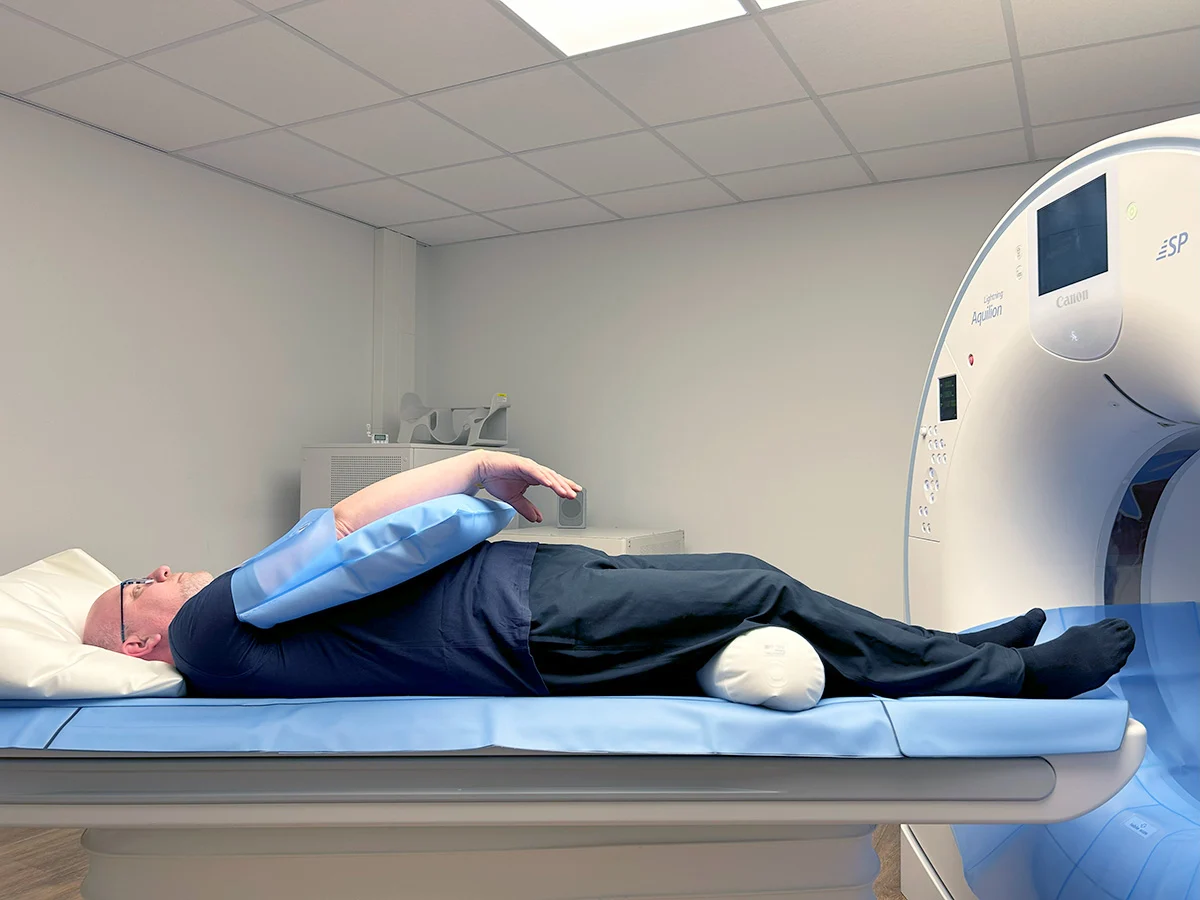
كيفية التحضير للرنين المغناطيسي للبطن
التحضير السليم يُحسّن جودة الصورة والدقة التشخيصية. خطوات التحضير القياسية:
- الصيام: لا تأكل أو تشرب 4-6 ساعات قبل الفحص. يُقلّل الصيام نشاط الأمعاء ويضمن امتلاء المرارة.
- الدواء: استمر في الأدوية المعتادة ما لم يُوجَّه لخلاف ذلك. إذا كنت تتناول دواء السكري (خاصةً الميتفورمين)، أخبر فريق الأشعة.
- وظائف الكلى: إن خُطط لحقن التباين، يُشترط اختبار وظائف الكلى حديثًا.
- فحص المعادن: أكمل استبيان سلامة الرنين المغناطيسي وأزِل جميع المعادن قبل دخول غرفة الفحص.
- الحضور مبكرًا: احضر قبل 15-20 دقيقة من موعدك لإكمال الأوراق والتحضير.
احجز رنينك المغناطيسي للبطن في DCDC
في Doctors Clinic Diagnostic Center بـ Dubai Healthcare City، نُقدم فحوصات الرنين المغناطيسي للبطن بما فيها MRCP وبروتوكولات التباين الخاص بالكبد. تُسلَّم التقارير خلال 24-48 ساعة.
خدمات ذات صلة في DCDC
رعاية متخصصة وتشخيص متقدم في مدينة دبي الطبية
الأسئلة الشائعة
الخلاصة
الرنين المغناطيسي للبطن هو أدق وأشمل أداة تصوير لتقييم الكبد والكلى والبنكرياس والقنوات الصفراوية والغدد الكظرية والطحال. تباينه الفائق للأنسجة الرخوة يجعله الفحص القاطع لتوصيف آفات الأعضاء وتحديد مرحلة أمراض الكبد وتقييم الجهاز الصفراوي عبر MRCP.
في Doctors Clinic Diagnostic Center بـ Dubai Healthcare City، نُقدم الرنين المغناطيسي للبطن بجميع البروتوكولات ذات الصلة. يُسلّم استشاريو الأشعة تقارير شاملة باستخدام أنظمة التصنيف المعترف بها دوليًا.
المصادر والمراجع
تمت مراجعة هذا المقال من قبل فريقنا الطبي ويستند إلى المصادر التالية:
- American College of Radiology - LI-RADS (Liver Imaging Reporting)
- Radiological Society of North America - Abdominal MRI
- European Association for the Study of the Liver - HCC Surveillance
- Society of Abdominal Radiology - MRCP Guidelines
- Dubai Health Authority - Diagnostic Imaging Standards
يتم مراجعة المحتوى الطبي على هذا الموقع من قبل أطباء مرخصين من هيئة الصحة. اطلع على سياستنا التحريرية لمزيد من المعلومات.

كتبه
Dr. Osama Elzamzami
أشعة تشخيصية
دكتوراه في الطب، FRCR
Dr. Osama Elzamzami استشاري أشعة تشخيصية متخصص في التصوير التشخيصي بما فيه الرنين المغناطيسي والأشعة المقطعية والموجات فوق الصوتية في DCDC Dubai Healthcare City.
مقالات ذات صلة
الرنين المغناطيسي في دبي: دليل شامل للأنواع والتكلفة

رنين الحوض في دبي: ما يكشفه والتكلفة والتحضير
تكلفة الرنين الكامل للجسم في دبي: 5,000-15,000 درهم
الرنين مع التباين: ما تتوقعه والسلامة ومتى يُحتاج إليه
رنين الثدي في دبي: متى تحتاجه بعد الماموغرام
المزيد في Diagnostic Imaging
CT Scan Cost Dubai: Prices & Types (2026)
اقرأ المزيد
DEXA Scan Dubai: Cost, T-Score & Who Needs It (2026)
اقرأ المزيدFull Body MRI Cost Dubai: AED 5,000-15,000 [2026]
اقرأ المزيد
MRI Preparation Dubai: Complete Guide (2026)
اقرأ المزيد
CBCT Scan: 3D Dental Imaging Guide Dubai (2026)
اقرأ المزيد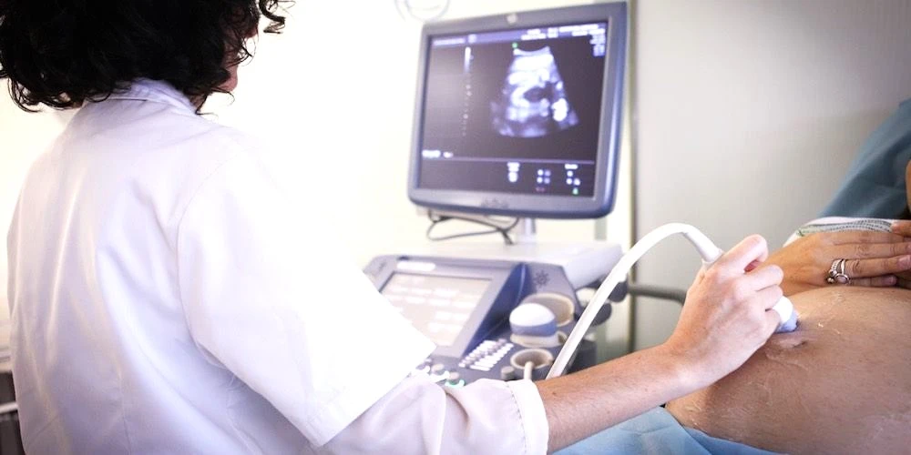
Ultrasound Preparation Dubai: What to Expect (2026)
اقرأ المزيد© 2026 Doctors Clinic Diagnostic Center (DCDC), Dubai Healthcare City. Originally published at https://doctorsclinicdubai.ae/blog/abdominal-mri-dubai. All rights reserved. Unauthorized reproduction is prohibited.