
اہم نکات
- MRI uses magnetic fields for detailed soft tissue imaging (joints, brain, spine); ultrasound uses sound waves for real-time imaging (organs, pregnancy, blood flow)
- Neither MRI nor ultrasound uses ionizing radiation, making both safer than CT or X-ray for repeated imaging
- MRI is the gold standard for brain, spine, and joint injuries; ultrasound is preferred for abdominal organs, pregnancy, and thyroid evaluation
- Ultrasound costs from AED 399 and takes 15-30 minutes; MRI costs from AED 900 and takes 30-60 minutes at DCDC
- Some conditions benefit from both scans used together for a more complete diagnostic picture
- DCDC uses a Siemens 1.5T wide-bore MRI scanner with subspecialty radiologist interpretation for every scan
When your doctor suspects an internal injury, a mass, or a structural problem, the choice between an MRI scan and an ultrasound depends on the body part being examined, the clinical question, and your individual circumstances. Both are radiation-free imaging technologies, but they work in fundamentally different ways and excel at visualizing different structures. Understanding these differences helps you know what to expect and why your doctor chose one over the other.
This comprehensive comparison explains how MRI and ultrasound technology differ, when each test is the better choice, what each costs in Dubai, and how to decide which scan you need. Whether you have been referred for imaging or want to understand your options, this guide covers everything patients need to know about MRI versus ultrasound in 2026.


MRI vs Ultrasound: Side-by-Side Comparison
Before exploring each technology in detail, here is a quick-reference comparison table covering the most important differences between MRI and ultrasound scans.
| Feature | MRI Scan | Ultrasound Scan |
|---|---|---|
| Technology | Magnetic field + radio waves | High-frequency sound waves |
| Radiation | None | None |
| Scan duration | 30-60 minutes | 15-30 minutes |
| Noise level | Loud (rhythmic banging; ear protection provided) | Silent |
| Scanner design | Enclosed tunnel (wide-bore or open options available) | Handheld probe moved over skin surface |
| Patient comfort | Must lie still inside scanner; can trigger claustrophobia | Comfortable; patient lies on exam table |
| Soft tissue detail | Excellent — superior for deep structures | Good for superficial structures; limited for deep tissues |
| Bone imaging | Good for bone marrow; limited for cortical bone | Limited — sound waves reflect off bone surfaces |
| Real-time imaging | No — images acquired and processed | Yes — live moving images |
| Blood flow assessment | Yes (MR angiography) | Yes (Doppler ultrasound) — the standard |
| Pregnancy safe | Generally avoided in first trimester (precautionary) | Yes — the standard for all prenatal imaging |
| Metal implant restriction | Yes — some implants are not MRI-safe | No restrictions |
| Contrast agent | Gadolinium-based IV contrast (when needed) | Usually none required |
| Best for | Brain, spine, joints, ligaments, tumors | Abdomen, thyroid, pregnancy, blood flow, guided biopsies |
| Cost at DCDC (AED) | From AED 900 | From AED 399 |
| Market range Dubai (AED) | 900 - 5,000 | 300 - 1,500 |
Both MRI and ultrasound are radiation-free. The choice depends on which body part needs evaluation and what clinical information your doctor needs.
How MRI Technology Works
MRI (Magnetic Resonance Imaging) uses a powerful superconducting magnet, gradient coils, and radiofrequency pulses to create highly detailed images of internal structures. When you enter the scanner, the magnetic field aligns hydrogen atoms (protons) in your body. Radiofrequency pulses then temporarily disturb this alignment. As the protons return to their resting state, they emit signals that the scanner detects. A computer converts these signals into cross-sectional images with extraordinary soft tissue contrast.
Different tissues contain different amounts of water and therefore different concentrations of hydrogen atoms. This is why MRI produces such exceptional contrast between the brain's grey and white matter, between healthy and damaged cartilage, and between normal tissue and tumors. The scanner can also be programmed with different pulse sequences — T1-weighted, T2-weighted, FLAIR, diffusion-weighted, and more — each highlighting different tissue properties. This versatility is one of MRI's greatest strengths.
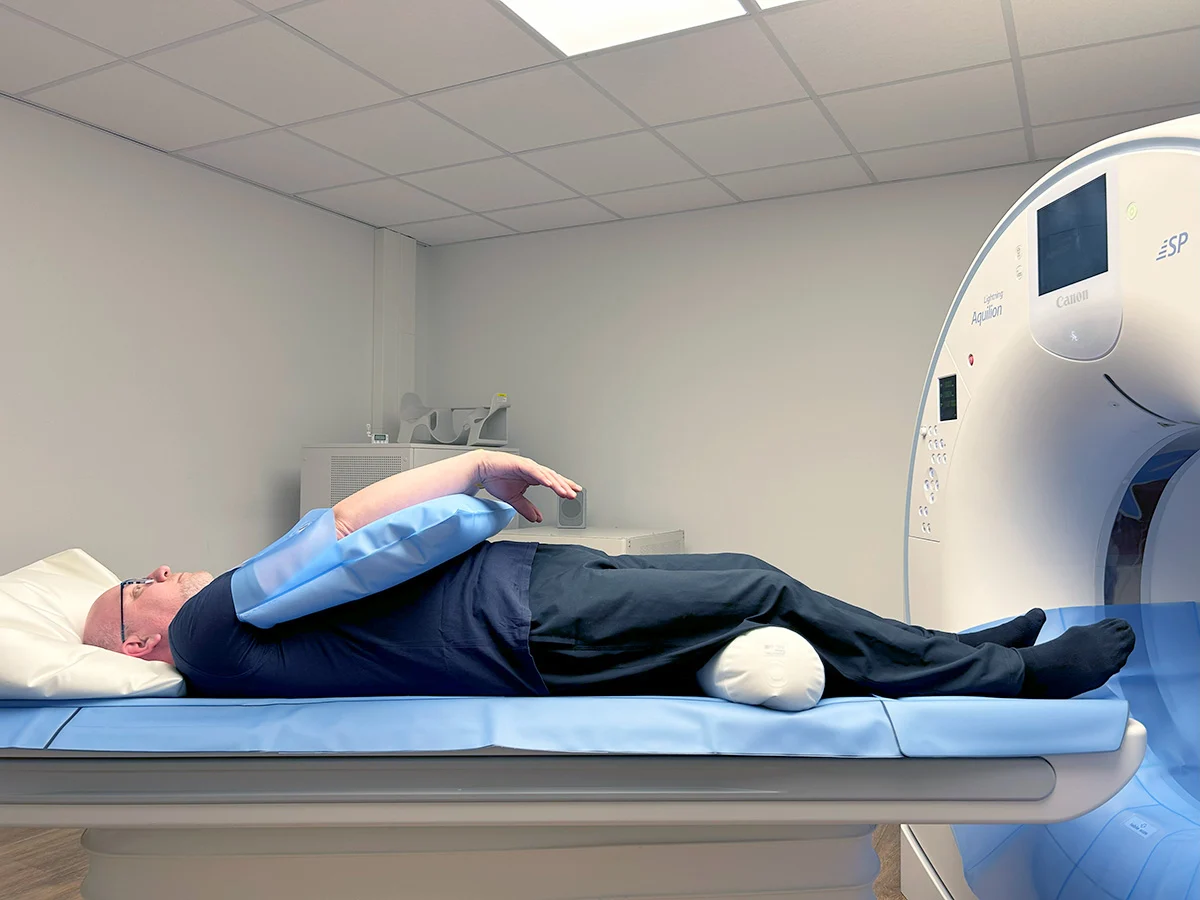
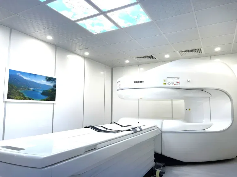
At DCDC, we use a Siemens 1.5T wide-bore MRI scanner with a 70 cm opening, which is 10 cm wider than standard scanners. This makes the experience significantly more comfortable, particularly for patients who are larger, anxious, or prone to claustrophobia. An open MRI option is also available for patients who cannot tolerate a closed scanner.
How Ultrasound Technology Works
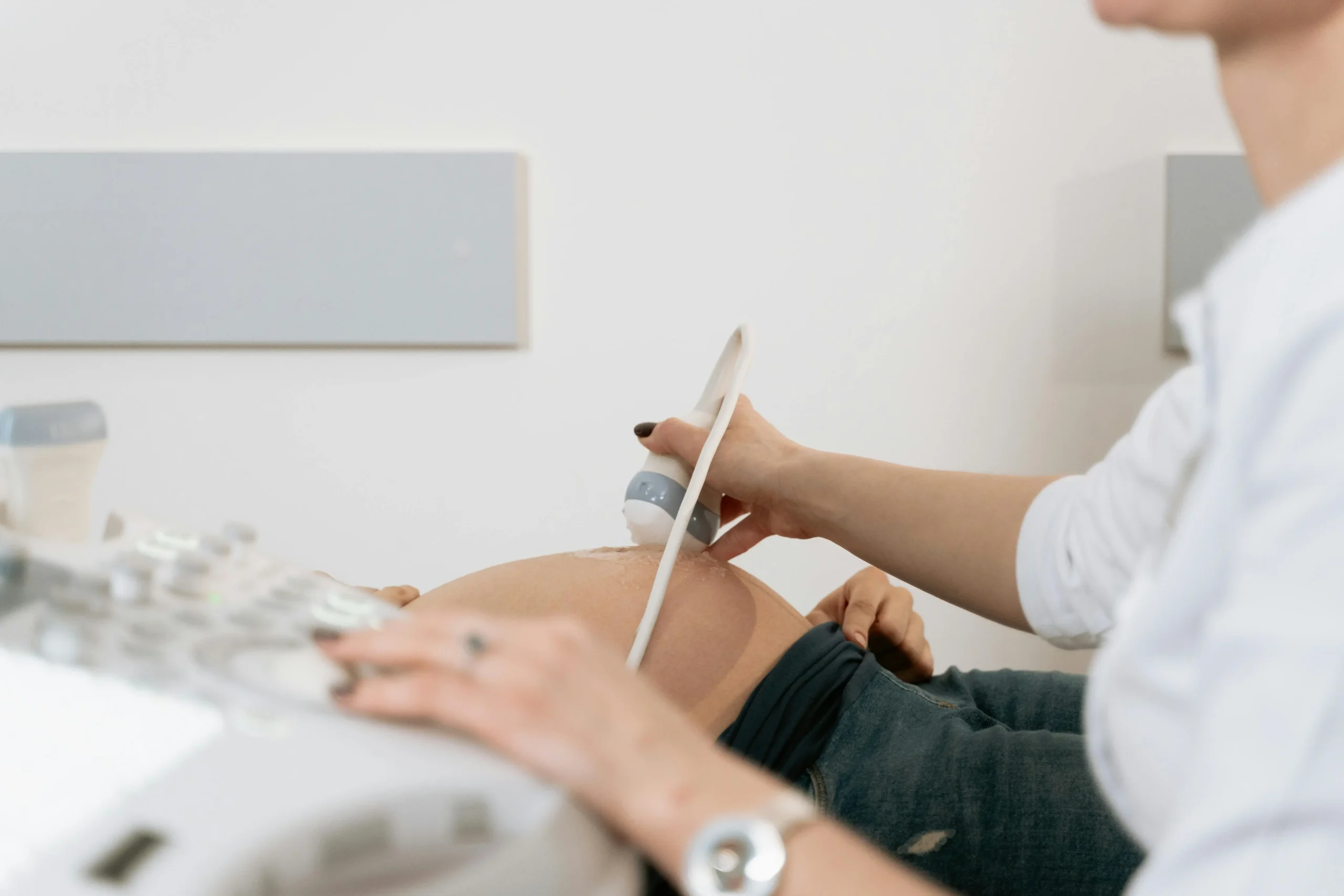
Ultrasound imaging, also called sonography, uses high-frequency sound waves (typically 2-18 MHz) to produce images. A handheld transducer (probe) is placed against the skin, often with a water-based gel to eliminate air gaps. The probe sends sound waves into the body and listens for the echoes that bounce back from internal structures. Different tissues reflect sound waves differently: fluid-filled structures appear black, dense structures appear bright white, and soft tissues appear in varying shades of grey.
One of ultrasound's unique advantages is real-time imaging. The images update continuously as the sonographer moves the probe, allowing assessment of movement — a baby kicking, blood flowing through an artery, or a tendon sliding as you move your finger. This dynamic capability makes ultrasound indispensable for pregnancy monitoring, cardiac assessment (echocardiography), and vascular evaluation. Doppler ultrasound adds colour-coded blood flow information, showing the direction and speed of blood movement through vessels.
Ultrasound has important limitations. Sound waves cannot pass through bone or gas-filled structures effectively, so ultrasound is poor at imaging the brain (in adults), lungs, and bowel. Image quality also depends heavily on the operator's skill and the patient's body habitus — deeper structures in larger patients can be difficult to visualize. For a detailed exploration of ultrasound applications, see our ultrasound costs in Dubai guide.
When MRI Is the Better Choice
MRI is the gold standard imaging modality for several body systems where its superior soft tissue contrast and multiplanar capability provide diagnostic information that ultrasound simply cannot match.
Brain and Neurological Conditions
Brain MRI is the definitive test for evaluating headaches, seizures, memory loss, suspected tumors, multiple sclerosis, and other neurological conditions. The skull blocks ultrasound waves in adults, making brain ultrasound impossible except in newborns (whose fontanelles provide an acoustic window). MRI's ability to distinguish grey matter from white matter, detect subtle lesions, and characterize tumors makes it irreplaceable for neuroimaging.
Spine and Spinal Cord
For disc herniations, spinal cord compression, degenerative disc disease, and spinal tumors, MRI provides the definitive diagnostic images. Ultrasound cannot penetrate the bony vertebral column to image the spinal cord or nerve roots. MRI clearly shows disc material pressing on nerves, spinal cord signal changes indicating myelopathy, and the relationship between spinal structures.
Joint and Ligament Injuries
MRI is the gold standard for evaluating internal joint structures: ACL and meniscus tears in the knee, rotator cuff tears in the shoulder, labral tears in the hip, and ligament injuries in the ankle and wrist. While ultrasound can assess some superficial tendons and detect joint fluid, it cannot visualize deep intra-articular structures like the cruciate ligaments, menisci, or labrum.
Tumor Characterization
When a mass is detected — whether by physical examination, blood tests, or another imaging modality — MRI with contrast often provides the most specific characterization. Liver MRI, breast MRI, and pelvic MRI can often determine whether a lesion is benign or malignant based on its enhancement pattern, signal characteristics, and morphology.
When Ultrasound Is the Better Choice
Ultrasound excels in situations requiring real-time assessment, evaluation of superficial structures, and conditions where its unique capabilities — portability, no radiation, no contrast, and no claustrophobia risk — make it the ideal first-line test.
Pregnancy and Prenatal Monitoring
Ultrasound is the standard imaging modality throughout pregnancy. It confirms pregnancy dating, monitors fetal growth and development, detects anomalies, assesses placental position, measures amniotic fluid, and evaluates fetal wellbeing — all without any radiation exposure. Doppler ultrasound during pregnancy assesses blood flow through the umbilical cord and fetal vessels. MRI is used in pregnancy only when ultrasound findings are inconclusive and additional detail is needed.
Abdominal Organ Assessment
For evaluating the liver, gallbladder, kidneys, pancreas, and spleen, ultrasound is typically the first-line imaging test. It readily detects gallstones, kidney stones, liver cysts, and organ enlargement. For further reading on this topic, see our abdominal ultrasound guide. MRI is reserved for cases where ultrasound findings need further characterization — such as distinguishing between types of liver lesions or evaluating the bile ducts in detail (MRCP).
Thyroid and Superficial Structures
Thyroid nodules, lymph nodes, breast lumps, and other superficial structures are best evaluated with ultrasound, which provides excellent resolution for structures close to the skin surface. Ultrasound can also guide fine-needle aspiration biopsies of thyroid nodules and suspicious lymph nodes in real time.
Blood Flow and Vascular Assessment
Doppler ultrasound is the standard test for evaluating blood flow in arteries and veins. It detects deep vein thrombosis (DVT), assesses carotid artery disease, evaluates varicose veins, and monitors blood flow in transplanted organs. While MR angiography also evaluates vessels, Doppler ultrasound is faster, more accessible, and does not require contrast.
Cost Comparison: MRI vs Ultrasound in Dubai
Cost is often a significant factor for patients choosing between imaging modalities. Here is a comparison of typical pricing in Dubai for common scan types.
| Scan Type | Ultrasound Cost (AED) | MRI Cost (AED) | When Each Is Preferred |
|---|---|---|---|
| Abdomen | 399 - 800 | 2,000 - 4,000 | Ultrasound first; MRI for lesion characterization |
| Knee / Shoulder | 399 - 700 (limited to superficial tendons) | 900 - 3,000 | MRI strongly preferred for internal joint structures |
| Brain | Not applicable in adults | 900 - 3,000 | MRI is the only option |
| Spine | Not applicable | 900 - 3,500 | MRI is the only option |
| Thyroid | 399 - 600 | 1,500 - 3,000 | Ultrasound is first-line; MRI rarely needed |
| Breast | 399 - 800 | 2,000 - 4,500 | Ultrasound for initial assessment; MRI for high-risk screening |
| Pelvic / Obstetric | 399 - 1,000 | 2,000 - 4,000 | Ultrasound for pregnancy; MRI for complex pathology |
| Vascular (Doppler) | 500 - 1,500 | 2,500 - 5,000 (MR angiography) | Doppler ultrasound is the standard first-line test |
| Full body screening | Not applicable | 3,850 - 15,000 | MRI only — ultrasound cannot cover all regions |
At DCDC, ultrasound scans start from AED 399 and MRI scans from AED 900. Actual cost depends on the body part, whether contrast is needed, and insurance coverage. Most insurance plans cover both when medically indicated.
At DCDC, we offer direct billing with 20+ insurance providers, so most patients with insurance coverage pay little or nothing out of pocket for medically indicated imaging. For self-pay patients, our pricing starts from AED 399 for ultrasound and from AED 900 for MRI — competitive rates for a MOHAP-licensed facility in Dubai Healthcare City with subspecialty radiologist interpretation.
Which Scan Do You Need? A Decision Guide
While your doctor will make the final recommendation based on your specific clinical situation, here is a general framework to help you understand why one modality may be chosen over the other.
Choose MRI when:
- You have a suspected brain or spinal cord problem (headaches, seizures, numbness, disc herniation)
- You have a joint injury that may involve internal ligaments, cartilage, or meniscus
- A mass or tumor has been found and needs further characterization
- You need detailed evaluation of the pelvic organs for complex conditions (endometriosis, rectal cancer staging)
- Ultrasound was inconclusive and more detail is needed for the same body region
- Your doctor wants to monitor a known condition over time without repeated radiation exposure
Choose ultrasound when:
- You are pregnant or need prenatal imaging
- You have abdominal pain and your doctor needs a quick assessment of your organs
- You have a palpable lump in the thyroid, breast, or another superficial location
- You need blood flow assessment (suspected DVT, carotid disease, varicose veins)
- You are claustrophobic and cannot tolerate an MRI scanner
- Your doctor needs a fast, initial screening test before deciding on further imaging
- A guided biopsy or fluid drainage is planned
"I often explain to patients that choosing between MRI and ultrasound is not a matter of one being better than the other — they are different tools designed for different jobs," says Dr. Osama Elzamzami. "Ultrasound is like a flashlight that gives you a quick, focused look at structures near the surface. MRI is like turning on all the lights in the room — you see everything in extraordinary detail, especially deep structures, but it takes more time and costs more. The key is matching the right tool to the clinical question."
Can You Need Both MRI and Ultrasound?
Yes. In many clinical scenarios, ultrasound and MRI serve complementary roles, and your doctor may order both to build a complete diagnostic picture. This is not unnecessary duplication — each test provides different information. For a related comparison of imaging modalities, see our CT vs MRI comparison.
Common scenarios where both scans are used include:
- Liver lesion workup: Ultrasound detects a lesion, then MRI with liver-specific contrast (Primovist) characterizes it as benign (haemangioma, focal nodular hyperplasia) or potentially malignant
- Breast assessment: Ultrasound evaluates a palpable lump or mammographic finding, then breast MRI provides a comprehensive survey in high-risk patients or complex cases
- Pelvic pathology: Pelvic ultrasound provides initial assessment of a mass or abnormal bleeding, then pelvic MRI maps the extent of disease (endometriosis, fibroids, ovarian masses)
- Musculoskeletal evaluation: Ultrasound assesses a superficial tendon (e.g., Achilles tendon) dynamically, while MRI evaluates the deeper joint structures and bone marrow that ultrasound cannot reach
- Prenatal follow-up: Routine ultrasound identifies a suspected fetal anomaly, then fetal MRI provides additional detail to guide management decisions
What to Expect at DCDC for Each Scan
Your MRI Experience at DCDC
When you arrive for your MRI at DCDC in Dubai Healthcare City (Building 64), you will be asked to complete a safety screening questionnaire to ensure there are no contraindications to entering the magnetic field. You will change into a gown and remove all metal objects including jewellery, watches, and belts. Our Siemens 1.5T wide-bore scanner has a 70 cm opening — 10 cm wider than standard MRI scanners — which most patients find noticeably more comfortable. An open MRI option is available for patients with severe claustrophobia.
During the scan, you will lie on a padded table that slides into the scanner. Ear protection is provided because MRI machines produce loud rhythmic sounds. Most scans take 30-45 minutes, during which you must remain as still as possible. Some scans require gadolinium contrast, which is injected through a small IV line. Our technologists communicate with you throughout via an intercom system, and you will have a call button to press if you need anything.
Your Ultrasound Experience at DCDC
Ultrasound is generally the simpler experience. You may need to prepare depending on the type of scan — for example, abdominal ultrasound requires fasting for 6-8 hours, while pelvic ultrasound requires a full bladder. You will lie on a comfortable examination table, and the sonographer applies warm gel to the area being examined. The transducer is pressed gently against your skin and moved to capture images from different angles.
Most ultrasound scans take 15-30 minutes. You can usually see the screen during your scan, and the sonographer may explain some findings in real time (particularly during obstetric scans). There is no pain, no noise, no enclosed space, and no injection. Ultrasound is one of the most patient-friendly imaging modalities available.
Results and Reporting
For both MRI and ultrasound at DCDC, your images are interpreted by a subspecialty radiologist. Brain MRIs are read by neuroradiologists, joint MRIs by musculoskeletal radiologists, and each subspecialist brings focused expertise that catches findings a generalist might miss. Results are typically available within 18-24 hours, with same-day reporting for urgent cases. You can access your report and images through our patient portal for digital convenience.
Not Sure Which Scan You Need?
Our radiologists can help determine the right imaging for your condition. Contact DCDC Dubai Healthcare City to discuss your imaging needs — we offer both MRI and ultrasound with expert interpretation.
MRI and Ultrasound Safety Compared
Both MRI and ultrasound are considered very safe imaging modalities. Neither uses ionizing radiation (unlike CT scans and X-rays), which means they do not increase cancer risk and can be repeated as clinically needed. However, each has specific safety considerations.
MRI safety considerations: The powerful magnetic field means that ferromagnetic objects (certain metal implants, pacemakers, cochlear implants, metallic foreign bodies) can be dangerous inside the scanner. All patients complete a detailed screening questionnaire before their scan. Some patients experience claustrophobia inside the tunnel, though DCDC's wide-bore scanner and open MRI option reduce this significantly. Gadolinium contrast, when used, has a small risk of allergic reaction and is avoided in patients with severe kidney disease (risk of nephrogenic systemic fibrosis).
Ultrasound safety considerations: Ultrasound has no known risks at diagnostic intensity levels and has been used safely in pregnancy for decades. It has no contraindications related to metal implants or pacemakers. The only limitation is diagnostic — if ultrasound cannot adequately visualize the area of interest, another modality (MRI or CT) may be needed.
Accuracy and Limitations: An Honest Comparison
No imaging modality is perfect. Understanding the limitations of each helps set realistic expectations and explains why your doctor sometimes orders additional tests.
MRI limitations: MRI is poor at imaging lungs (air causes signal loss) and cortical bone detail (CT is better). Scans take longer and cost more. Image quality can be degraded by patient movement — holding still for 30-60 minutes is difficult for some patients, especially children and elderly individuals. Certain implants prevent MRI scanning entirely. Motion artifacts from breathing and heartbeat can also affect image quality in body imaging.
Ultrasound limitations: Ultrasound cannot penetrate bone or air effectively, making it useless for the brain (in adults), lungs, and deep pelvic structures obscured by bowel gas. Image quality is highly operator-dependent — an experienced sonographer produces significantly better diagnostic images than a novice. Deeper structures in patients with a larger body habitus can be difficult to evaluate. Ultrasound provides limited field of view compared to MRI's comprehensive cross-sectional imaging.
"Subspecialty interpretation makes a real difference in imaging accuracy," notes Dr. Osama Elzamzami. "At DCDC, brain MRIs are reported by neuroradiologists and joint MRIs by musculoskeletal radiologists who have spent years focusing on their specific area. This focused expertise consistently catches subtle findings that might be overlooked in a general read."
How to Prepare for Your Scan
MRI Preparation
- Clothing: Wear comfortable clothes without metal zippers, buttons, or underwire. You will be given a gown to change into
- Metal objects: Remove all jewellery, watches, hairpins, hearing aids, and removable dental work before entering the scan room
- Screening questionnaire: Complete the MRI safety form honestly — inform staff about all implants, surgical history, and any metal in your body
- Fasting: Required for abdominal and pelvic MRI (typically 4-6 hours). Not required for brain, spine, or joint MRI
- Claustrophobia: Inform your booking coordinator if you are anxious about enclosed spaces — DCDC offers a wide-bore scanner and open MRI option
- Contrast scans: If contrast MRI is ordered, bring recent blood test results showing kidney function (creatinine/eGFR)
Ultrasound Preparation
- Abdominal ultrasound: Fast for 6-8 hours before the scan (water is usually permitted). This allows the gallbladder to fill and reduces bowel gas for better image quality
- Pelvic ultrasound: Drink 1 litre of water 1 hour before the scan and do not empty your bladder. A full bladder acts as an acoustic window for pelvic organs
- Thyroid, breast, MSK ultrasound: No preparation required
- Doppler ultrasound: Preparation varies by type — your booking coordinator will provide specific instructions
- Obstetric ultrasound: May require a full bladder for early pregnancy scans; no preparation for later trimesters
Why Choose DCDC for Your MRI or Ultrasound
Doctors Clinic Diagnostic Center (DCDC) in Dubai Healthcare City is a MOHAP-licensed facility with a 4.8 out of 5 Google rating from over 1,000 reviews and 98% patient satisfaction. For a complete overview of our MRI services and pricing, read our complete MRI guide.
- Advanced MRI technology: Siemens 1.5T wide-bore scanner with 70 cm opening for greater comfort, plus an open MRI option for claustrophobic patients
- Subspecialty radiologist reads: Every scan is interpreted by a radiologist with subspecialty expertise — neuroradiology for brain scans, musculoskeletal radiology for joint scans, and so on
- Competitive pricing: MRI from AED 900 and ultrasound from AED 399 with direct billing for 20+ insurance providers
- Fast turnaround: Results within 18-24 hours, with same-day reporting for urgent cases and digital access via patient portal
- Extended hours: Saturday to Thursday 8 AM to 10 PM, Friday 9 AM to 9 PM — convenient scheduling for working professionals
- Accessible location: Building 64, Dubai Healthcare City, with free parking available
Book Your MRI or Ultrasound at DCDC
Whether you need an MRI scan from AED 900 or an ultrasound from AED 399, DCDC offers expert imaging with subspecialty interpretation in Dubai Healthcare City. Call or WhatsApp to book your appointment.
DCDC میں متعلقہ خدمات
دبئی ہیلتھ کیئر سٹی میں ماہرانہ دیکھ بھال اور جدید تشخیص
Frequently Asked Questions
Final Thoughts
MRI and ultrasound are both radiation-free imaging technologies that serve different but sometimes overlapping diagnostic roles. Ultrasound is the faster, more accessible, and less expensive option, ideal for pregnancy, abdominal organs, thyroid, and blood flow assessment. MRI provides unmatched detail for the brain, spine, joints, and deep soft tissue pathology. The choice between them is not about which is better overall, but which answers your specific clinical question most accurately.
Cost should not be the primary driver when choosing between MRI and ultrasound — the right test is the one that gives your doctor the diagnostic information needed to guide your treatment. An ultrasound ordered when MRI is needed may save money initially but could delay diagnosis, while an MRI ordered when ultrasound would suffice adds unnecessary cost and wait time.
At Doctors Clinic Diagnostic Center in Dubai Healthcare City, our subspecialty radiologists ensure you receive expert interpretation regardless of which modality is used. With competitive pricing (ultrasound from AED 399, MRI from AED 900), a wide-bore and open MRI scanner, and results within 18-24 hours, DCDC provides a complete diagnostic imaging experience under one roof.
ذرائع اور حوالہ جات
یہ مضمون ہماری طبی ٹیم نے جائزہ لیا ہے اور درج ذیل ذرائع کا حوالہ دیتا ہے:
- American College of Radiology - Appropriateness Criteria for Imaging Selection
- Radiological Society of North America - MRI and Ultrasound Patient Information
- European Society of Radiology - Imaging Referral Guidelines
- NHS - MRI Scan and Ultrasound Scan Information
- Mayo Clinic - MRI and Ultrasound Overview
اس سائٹ پر طبی مواد کا جائزہ DHA لائسنس یافتہ ڈاکٹرز نے لیا ہے۔ ہماری دیکھیں تحریری پالیسی مزید معلومات کے لیے۔

تحریر
Dr. Osama Elzamzami
Diagnostic Radiologist
MD, FRCR
Dr. Osama Elzamzami is a Diagnostic Radiologist at Doctors Clinic Diagnostic Center (DCDC) in Dubai Healthcare City.
Related Articles
CT Scan vs MRI: Which Do You Need? Complete Comparison (2026)
![Ultrasound Cost Dubai: AED 300-800 by Type [2026 Prices]](/wp-media/blog/ultrasound-scan-guide.webp)
Ultrasound Cost Dubai: AED 300-800 by Type [2026 Prices]
MRI Cost Dubai: AED 900-15,000 by Type (2026)
Abdominal Ultrasound: What It Shows & Preparation (2026)

Mammogram vs Breast Ultrasound Dubai (2026)
More in Diagnostic Imaging

3D vs 2D Mammogram Dubai: Key Differences (2026)
مزید پڑھیں
X-Ray at Home Dubai: Cost & Guide (2026)
مزید پڑھیں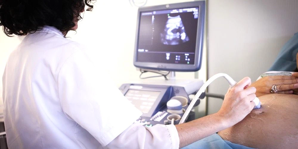
Ultrasound at Home Dubai: From AED 599 (2026)
مزید پڑھیں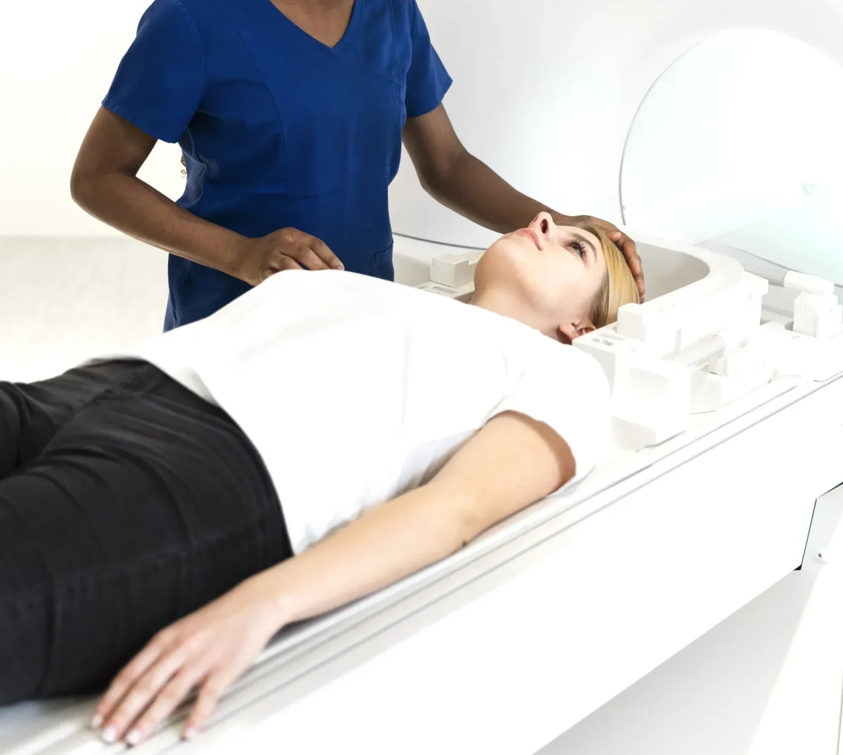
Neck MRI Dubai: What It Shows & Cost (2026)
مزید پڑھیں
X-Ray Scan Dubai: Complete Guide (2026)
مزید پڑھیں
MRI vs X-Ray Dubai: Which Scan Do You Need? (2026)
مزید پڑھیں© 2026 Doctors Clinic Diagnostic Center (DCDC), Dubai Healthcare City. Originally published at https://doctorsclinicdubai.ae/blog/mri-vs-ultrasound-dubai. All rights reserved. Unauthorized reproduction is prohibited.