Ключевые выводы
- A neck MRI in Dubai costs from AED 900 to AED 2,500, depending on whether the study focuses on the cervical spine, soft tissues, or both, and whether contrast is required
- Neck MRI evaluates the cervical spine, spinal cord, nerve roots, intervertebral discs, thyroid gland, salivary glands, lymph node chains, and major neck vessels in a single radiation-free study
- A cervical spine-focused neck MRI is the gold standard for diagnosing disc herniation, foraminal stenosis, cervical myelopathy, and nerve root compression causing arm pain or numbness
- A soft tissue neck MRI is essential for evaluating neck masses, thyroid nodules, salivary gland tumors, enlarged lymph nodes, and brachial plexus injuries
- At DCDC in Dubai Healthcare City, neck MRIs are performed on a Siemens 1.5T wide-bore scanner (70 cm opening) and interpreted by subspecialty neuroradiologists, with results delivered within 18-24 hours
- Most health insurance plans in the UAE cover neck MRI when ordered by a licensed physician with clinical justification, and DCDC offers direct billing with 20+ insurance partners
A neck MRI scan is the most detailed imaging tool available for evaluating the complex anatomy of the cervical region. The neck contains critical structures packed into a remarkably small space: the cervical spinal cord, seven vertebrae and their discs, nerve roots supplying the arms, the thyroid and parathyroid glands, salivary glands, lymph node chains, carotid and vertebral arteries, and the deep soft tissues of the pharynx and larynx. When any of these structures are affected by disease or injury, neck MRI provides the diagnostic clarity that physicians need to determine the right course of treatment. At Doctors Clinic Diagnostic Center (DCDC) in Dubai Healthcare City, neck MRI starts from AED 900 and is interpreted by subspecialty radiologists.
Whether you have been referred for persistent neck pain with arm symptoms, a suspicious lump in the neck, or unexplained neurological changes, this guide explains what a neck MRI shows, how much it costs in Dubai, and what the experience is like at DCDC.



What Is a Neck MRI and What Does It Show?
A neck MRI uses powerful magnetic fields and radiofrequency pulses to generate detailed cross-sectional images of the cervical region without any ionizing radiation. Unlike X-rays or CT scans, which primarily show bone, MRI excels at visualizing the soft tissue structures that are responsible for the majority of neck symptoms. The term "neck MRI" actually encompasses two distinct but overlapping studies, each with a different clinical focus.
Cervical Spine MRI (Spine-Focused)
This protocol targets the seven cervical vertebrae (C1-C7), intervertebral discs, spinal cord, nerve roots, facet joints, and spinal ligaments. It is the study of choice when patients present with neck pain radiating to the arms, numbness or tingling in the hands, weakness, or signs of spinal cord compression. The dedicated cervical spine protocol uses thin-slice sagittal and axial imaging to evaluate each disc level and neural foramen with precision.
Soft Tissue Neck MRI
This protocol evaluates the non-spinal structures of the neck, including the thyroid and parathyroid glands, parotid and submandibular salivary glands, lymph node chains (cervical, supraclavicular, retropharyngeal), deep neck spaces, pharynx, larynx, and major vascular structures. It is ordered when patients present with a palpable neck mass, suspected thyroid pathology, salivary gland swelling, or unexplained lymphadenopathy. Gadolinium contrast is frequently used in this protocol to characterize masses and differentiate benign from potentially malignant lesions.
At DCDC, a dedicated neck coil is used for both protocols to optimize image quality. Diffusion-weighted imaging (DWI) sequences are routinely added for lymph node evaluation and tumor characterization, providing additional diagnostic information beyond standard anatomical imaging.
When Do You Need a Neck MRI?
Neck MRI is not a routine screening test. It is ordered by a physician when clinical symptoms or examination findings suggest a specific neck pathology that requires detailed imaging for diagnosis or treatment planning. The following are the most common clinical scenarios that lead to a neck MRI referral in Dubai:
- Neck pain with arm symptoms (radiculopathy): Pain, numbness, tingling, or weakness radiating from the neck into one or both arms is the hallmark of cervical radiculopathy. This pattern strongly suggests nerve root compression by a herniated disc or foraminal stenosis, and MRI is the definitive investigation to confirm the diagnosis and identify the exact level of compression.
- Signs of cervical myelopathy: Difficulty with fine motor tasks (buttoning shirts, writing), gait instability, leg stiffness, or bladder dysfunction may indicate spinal cord compression in the neck. Cervical myelopathy is a serious condition that requires urgent MRI to assess the degree of cord compression and guide surgical decision-making.
- Persistent neck pain unresponsive to treatment: Neck pain lasting more than 6-8 weeks despite conservative management (physiotherapy, medication) warrants MRI evaluation to identify structural causes such as disc degeneration, facet arthropathy, or occult fracture.
- Neck mass or lump: Any new or growing mass in the neck requires imaging to determine its origin, size, and characteristics. MRI with contrast is the preferred modality for evaluating deep-seated neck masses, parapharyngeal tumors, salivary gland lesions, and suspicious lymph nodes.
- Thyroid nodule evaluation: While ultrasound is the first-line imaging for thyroid nodules, MRI is used for large or retrosternal goiters, suspected thyroid malignancy with potential invasion of surrounding structures, and surgical planning when the full extent of thyroid disease needs to be mapped.
- Brachial plexus injury: Trauma, traction injuries, or suspected tumors involving the brachial plexus (the nerve network that controls arm function) require dedicated MRI with specialized protocols to visualize these complex neural structures.
- Post-surgical evaluation: Patients who have undergone cervical spine surgery (discectomy, fusion) or neck surgery (thyroidectomy, lymph node dissection) may require follow-up MRI to assess surgical outcomes, detect recurrence, or investigate new symptoms.
- TMJ dysfunction referral: Although TMJ has its own dedicated MRI protocol, some neck MRI referrals originate from temporomandibular joint problems that are causing secondary neck symptoms.
Neck MRI vs Cervical Spine MRI: What's the Difference?
This is one of the most common sources of confusion among patients, and the distinction matters because ordering the wrong study can result in a non-diagnostic scan. A cervical spine MRI and a soft tissue neck MRI use different imaging protocols, coil configurations, and field-of-view settings that are optimized for their respective anatomical targets.
| Feature | Cervical Spine MRI | Soft Tissue Neck MRI |
|---|---|---|
| Primary focus | Vertebrae, discs, spinal cord, nerve roots | Thyroid, salivary glands, lymph nodes, deep neck spaces |
| Common indications | Disc herniation, stenosis, myelopathy, radiculopathy | Neck mass, thyroid pathology, lymphadenopathy, tumor staging |
| Contrast use | Usually without contrast (added for tumors, infection) | Frequently with gadolinium contrast |
| Scan duration | 30-40 minutes | 35-45 minutes |
| Approximate cost | From AED 900-1,500 | From AED 1,000-1,800 |
| Ordering physician | Orthopedic surgeon, neurosurgeon, neurologist | ENT specialist, endocrinologist, oncologist |
A combined cervical spine + soft tissue neck MRI (from AED 1,500-2,500) is sometimes ordered when both spinal and soft tissue pathology need evaluation in a single session.
The critical point is that your referring physician must specify the clinical question so the radiologist can select the appropriate protocol. A patient with arm numbness needs a cervical spine protocol, while a patient with a thyroid nodule needs a soft tissue protocol. When in doubt, the radiologist at DCDC will contact the referring physician to clarify the clinical question before proceeding. For more on how spinal imaging costs are structured, see our guide on spine MRI cost in Dubai.
How Much Does a Neck MRI Cost in Dubai?
The cost of a neck MRI in Dubai varies depending on whether the study focuses on the cervical spine, the soft tissues, or both, and whether intravenous contrast is required. At Doctors Clinic Diagnostic Center, all neck MRI pricing includes the scan, a comprehensive subspecialty radiologist report, and digital access to images and reports through the patient portal.
| Neck MRI Type | Approximate Cost (AED) |
|---|---|
| Neck MRI — cervical spine focus (without contrast) | From 900 – 1,500 |
| Neck MRI — soft tissue focus (without contrast) | From 1,000 – 1,800 |
| Combined cervical spine + soft tissue neck MRI | From 1,500 – 2,500 |
| Contrast (gadolinium) add-on | From 300 – 500 additional |
Prices include subspecialty radiologist report and digital image access. Contact DCDC for exact pricing and insurance coverage verification.
Several factors influence the final cost. Contrast-enhanced studies cost more because of the gadolinium agent and the additional imaging sequences required after contrast injection. Combined studies covering both the cervical spine and soft tissues take longer and are priced accordingly. Some facilities in Dubai charge separately for the radiologist report — at DCDC, the report by a subspecialty radiologist is always included in the scan price.
Compared to other imaging centers in Dubai, DCDC offers competitive pricing with the advantage of subspecialty interpretation. A cervical MRI read by a neuroradiologist rather than a generalist provides significantly more detailed and clinically relevant findings, particularly for complex cases involving multilevel disease, cord signal changes, or post-surgical anatomy.
What Conditions Can Neck MRI Detect?
The neck is anatomically one of the most complex regions in the body, and neck MRI can detect a wide range of conditions affecting its various structures. The following conditions are among the most important findings on neck MRI:
- Cervical disc herniation: The most common reason for cervical spine MRI referrals. A herniated disc in the neck compresses adjacent nerve roots, causing pain, numbness, and weakness radiating into the arm and hand. MRI shows the exact location and size of the herniation, whether it is compressing the spinal cord (central herniation) or a nerve root (foraminal herniation), and guides the decision between conservative treatment and surgery.
- Foraminal stenosis: Narrowing of the neural foramina — the bony channels through which nerve roots exit the spinal canal — is a common cause of cervical radiculopathy, especially in patients over 50. MRI demonstrates whether the narrowing is caused by disc material, bone spurs (osteophytes), ligament hypertrophy, or a combination of factors.
- Cervical myelopathy: Compression of the spinal cord in the cervical region is a potentially serious condition that can cause progressive neurological deterioration if left untreated. MRI not only shows the cause and level of compression but also detects signal changes within the spinal cord itself (myelomalacia), which help predict surgical outcomes and urgency.
- Neck masses and tumors: Soft tissue neck MRI is essential for characterizing neck masses, determining their tissue of origin, and assessing their relationship to critical structures such as the carotid artery, jugular vein, and cranial nerves. This information is vital for surgical planning and biopsy guidance.
- Lymph node pathology: MRI with diffusion-weighted imaging can help differentiate reactive (benign) lymph nodes from potentially malignant ones. This is particularly important in head and neck cancer staging and in patients with unexplained cervical lymphadenopathy.
- Thyroid and parathyroid pathology: While ultrasound remains the primary imaging tool for thyroid evaluation, MRI provides complementary information for large goiters, substernal extension, suspected invasion of surrounding structures, and recurrent thyroid cancer.
- Salivary gland disorders: Parotid and submandibular gland tumors (both benign and malignant), inflammatory conditions, and ductal pathology are well evaluated with MRI. The multiplanar imaging capability of MRI helps define the relationship of salivary gland lesions to the facial nerve, which is crucial for surgical planning.
- Brachial plexus injuries: MRI is the only imaging modality that can directly visualize the brachial plexus nerve roots and trunks. It is essential for evaluating traction injuries, nerve sheath tumors (schwannomas, neurofibromas), and thoracic outlet syndrome.
Neck MRI With and Without Contrast
Whether your neck MRI requires contrast depends entirely on the clinical question being asked. Gadolinium-based contrast agents are injected intravenously during the scan to highlight areas of increased blood flow, inflammation, or abnormal tissue enhancement. Understanding when contrast is needed helps patients prepare for their appointment and understand the cost implications.
When Contrast Is Typically Not Needed
- Evaluation of cervical disc herniation and foraminal stenosis
- Assessment of degenerative disc disease and spondylosis
- Initial evaluation of cervical myelopathy
- Follow-up of known stable cervical spine conditions
When Contrast Is Recommended
- Evaluation of neck masses and suspected tumors
- Characterization of lymph node pathology
- Suspected spinal infection (discitis, epidural abscess)
- Post-operative evaluation after cervical spine surgery or neck tumor resection
- Suspected vascular malformation or tumor invasion of vessels
- Salivary gland mass evaluation
At DCDC, the decision to use contrast is made by the radiologist based on the clinical information provided by the referring physician. Gadolinium contrast has an excellent safety profile in patients with normal kidney function. Patients with impaired kidney function or a history of severe allergic reactions are assessed individually, and alternative protocols may be used. The contrast add-on costs from AED 300-500 and extends the scan time by approximately 10-15 minutes. If you have concerns about claustrophobia during the extended scan, our MRI claustrophobia guide covers strategies that help patients complete their scan comfortably.
How to Prepare for Your Neck MRI
Neck MRI requires minimal preparation, but following these guidelines ensures a smooth, efficient experience and optimal image quality:
- Metal safety screening: You will complete an MRI safety questionnaire before your scan. Inform the team about any metal implants, surgical hardware, pacemakers, cochlear implants, or metallic foreign bodies. Most modern orthopedic implants are MRI-compatible, but this must be verified before entering the scanner room.
- Remove all metal: Leave jewelry, watches, hairpins, belt buckles, coins, and any clothing with metal components at home or in the locker provided. Hearing aids, dentures with metal clasps, and body piercings must be removed.
- Clothing: Wear comfortable clothing without metal zippers, snaps, or underwire. You may be asked to change into a gown. Avoid clothing with metallic threads or prints.
- Eating and drinking: No fasting is required for a standard neck MRI without contrast. If contrast is being used, you may be asked to avoid eating for 2 hours before the scan as a precaution against nausea (though this is uncommon with modern gadolinium agents).
- Bring your referral and previous imaging: Bring the physician's referral letter detailing the clinical question. If you have previous neck imaging (MRI, CT, or X-rays) from another facility, bring copies or have them sent to DCDC in advance so the radiologist can compare with prior studies.
- Kidney function test: If contrast is planned, a recent blood creatinine or eGFR result (within the past 3 months) is required to confirm normal kidney function. DCDC can perform this blood test on-site if needed.
- Claustrophobia management: If you experience claustrophobia, inform the team when booking. DCDC's Siemens 1.5T wide-bore scanner has a 70 cm opening — significantly wider than the standard 60 cm — which substantially reduces claustrophobic reactions. Additional measures such as relaxation techniques, music through headphones, and a companion in the room may be arranged.
What to Expect at DCDC During Your Neck MRI
The patient journey at Doctors Clinic Diagnostic Center in Dubai Healthcare City is designed to be straightforward and efficient. Here is a step-by-step overview of what happens during your visit for a neck MRI:
- Step 1 — Check-in and registration: Arrive at Building 64, Block A, Al Razi Medical Complex in Dubai Healthcare City. Free parking is available. The reception team will verify your referral, insurance details (DCDC offers direct billing with 20+ insurance partners), and complete the registration process.
- Step 2 — MRI safety screening: A radiographer will review the MRI safety questionnaire with you, confirming the absence of any MRI contraindications. If contrast is needed, an IV line will be placed in your arm.
- Step 3 — Positioning and coil placement: You will lie on your back on the MRI table. A dedicated neck coil — a specialized device shaped to fit closely around the neck — is positioned to maximize image quality. Foam pads help keep you comfortable and minimize movement.
- Step 4 — The scan: The table slides into the Siemens 1.5T wide-bore scanner. The 70 cm opening provides noticeably more space than standard MRI machines, and your head typically remains close to the front of the bore, which helps reduce enclosed feelings. You will hear rhythmic knocking, buzzing, and clicking sounds — these are normal. Earplugs or headphones with music are provided. A radiographer monitors you continuously and communicates through an intercom. A squeeze ball alarm is placed in your hand.
- Step 5 — Scan duration: A cervical spine-focused study takes approximately 30-40 minutes. A soft tissue neck study takes 35-45 minutes. If both protocols are combined, the total scan time is approximately 45-55 minutes. If contrast is used, there is a brief pause mid-scan for the injection, after which additional sequences are acquired.
- Step 6 — After the scan: You can get up and leave immediately. There is no recovery period, and you can drive, eat, and resume all normal activities. If contrast was used, drink extra water throughout the day to help flush the agent from your system.
- Step 7 — Results: Your images are interpreted by a subspecialty neuroradiologist — not a generalist radiologist. The detailed report and images are available within 18-24 hours through DCDC's patient portal. For urgent cases (suspected cord compression, for example), same-day results are provided directly to the referring physician.
DCDC operates extended hours (Saturday to Thursday, 8 AM to 10 PM; Friday, 9 AM to 9 PM), making it convenient to schedule neck MRI appointments around work and family commitments. The clinic maintains a Google rating of 4.8 out of 5 from over 1,000 verified reviews, with a 98% patient satisfaction rate.
Book Your Neck MRI at DCDC
At Doctors Clinic Diagnostic Center in Dubai Healthcare City, neck MRI is performed on a Siemens 1.5T wide-bore scanner and interpreted by subspecialty neuroradiologists. Reports are delivered within 18-24 hours. MOHAP licensed, 20+ insurance partners with direct billing. Learn more about our neck MRI service.
Call, WhatsApp, or book online. Neck MRI starts from AED 900.
Understanding Your Neck MRI Results
After your scan is completed and interpreted, you will receive a structured radiologist report. Understanding the terminology used in neck MRI reports helps you have a more productive discussion with your referring physician about your findings and treatment options.
Common Cervical Spine MRI Findings
- Disc bulge vs. disc herniation: A bulge is a broad-based, circumferential extension of the disc margin — it is extremely common with aging and is often incidental. A herniation is a focal protrusion of disc material beyond the normal margin, and it becomes clinically significant when it compresses a nerve root or the spinal cord.
- Foraminal stenosis grading: Neural foraminal narrowing is typically graded as mild, moderate, or severe. Mild stenosis rarely causes symptoms. Moderate to severe stenosis, especially when it contacts or displaces a nerve root, correlates with radiculopathy symptoms.
- Spinal cord signal changes: High signal within the spinal cord on T2-weighted images (termed "myelopathy signal") indicates chronic compression and potential cord damage. This finding increases the urgency for surgical decompression.
- Degenerative changes: Terms like "spondylosis," "osteophytes," "disc desiccation," and "facet hypertrophy" describe age-related wear-and-tear changes. These are nearly universal after age 40 and are clinically significant only when they cause neural compression.
Common Soft Tissue Neck MRI Findings
- Lymph node characterization: The report will describe lymph node size, morphology, enhancement pattern, and diffusion characteristics. Nodes exceeding 1 cm in short axis, those with irregular margins or central necrosis, or those showing restricted diffusion may require further evaluation.
- Thyroid findings: MRI can identify thyroid nodules, goiter extension, and features suspicious for malignancy. However, ultrasound-guided fine-needle aspiration remains the standard for definitive thyroid nodule characterization.
- Mass characterization: The report will describe the mass location, size, margins, signal characteristics, and enhancement pattern, which together help narrow the differential diagnosis. The radiologist will often suggest the most likely diagnoses and recommend next steps.
At DCDC, subspecialty neuroradiologists read cervical spine MRIs, which means the reports include detailed disc-level analysis, neural compression grading, and direct clinical correlation — not just anatomical descriptions. Your report is accessible digitally through the patient portal, and you can share it directly with your physician. For patients undergoing neck MRI for pain-related conditions, our article on neck pain physiotherapy in Dubai explains the non-surgical treatment options that may be recommended based on your results.
"Neck MRI is unique because the cervical region packs so many critical structures into a small space — the spinal cord, nerve roots, thyroid, lymph nodes, and major vessels," explains Dr. Osama Elzamzami, Diagnostic Radiologist at DCDC. "As a subspecialty radiologist, I use dedicated protocols for each clinical question. A patient with arm numbness needs a cervical spine-focused study, while someone with a thyroid nodule needs a soft tissue neck protocol. The right protocol makes the difference between a diagnostic and a non-diagnostic scan."
Neck MRI vs CT Scan: Which Is Better?
Both MRI and CT scan can image the neck, but they have fundamentally different strengths. Understanding these differences helps patients and physicians select the most appropriate modality for each clinical scenario.
| Factor | Neck MRI | Neck CT Scan |
|---|---|---|
| Radiation | None (zero radiation) | Yes (approximately 3-5 mSv) |
| Soft tissue detail | Excellent (gold standard) | Moderate |
| Bone detail | Good | Excellent |
| Spinal cord visualization | Excellent (directly visible) | Limited (inferred from canal size) |
| Scan time | 30-55 minutes | 5-10 minutes |
| Cost | From AED 900-2,500 | From AED 500-1,200 |
| Best for | Disc herniation, cord compression, soft tissue masses, tumors | Fractures, bony stenosis, acute trauma, calcified lesions |
| Claustrophobia risk | Present (mitigated by wide-bore scanners) | Minimal (open, fast scanner) |
MRI is preferred for most neck soft tissue and spinal cord conditions. CT is preferred for bony pathology and acute trauma.
In practice, MRI and CT are often complementary rather than competing. A patient with acute neck trauma may receive a CT scan in the emergency department to rapidly rule out fractures, followed by an MRI to evaluate the spinal cord, discs, and ligaments. For non-urgent conditions such as chronic neck pain, radiculopathy, or neck masses, MRI is almost always the preferred first-line advanced imaging study because of its superior soft tissue resolution and absence of radiation.
Insurance Coverage for Neck MRI in Dubai
Most health insurance plans in the UAE cover neck MRI when it is medically indicated and ordered by a licensed physician. However, coverage policies vary between insurers and plans, and understanding the process helps avoid unexpected costs.
- Prior authorization: Most insurance companies require pre-approval for MRI scans. Your referring physician submits a request with the clinical indication, and the insurer typically responds within 24-48 hours. DCDC's insurance coordination team handles this process on behalf of patients.
- Clinical justification: Insurers approve neck MRI for documented clinical indications such as radiculopathy, myelopathy, neck mass evaluation, post-surgical assessment, or persistent pain unresponsive to conservative treatment. A clear clinical history from the referring physician facilitates smooth approval.
- Direct billing: DCDC partners with over 20 insurance companies for direct billing, meaning you do not need to pay upfront and claim reimbursement later. Covered patients pay only the applicable co-pay or deductible at the time of the scan.
- Self-pay patients: For patients without insurance or those whose plans do not cover MRI, DCDC offers transparent self-pay pricing starting from AED 900 for cervical spine MRI. Payment plans may be available for combined or higher-cost studies.
DCDC is MOHAP licensed, which means all major UAE insurance networks recognize and accept the clinic's imaging services. Contact the DCDC insurance desk before your appointment to verify your specific plan's coverage for neck MRI.
Check Your Insurance Coverage for Neck MRI
DCDC partners with 20+ insurance companies for direct billing. Our insurance coordination team can verify your coverage and handle pre-authorization before your appointment. Contact us to confirm your plan covers neck MRI at DCDC.
Self-pay neck MRI available from AED 900. Extended hours: Sat-Thu 8AM-10PM, Fri 9AM-9PM.
Связанные услуги в DCDC
Квалифицированная помощь и современная диагностика в Dubai Healthcare City
Frequently Asked Questions
Final Thoughts
Neck MRI is an indispensable diagnostic tool for evaluating the full spectrum of cervical pathology — from disc herniations and spinal cord compression to thyroid nodules, neck masses, and lymph node disease. The key to a diagnostic neck MRI is matching the imaging protocol to the clinical question, which is why subspecialty radiologist expertise and appropriate protocol selection are as important as the scanner hardware itself.
At Doctors Clinic Diagnostic Center in Dubai Healthcare City, neck MRI is performed on a Siemens 1.5T wide-bore scanner with a dedicated neck coil, interpreted by subspecialty neuroradiologists, and reported within 18-24 hours. With competitive pricing starting from AED 900, direct billing with 20+ insurance partners, and extended operating hours, DCDC makes expert-level neck MRI accessible and convenient. Contact us to schedule your neck MRI or to discuss whether this study is appropriate for your symptoms.
Источники и ссылки
Эта статья проверена нашей медицинской командой и ссылается на следующие источники:
- NHS - MRI Scan: What Happens, Uses and Safety
- Mayo Clinic - MRI: Overview, Uses and Risks
- RadiologyInfo.org - Spine MRI: What to Expect
- Cleveland Clinic - Cervical Disc Disease: Causes and Treatment
- WHO - Diagnostic Imaging Fact Sheet
Медицинский контент на этом сайте проверяется врачами, лицензированными DHA. См. нашу редакционную политику для получения дополнительной информации.

Автор
Dr. Osama Elzamzami
Diagnostic Radiologist
MD, FRCR
Dr. Osama Elzamzami is a Diagnostic Radiologist at Doctors Clinic Diagnostic Center (DCDC) in Dubai Healthcare City.
Related Articles
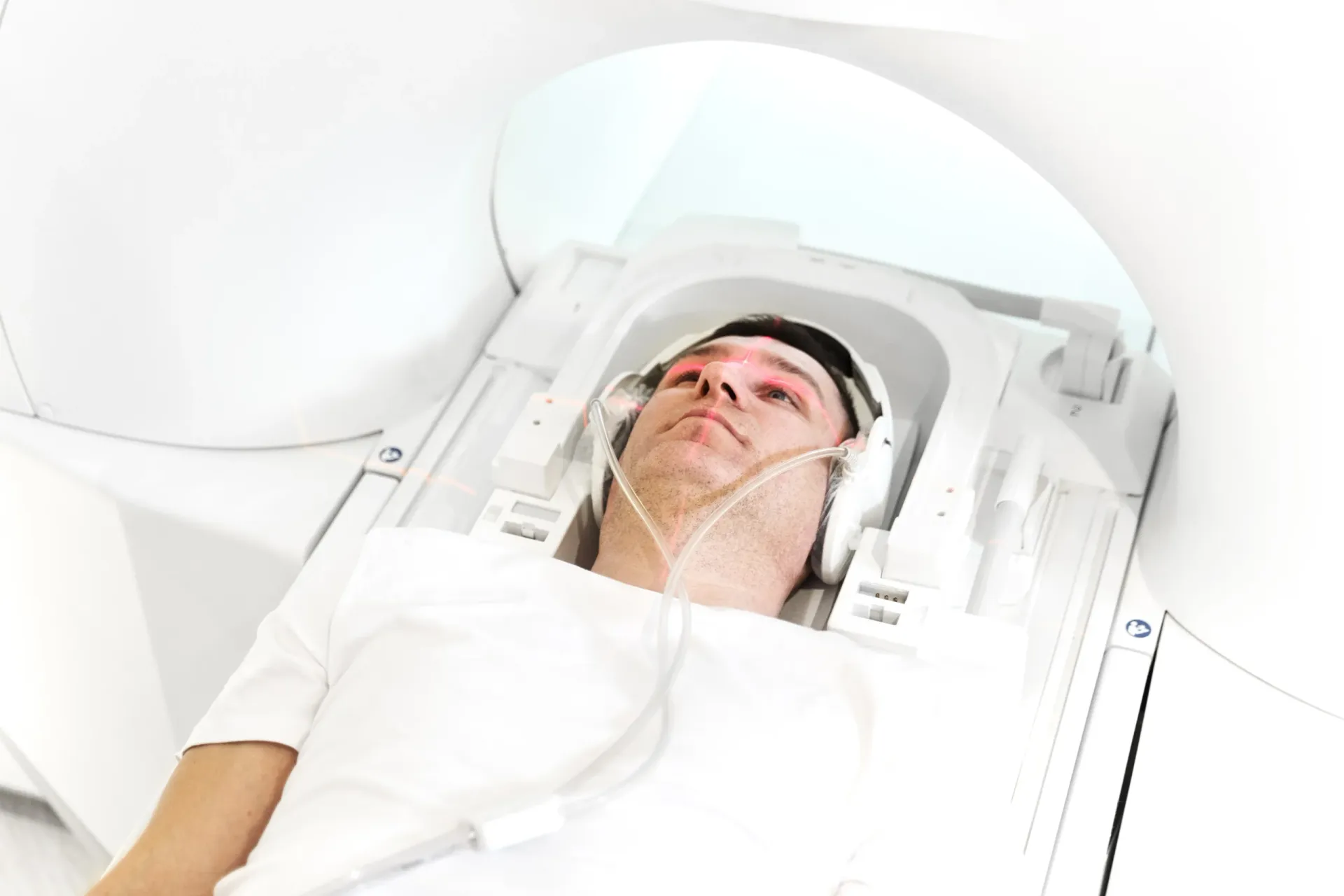
Brain MRI Dubai: Complete Guide & Cost (2026)
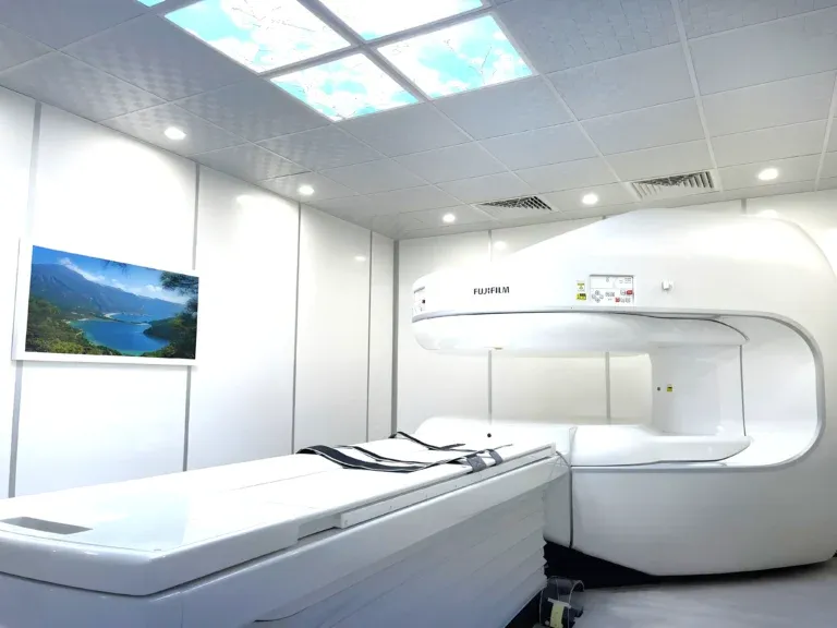
Complete Guide to MRI Scan in Dubai (2026)
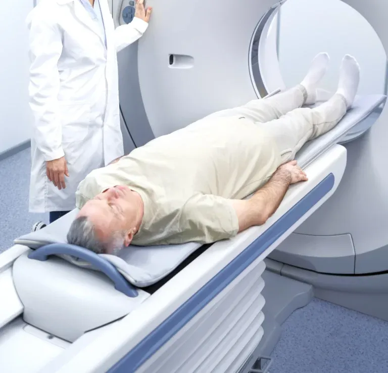
MRI Scan Preparation Dubai: What to Know (2026)
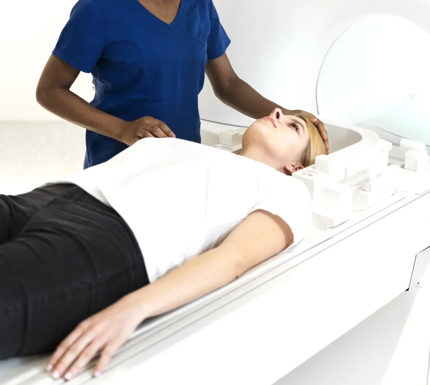
Spine MRI Dubai: Cost, Uses & When You Need One (2026)
More in Diagnostic Imaging

X-Ray Scan Dubai: Complete Guide (2026)
Читать далееMRI vs Ultrasound Dubai: Which Scan? (2026)
Читать далее
MRI vs X-Ray Dubai: Which Scan Do You Need? (2026)
Читать далее
Kidney Ultrasound vs CT Dubai: Which Test? (2026)
Читать далееMRI Preparation Dubai: Complete Guide (2026)
Читать далее
Penile Doppler Dubai: ED Diagnosis Guide (2026)
Читать далее© 2026 Doctors Clinic Diagnostic Center (DCDC), Dubai Healthcare City. Originally published at https://doctorsclinicdubai.ae/blog/neck-mri-dubai. All rights reserved. Unauthorized reproduction is prohibited.