Mga Pangunahing Punto
- Ang abdominal ultrasound ay isang ligtas, walang sakit, at walang radiation na imaging exam na gumagamit ng high-frequency na sound waves upang makagawa ng real-time na mga larawan ng mga organ sa loob ng tiyan, kabilang ang atay, apdo, bato, pancreas, at pali
- Ang eksaminasyon ay nakakakita ng gallstones, kidney stones, sakit sa atay, abdominal aortic aneurysms, mga tumor, mga cyst, mga koleksyon ng likido, at malawak na hanay ng iba pang kondisyon na nakakaapekto sa mga organ ng tiyan
- Ang paghahanda ay karaniwang nangangailangan ng pag-aayuno ng 8 hanggang 12 oras bago ang scan upang manatiling distended ang apdo at ang mga larawan ay hindi matakpan ng intestinal gas mula sa pagtunaw
- Ang prosedyur ay tumatagal ng 20 hanggang 30 minuto, walang kasamang mga karayom o contrast dye, at ang mga resulta ay karaniwang available sa parehong araw o sa loob ng 24 oras sa DCDC sa Dubai Healthcare City
- Ang abdominal ultrasound ay kadalasang ang unang imaging test na ini-order para sa mga pasyenteng may pananakit ng tiyan, abnormal na mga blood work, o pinaghihinalaang sakit sa organ dahil ito ay mabilis, abot-kaya, at lubos na nagbibigay-impormasyon
Ang abdominal ultrasound ay isang non-invasive na diagnostic imaging examination na gumagamit ng high-frequency na sound waves upang lumikha ng real-time na mga larawan ng mga organ at istruktura sa loob ng tiyan. Hindi tulad ng CT scans o X-rays, ang ultrasound ay hindi gumagamit ng ionizing radiation, na ginagawa itong isa sa mga pinakaligtas na imaging modalities na available para sa mga pasyente sa lahat ng edad, kabilang ang mga buntis na babae at mga bata. Ang eksaminasyon ay nagbibigay ng detalyadong visualization ng atay, apdo, bile ducts, pancreas, pali, mga bato, abdominal aorta, at nakapalibot na mga tisyu, na nagpapahintulot sa mga doktor na mag-diagnose ng malawak na spectrum ng mga kondisyon mula sa gallstones at kidney stones hanggang sa liver cirrhosis at abdominal masses.
Ang komprehensibong gabay na ito ay nagpapaliwanag kung ano ang abdominal ultrasound, ano ang kayang ipakita at makita nito, sino ang nangangailangan nito at bakit, paano maghanda para sa eksaminasyon, ano ang nangyayari sa panahon ng prosedyur, paano maunawaan ang iyong mga resulta, at magkano ang gastos ng abdominal ultrasound sa Dubai. Kung ang iyong doktor ay nag-order ng abdominal scan o kung isinasaalang-alang mo ito para sa hindi maipaliwanag na mga sintomas, sinasaklaw ng artikulong ito ang lahat ng kailangan mong malaman bago ang iyong appointment sa Doctors Clinic Diagnostic Center (DCDC) sa Dubai Healthcare City.


Ano ang Abdominal Ultrasound?
Ang abdominal ultrasound ay isang medical imaging procedure na gumagamit ng isang handheld na aparato na tinatawag na transducer upang magpadala ng high-frequency na sound waves (karaniwang 2 hanggang 5 megahertz) sa katawan. Ang mga sound waves na ito ay tumatalbog sa mga internal na organ, tisyu, at fluid-filled na mga istruktura, at ang mga bumabalik na echoes ay kinukuha ng transducer at kino-convert ng computer sa detalyadong grayscale na mga larawan na ipinapakita sa monitor nang real-time. Ang teknik ay kilala rin bilang abdominal sonography o abdominal scan.
Ang pisika sa likod ng ultrasound imaging ay diretso: ang iba't ibang mga tisyu ay nagre-reflect ng sound waves nang iba-iba batay sa kanilang density at komposisyon. Ang solid na mga organ tulad ng atay ay gumagawa ng moderate na mga echoes at lumilitaw sa iba't ibang shade ng gray, ang fluid-filled na mga istruktura tulad ng apdo at pantog ay lumilitaw na itim (anechoic), at ang matataas na density na istruktura tulad ng gallstones ay gumagawa ng maliwanag na puting echoes na may katangiang acoustic shadows sa likod nila. Ang pagkakaiba na ito sa pagitan ng mga uri ng tisyu ang nagpapahintulot sa sonographer at radiologist na matukoy ang normal na anatomy at makita ang mga abnormalidad na may kapansin-pansing katumpakan.
May dalawang pangunahing uri ng abdominal ultrasound. Ang kumpletong abdominal ultrasound ay sinusuri ang lahat ng pangunahing organ ng tiyan kabilang ang atay, apdo, bile ducts, pancreas, pali, mga bato, at abdominal aorta. Ang limitado o nakatutok na abdominal ultrasound ay nagta-target sa isang partikular na organ o lugar, tulad ng right upper quadrant ultrasound na nakatutok sa apdo o renal ultrasound na nakatutok sa mga bato. Ang iyong doktor ang magtutukoy kung aling uri ang kailangan batay sa iyong mga sintomas at klinikal na tanong.
"Ang abdominal ultrasound ang workhorse ng diagnostic imaging," sabi ni Dr. Osama Elzamzami, Head of Radiology sa DCDC. "Ito ang unang test na kinukuha namin sa napakaraming kaso ng abdominal complaints dahil ito ay mabilis, ligtas, walang radiation, at nagbibigay sa amin ng napakalaking dami ng diagnostic na impormasyon nang walang anumang diskomfort sa pasyente."
Dahil ang ultrasound ay gumagawa ng mga larawan nang real-time, ang radiologist ay maaaring obserbahan ang mga organ habang gumagalaw sa paghinga, panoorin ang daloy ng dugo sa mga daluyan ng dugo gamit ang Doppler ultrasound, at dynamic na i-adjust ang eksaminasyon upang tumuon sa mga lugar ng alalahanin. Ang real-time na kakayahan na ito ay isang makabuluhang kalamangan sa mga static na imaging modalities tulad ng CT at MRI, dahil pinapahintulutan nito ang examiner na i-tailor ang scan sa partikular na klinikal na sitwasyon ng bawat pasyente.
Ano ang Ipinapakita ng Abdominal Ultrasound?
Ang abdominal ultrasound ay nagbibigay ng detalyadong imaging ng maramihang organ system sa loob ng tiyan. Sinusuri ng eksaminasyon ang laki, hugis, texture, at daloy ng dugo ng bawat organ, at maaari itong magtukoy ng malawak na hanay ng structural na abnormalidad, sakit, at acute na kondisyon. Ang sumusunod na talahanayan ay nagbubuod ng mga pangunahing organ na sinusuri at ang mga kondisyon na maaaring makita sa bawat isa.
| Organ na Sinusuri | Mga Kondisyong Nakikita |
|---|---|
| Atay | Fatty liver disease, hepatitis, cirrhosis, liver cysts, hemangiomas, liver tumors (benign at malignant), liver abscess, portal hypertension, hepatomegaly |
| Apdo | Gallstones, gallbladder polyps, acute at chronic cholecystitis, pagkapal ng pader ng apdo, biliary sludge, gallbladder tumors |
| Bile Ducts | Pagpapalaki ng bile duct (obstruksyon), common bile duct stones (choledocholithiasis), biliary strictures |
| Pancreas | Pancreatitis (acute at chronic), pancreatic cysts, pancreatic pseudocysts, pancreatic tumors, pancreatic duct dilation |
| Pali | Splenomegaly (pinalaking pali), splenic cysts, splenic infarction, accessory spleen |
| Mga Bato | Kidney stones, hydronephrosis (namamagang bato), renal cysts, kidney tumors, renal artery stenosis, kidney infection (pyelonephritis), kidney size asymmetry |
| Abdominal Aorta | Abdominal aortic aneurysm (AAA), aortic dissection, aortic calcification |
| Pantog (kung kasama) | Bladder stones, pagkapal ng pader ng pantog, post-void residual volume, bladder tumors |
| Peritoneal Cavity | Ascites (libreng likido), lymphadenopathy (pinalaking lymph nodes), peritoneal masses |
Mga organ na sinusuri sa panahon ng kumpletong abdominal ultrasound at ang mga kondisyon na maaaring makita ng bawat assessment.
Bukod sa pagtukoy ng structural na mga abnormalidad, ang abdominal ultrasound na may Doppler imaging ay sinusuri ang daloy ng dugo sa mga pangunahing daluyan kabilang ang portal vein, hepatic veins, renal arteries, at abdominal aorta. Ito ay partikular na mahalaga para sa pagsusuri ng liver cirrhosis na may portal hypertension, renal artery stenosis, deep vein thrombosis, at vascular na mga abnormalidad. Ang Color Doppler at spectral Doppler ay nagbibigay ng parehong visual at quantitative na impormasyon tungkol sa direksyon, bilis, at pattern ng daloy ng dugo.
Mahalagang tandaan na ang ultrasound ay may mga limitasyon. Ang bowel gas ay maaaring makatakip sa pancreas at retroperitoneal na mga istruktura sa ilang pasyente, at ang napakalalim na mga istruktura ay maaaring mahirap makita sa mga pasyenteng may mas malaking katawan. Sa mga ganitong kaso, ang CT scan o MRI ay maaaring irekomenda bilang complementary na pag-aaral. Gayunpaman, para sa paunang pagsusuri ng karamihan ng mga sintomas ng tiyan, ang ultrasound ay nananatiling pinakaangkop at cost-effective na first-line na pagsisiyasat.
Sino ang Nangangailangan ng Abdominal Ultrasound?
Ang mga doktor ay nag-oorder ng abdominal ultrasounds para sa malawak na hanay ng klinikal na indikasyon. Ang eksaminasyon ay kadalasang ang unang imaging study na hinihiling kapag ang pasyente ay nagpresenta ng mga sintomas sa tiyan o kapag ang mga blood tests ay nagpapahiwatig ng abnormalidad sa isa sa mga organ ng tiyan. Ang mga sumusunod ay ang mga pinakakaraniwang dahilan kung bakit ang doktor ay maaaring magrekomenda ng abdominal ultrasound.
Pananakit ng Tiyan
Ang pananakit ng tiyan ang pinakakaraniwang dahilan para sa pag-order ng abdominal ultrasound. Ang lokasyon at katangian ng pananakit ay tumutulong sa doktor na matukoy kung aling mga organ ang pagtutuunan. Ang pananakit sa kanang itaas na bahagi ay madalas na tumuturo sa sakit sa apdo tulad ng gallstones o cholecystitis. Ang pananakit sa tagiliran ay maaaring magpahiwatig ng kidney stones o hydronephrosis. Ang epigastric na pananakit ay maaaring magpahiwatig ng pancreatitis o gastric na patolohiya. Ang diffuse na pananakit ng tiyan ay maaaring may kaugnayan sa ascites, bowel obstruction, o peritonitis. Tinutulungan ng ultrasound na paliitin ang differential diagnosis nang mabilis at gabayan ang mga susunod na hakbang sa paggamot.
Abnormal na Mga Blood Tests
Ang mataas na liver enzymes (ALT, AST, GGT, ALP), mataas na bilirubin levels, abnormal na kidney function tests (creatinine, BUN), mataas na amylase o lipase, at abnormal na tumor markers tulad ng AFP (alpha-fetoprotein) o CA 19-9 ay lahat nag-uudyok sa mga doktor na mag-order ng abdominal ultrasound. Tinutulungan ng imaging na matukoy ang structural na dahilan sa likod ng abnormal na laboratory values, maging ito man ay fatty liver, bile duct obstruction, kidney mass, o pancreatic inflammation.
Screening at Surveillance
Ang abdominal ultrasound ay ginagamit bilang screening tool sa ilang mahalagang klinikal na senaryo. Ang mga lalaking higit sa 65 taong gulang na nakapanigarilyo kailanman ay inirerekomendahan na magkaroon ng one-time screening ultrasound para sa abdominal aortic aneurysm (AAA), isang potensyal na banta sa buhay na kondisyon na madalas na walang sintomas hanggang sa pumutok. Ang mga pasyenteng may chronic hepatitis B o C o liver cirrhosis ay sumasailalim sa regular na anim-na-buwan na ultrasound surveillance para sa hepatocellular carcinoma (kanser sa atay). Ang mga pasyenteng may kilalang kidney cysts, liver hemangiomas, o iba pang benign na lesions ay maaaring magkaroon ng periodic na follow-up ultrasounds upang masubaybayan ang mga pagbabago sa laki.
Hindi Maipaliwanag na Mga Sintomas
Ang hindi maipaliwanag na pagbaba ng timbang, jaundice (paninilaw ng balat at mga mata), patuloy na pagduduwal at pagsusuka, bloating, nararamdamang masa sa tiyan, o dugo sa ihi (hematuria) ay lahat ng mga sintomas na nangangailangan ng abdominal ultrasound. Ang mga sintomas na ito ay maaaring dulot ng iba't ibang kondisyon, mula sa benign na gallstones hanggang sa malubhang malignancies, at ang ultrasound ay nagbibigay ng mabilis, non-invasive na paraan upang simulan ang diagnostic na workup.
Kwento ng Pasyente: Pag-diagnose ng Hindi Inaasahang Gallstones
Isang 38-taong-gulang na babae ang pumunta sa DCDC matapos maranasan ang paulit-ulit na matinding pananakit sa itaas ng kanyang tiyan sa loob ng ilang linggo, lalo na pagkatapos kumain ng matatabang pagkain. Sa simula ay inisip niya na ito ay indigestion at ginamot niya ito ng over-the-counter na antacids. Nang ang mga episode ay naging mas madalas at matindi, na kumakalat sa kanyang kanang balikat, ang kanyang general practitioner ay nag-order ng abdominal ultrasound.
Ang ultrasound ay nagpakita ng maramihang gallstones na may laki mula 5 hanggang 12 milimetro sa loob ng kanyang apdo, kasama ang banayad na pagkapal ng pader na pare-pareho sa chronic cholecystitis. Ang mga bile ducts ay normal sa caliber, na nag-rule out ng anumang stone migration. Sa tumpak na ultrasound findings, ang kanyang surgeon ay nakapagplano ng laparoscopic cholecystectomy (pagtatanggal ng apdo) nang may kumpiyansa.
"Ito ay isang klasikong presentasyon na regular naming nakikita," sabi ni Dr. Osama Elzamzami, Head of Radiology sa DCDC. "Ang gallstones ay isa sa mga pinakakaraniwang findings sa abdominal ultrasound, at ang kagandahan ng ultrasound ay nakakakita ito ng mga ito na may mahigit 95 porsyentong sensitivity. Isang limang-minutong scan ang nagbigay sa pasyenteng ito ng malinaw na diagnosis at direktang daan patungo sa paggamot, na iniiwasan ang mga linggo ng hindi kinakailangang mga pagsisiyasat."
Paano Maghanda para sa Abdominal Ultrasound
Ang tamang paghahanda para sa abdominal ultrasound ay mahalaga dahil direkta itong nakakaapekto sa kalidad ng mga larawan at katumpakan ng diagnosis. Ang paghahanda ay simple, ngunit ang maingat na pagsunod sa mga instruksyon ay gumagawa ng makabuluhang pagkakaiba sa kung ano ang makikita ng radiologist.
Mga Kinakailangan sa Pag-aayuno
Para sa isang kumpletong abdominal ultrasound, ang mga pasyente ay kinakailangang mag-ayuno ng 8 hanggang 12 oras bago ang eksaminasyon. Nangangahulugan ito na walang pagkain at walang inumin maliban sa maliliit na higop ng tubig para sa gamot. Ang pag-aayuno ay kinakailangan para sa dalawang dahilan. Una, pinapanatili nito ang apdo na distended ng bile, na ginagawang mas madaling makita ang mga gallstones, polyps, at wall abnormalities. Pagkatapos kumain, ang apdo ay kumokontrata at nagbabawas, nagiging maliit at mahirap suriin. Pangalawa, binabawasan ng pag-aayuno ang intestinal gas na nagagawa sa panahon ng pagtunaw, na maaaring makatakip sa pancreas at iba pang retroperitoneal na mga istruktura.
Kung ang iyong ultrasound ay naka-schedule sa umaga, ang pinakasimpleng paraan ay huminto sa pagkain at pag-inom pagkatapos ng hatinggabi ng gabi bago. Kung ang appointment ay sa hapon, maaari kang kumuha ng magaan na maagang almusal na hindi bababa sa 8 oras bago ang oras ng scan, pagkatapos ay mag-ayuno para sa natitirang panahon. Iwasan ang pagnguya ng gum at paninigarilyo bago ang eksaminasyon, dahil parehong nagpapasigla sa digestive system at nagpapataas ng intestinal gas.
Mga Eksepsiyon sa Pag-aayuno
Hindi lahat ng abdominal ultrasounds ay nangangailangan ng pag-aayuno. Ang renal ultrasound (mga bato lamang) o bladder ultrasound ay karaniwang hindi nangangailangan ng pag-aayuno, bagaman ang mga pasyente ay maaaring hilingang magkaroon ng punong pantog. Ang pelvic ultrasound ay nangangailangan ng punong pantog ngunit hindi ng pag-aayuno. Ang mga pasyenteng may diabetes na hindi maaaring ligtas na mag-ayuno nang mahabang panahon ay dapat ipaalam sa klinika kapag nagbu-book upang maayos ang maagang umaga na appointment upang mabawasan ang fasting window. Palaging sundin ang partikular na mga instruksyon sa paghahanda na ibinigay sa iyo kapag nagbu-book ng iyong appointment.
Gamot at Iba Pang Instruksyon
Maaari mong inumin ang iyong regular na mga gamot na may maliit na higop ng tubig sa umaga ng eksaminasyon maliban kung iba ang instruksyon ng iyong doktor. Magsuot ng maluwag, kumportableng damit na nagpapahintulot ng madaling access sa iyong tiyan, o maging handa na magpalit ng hospital gown. Dalhin ang iyong referral letter, anumang nakaraang imaging reports o CDs, at ang iyong Emirates ID o pasaporte para sa registration.
I-book ang Iyong Abdominal Ultrasound sa DCDC
Sa Doctors Clinic Diagnostic Center sa Dubai Healthcare City, ang aming radiology team ay nagbibigay ng expert na abdominal ultrasound examinations na may advanced equipment at same-day na mga resulta. Ang walk-ins ay tinatanggap, o mag-book nang maaga para sa garantisadong time slot.
Ano ang Nangyayari sa Panahon ng Eksaminasyon?
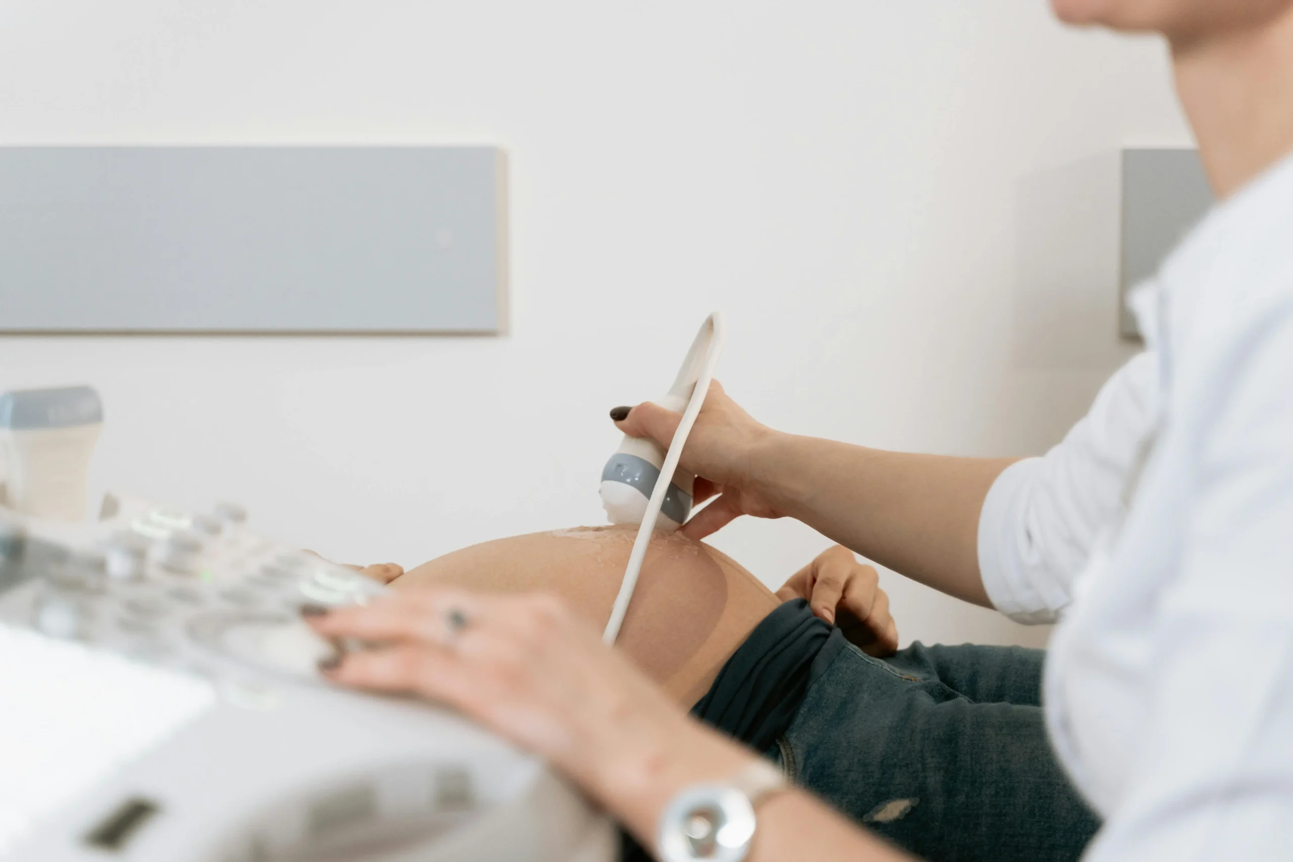
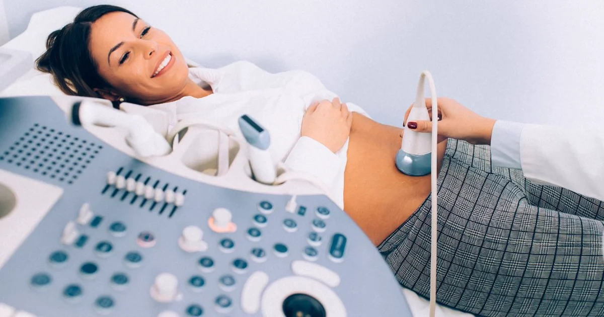
Ang abdominal ultrasound ay isang walang sakit, non-invasive na prosedyur na isinasagawa ng isang trained na sonographer o radiologist. Ang buong eksaminasyon ay karaniwang tumatagal ng 20 hanggang 30 minuto, depende sa mga organ na sinusuri at ang complexity ng mga findings. Narito ang isang step-by-step na overview ng kung ano ang aasahan.
Step-by-Step: Ang Abdominal Ultrasound Procedure
- Hakbang 1 - Check-in at paghahanda: Ire-register ka sa reception at hihilingin na kumpirmahin na nag-ayuno ka ayon sa instruksyon. Maaaring hilingin sa iyo na magpalit ng gown o basta itaas ang iyong damit upang ilantad ang tiyan mula sa ibabang dibdib hanggang sa pelvis.
- Hakbang 2 - Pagpoposisyon: Hihiga ka sa iyong likod (supine position) sa isang may padding na examination table. Ang kwarto ay bahagyang madilim upang malinaw na makita ng sonographer ang monitor. May ibibigay na unan para sa komport, at may ilalagay na tuwalya sa paligid ng iyong damit upang protektahan ito mula sa ultrasound gel.
- Hakbang 3 - Paglalagay ng gel: Isang mainit, water-based na gel ang ilalagay sa balat ng iyong tiyan. Inaalis ng gel na ito ang hangin sa pagitan ng transducer at ng iyong balat, tinitiyak na ang sound waves ay maayos na naita-transmit sa katawan. Ang gel ay hindi nagdadagdag ng mantsa at madaling matatanggal pagkatapos ng eksaminasyon.
- Hakbang 4 - Scanning: Inilalagay ng sonographer ang transducer sa iyong tiyan at sistematikong ginagalaw ito sa iba't ibang rehiyon, nag-a-apply ng mahinang pressure. Hihilingin sa iyo na huminga nang malalim at hawakan ang iyong hininga sa iba't ibang punto, dahil ito ay nagtutulak ng atay at pali pababa sa ilalim ng rib cage para sa mas magandang visualization. Maaari ka ring hilingan na gumulong sa iyong kaliwa o kanang gilid upang ma-optimize ang mga view ng ilang organ, lalo na ang mga bato.
- Hakbang 5 - Doppler assessment: Kung kinakailangan ang blood flow evaluation, ia-activate ng sonographer ang Doppler function. Maaari kang makarinig ng pulsating o whooshing na tunog mula sa makina, na kumakatawan sa tunog ng dugo na dumadaloy sa iyong mga daluyan ng dugo. Ito ay ganap na normal at bahagi ng routine na eksaminasyon.
- Hakbang 6 - Pagtatapos at paglilinis: Kapag lahat ng kinakailangang mga larawan ay nakuha na, pupunasan ng sonographer ang gel mula sa iyong tiyan gamit ang tuwalya. Maaari kang magbihis at umalis agad. Walang recovery time, walang side effects, at walang mga paghihigpit sa iyong mga aktibidad pagkatapos ng scan.
- Hakbang 7 - Reporting: Isang consultant radiologist ang nagsusuri ng lahat ng nakuhang mga larawan at mga sukat at naghahanda ng detalyadong nakasulat na ulat. Sa DCDC, ang mga resulta ay karaniwang available sa parehong araw o sa loob ng 24 oras at ibinahagi nang digital sa iyong referring physician.
Sa panahon ng eksaminasyon, maaari kang makaramdam ng banayad na pressure habang ang transducer ay pinapindot laban sa iyong tiyan, ngunit ang prosedyur ay hindi dapat magdulot ng pananakit. Kung nakakaranas ka ng tenderness kapag ang transducer ay inilagay sa isang partikular na lugar, mahalagang sabihin sa sonographer, dahil ang point tenderness ay maaaring maging isang diagnostic na finding sa sarili nito, tulad ng isang positive na sonographic Murphy sign sa acute cholecystitis.
"Palagi naming ipinapaliwanag ang bawat hakbang sa aming mga pasyente bago at sa panahon ng eksaminasyon," sabi ni Dr. Osama Elzamzami, Head of Radiology sa DCDC. "Ang isang may kaalamang pasyente ay isang nakarelaks na pasyente, at ang relaxation ay talagang nagpapabuti ng image quality dahil ang mga tensed na abdominal muscles ay maaaring gawing mas mahirap makita ang mas malalim na mga organ."
Pag-unawa sa Iyong mga Resulta ng Abdominal Ultrasound
Pagkatapos ng iyong abdominal ultrasound, ang consultant radiologist ay nagsusuri ng lahat ng nakuhang mga larawan at naghahanda ng pormal na ulat na ipinapadala sa iyong referring physician. Ang pag-unawa sa mga pangunahing termino at findings sa iyong ulat ay tumutulong sa iyo na magkaroon ng mas may kaalamang pag-uusap sa iyong doktor tungkol sa mga susunod na hakbang. Nasa ibaba ang paliwanag kung ano ang karaniwang kasama sa mga resulta at kung ano ang ibig sabihin ng mga karaniwang findings.
Mga Normal na Findings
Ang normal na abdominal ultrasound report ay karaniwang naglalarawan ng bawat organ bilang normal sa laki, hugis, at echotexture (ang pattern ng mga echoes na nagpapakita ng internal na istruktura ng organ). Halimbawa, ang normal na atay ay inilalarawan bilang may homogeneous na echotexture na may makinis na mga gilid at walang focal na lesions. Ang normal na apdo ay manipis ang pader, walang bato, at maayos na distended pagkatapos mag-ayuno. Ang normal na mga bato ay sumusukat ng humigit-kumulang 9 hanggang 12 sentimetro ang haba, may makinis na cortex, at walang hydronephrosis o calculi. Ang ganap na normal na ultrasound report ay nagbibigay ng matibay na katiyakan na ang mga pangunahing organ ng tiyan ay structurally na malusog.
Mga Karaniwang Abnormal na Findings
Maraming ultrasound findings ang karaniwan at madalas na benign. Ang fatty liver (hepatic steatosis) ay isa sa mga pinakakaraniwang findings at lumilitaw bilang increased na echogenicity (liwanag) ng atay kumpara sa kidney cortex. Ang simpleng kidney cysts at liver cysts ay napaka-karaniwan, lalo na sa pagtanda, at halos palaging benign at hindi nangangailangan ng paggamot. Ang gallstones ay lumilitaw bilang maliwanag na echogenic foci sa loob ng apdo na may posterior acoustic shadowing at matatagpuan sa humigit-kumulang 10 hanggang 15 porsyento ng populasyon ng mga adulto. Ang liver hemangiomas ay benign na vascular tumors na lumilitaw bilang well-defined na echogenic lesions at ang pinakakaraniwang benign na liver tumor.
Mga Findings na Nangangailangan ng Follow-Up
Ang ilang ultrasound findings ay nangangailangan ng karagdagang imaging o klinikal na follow-up. Ang dilated na common bile duct ay maaaring magpahiwatig ng obstructing na bato o tumor at madalas na nag-uudyok ng MRCP (magnetic resonance cholangiopancreatography) para sa karagdagang pagsusuri. Ang hydronephrosis (dilation ng kidney collecting system) ay nagpapahiwatig ng downstream na obstruksyon na kailangang matukoy at magamot. Ang abdominal aortic aneurysm na sumusukat ng 5.5 sentimetro o higit pa ay karaniwang tinutukoy para sa surgical evaluation. Ang solid na mga masa sa atay, bato, o pancreas ay nangangailangan ng karagdagang characterization gamit ang contrast-enhanced CT o MRI upang matukoy kung benign o malignant ang mga ito.
Ipapaliwanag ng iyong referring physician ang iyong partikular na mga resulta at irerekomenda ang anumang kinakailangang follow-up na mga pagsisiyasat o paggamot. Kung natanggap mo ang iyong ultrasound report bago mo makita ang iyong doktor, iwasan ang self-diagnosing batay sa mga indibidwal na termino sa ulat. Ang konklusyon ng radiologist at ang klinikal na interpretasyon ng iyong doktor ang magkasamang nagbibigay ng kumpletong larawan.
Gastos ng Abdominal Ultrasound sa Dubai
Ang gastos ng abdominal ultrasound sa Dubai ay nag-iiba depende sa uri ng eksaminasyon (kumpleto versus nakatutok), ang pasilidad, at kung may insurance coverage. Nasa ibaba ang isang pangkalahatang-ideya ng pricing landscape upang matulungan kang magplano.
Ang isang kumpletong abdominal ultrasound sa Dubai ay karaniwang nagkakahalaga ng AED 400 hanggang AED 1,200 depende sa healthcare facility. Ang hospital-based na radiology departments ay karaniwang nagcha-charge sa mas mataas na dulo ng range na ito, habang ang standalone na diagnostic centers at clinics ay may tendensyang mag-alok ng mas competitive na presyo. Ang isang nakatutok o limitadong ultrasound na nagta-target sa isang organ (tulad ng renal ultrasound o gallbladder ultrasound) ay karaniwang mas mababang presyo, karaniwang sa pagitan ng AED 300 at AED 800.
Sa DCDC sa Dubai Healthcare City, ang abdominal ultrasound pricing ay kasama ang mismong scan, ang buong radiologist report, at digital image storage. Tinatanggap ng center ang karamihan sa mga pangunahing insurance plans sa UAE, at ang mga pasyenteng may insurance ay dapat kumpirmahin ang coverage at anumang kinakailangang pre-authorization sa kanilang provider bago ang appointment. Ang mga self-pay na pasyente ay nakikinabang mula sa transparent, competitive na presyo na walang mga nakatagong bayarin.
Maraming mga salik ang maaaring makaapekto sa kabuuang gastos ng iyong ultrasound visit. Kung ang Doppler assessment ay idinagdag upang suriin ang daloy ng dugo, maaaring may karagdagang bayarin. Kung ang radiologist ay magtukoy ng mga findings na nangangailangan ng karagdagang imaging, tulad ng CT scan o MRI, ang mga ito ay iko-quote nang hiwalay. Gayunpaman, sa maraming kaso, ang abdominal ultrasound lamang ang nagbibigay ng lahat ng diagnostic na impormasyon na kailangan, na ginagawa itong isa sa mga pinakamurang imaging investigations na available.
Para sa kasalukuyang presyo at mga katanungan sa insurance, makipag-ugnayan sa DCDC nang direkta sa pamamagitan ng telepono, WhatsApp, o sa pamamagitan ng online booking form. Maaari mo ring i-explore ang aming detalyadong gabay sa gastos ng ultrasound sa Dubai para sa mas malawak na paghahambing sa iba't ibang uri ng ultrasound examinations.
Kunin ang Iyong Abdominal Ultrasound sa DCDC Dubai
Ang Doctors Clinic Diagnostic Center ay nag-aalok ng komprehensibong abdominal ultrasound services na may mga karanasang radiologist, advanced na ultrasound technology, at same-day na mga resulta. Matatagpuan sa Dubai Healthcare City na may madaling access mula sa buong UAE.
O tumawag sa amin para sa agarang tulong
Kaugnay na Serbisyo sa DCDC
Dalubhasang pangangalaga at advanced diagnostics sa Dubai Healthcare City
Frequently Asked Questions
Mga Pangwakas na Kaisipan
Ang abdominal ultrasound ay isa sa mga pinakamahalagang diagnostic tools sa modernong medisina. Ito ay ligtas, walang sakit, walang radiation, malawakang available, at kayang mag-diagnose ng malawak na hanay ng mga kondisyon na nakakaapekto sa atay, apdo, mga bato, pancreas, pali, at mga pangunahing daluyan ng dugo. Para sa mga pasyenteng nakakaranas ng pananakit ng tiyan, abnormal na blood tests, o hindi maipaliwanag na mga sintomas, ang ultrasound ay halos palaging ang lohikal na unang hakbang sa diagnostic journey dahil nagbibigay ito ng kayamanan ng impormasyon nang mabilis at abot-kaya. Ang paghahanda ay minimal, ang prosedyur ay tumatagal ng wala pang 30 minuto, at ang mga resulta ay mabilis na available.
Sa Doctors Clinic Diagnostic Center sa Dubai Healthcare City, ang aming radiology team ay nagsasagawa ng daan-daang abdominal ultrasound examinations bawat buwan, pinagsasama ang advanced na ultrasound technology sa expertise ng mga karanasang consultant radiologist upang maghatid ng tumpak, napapanahong mga resulta na nagbibigay-kapangyarihan sa iyong treating physician na gumawa ng pinakamahusay na mga desisyon para sa iyong pangangalaga. Kung kailangan mo ng kumpletong abdominal survey o nakatutok na scan ng isang partikular na organ, ang DCDC ay nag-aalok ng propesyonal, patient-centered na karanasan na may same-day reporting at transparent na presyo. Upang matuto pa tungkol sa aming ultrasound services, bisitahin ang aming gabay sa ano ang ultrasound scan, o makipag-ugnayan sa amin upang i-book ang iyong appointment.
Mga Sanggunian at Reperensya
Ang artikulong ito ay sinuri ng aming medikal na team at tumutukoy sa mga sumusunod na sanggunian:
- American College of Radiology (ACR) - ACR Appropriateness Criteria: Right Upper Quadrant Pain
- RadiologyInfo.org - Abdominal Ultrasound
- American Institute of Ultrasound in Medicine (AIUM) - Practice Parameter for the Performance of an Ultrasound Examination of the Abdomen
- National Institute for Health and Care Excellence (NICE) - Gallstone Disease: Diagnosis and Management
Ang medikal na nilalaman sa site na ito ay sinusuri ng mga DHA-licensed na manggagamot. Tingnan ang aming patakarang editorial para sa higit pang impormasyon.

Isinulat ni
Dr. Osama Elzamzami
Diagnostic Radiology
MD, FRCR
Si Dr. Osama Elzamzami ang Head of Radiology sa DCDC, na espesyalista sa diagnostic imaging kabilang ang ultrasound, CT, MRI, at interventional radiology sa Doctors Clinic Diagnostic Center sa Dubai Healthcare City.
Mga Kaugnay na Artikulo
Ano ang Ultrasound Scan? Kumpletong Gabay
Mga Resulta ng Ultrasound na Ipinaliwanag: Paano Basahin ang Iyong Report

Ano ang Doppler Ultrasound?
More in Diagnostic Imaging

3D vs 2D Mammogram Dubai: Key Differences (2026)
Basahin Pa
X-Ray at Home Dubai: Cost & Guide (2026)
Basahin Pa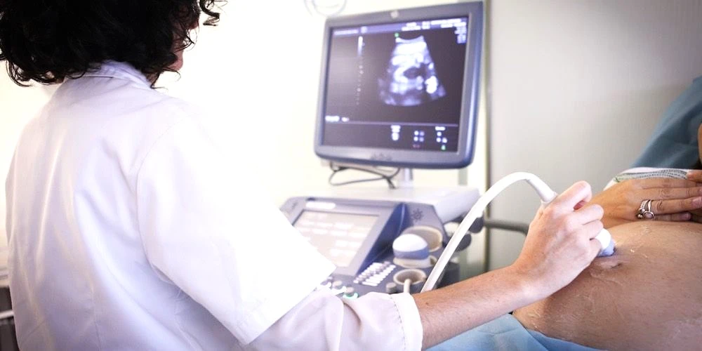
Ultrasound at Home Dubai: From AED 599 (2026)
Basahin Pa
Neck MRI Dubai: What It Shows & Cost (2026)
Basahin Pa
X-Ray Scan Dubai: Complete Guide (2026)
Basahin Pa
MRI vs Ultrasound Dubai: Which Scan? (2026)
Basahin Pa© 2026 Doctors Clinic Diagnostic Center (DCDC), Dubai Healthcare City. Originally published at https://doctorsclinicdubai.ae/blog/abdominal-ultrasound-complete-guide. All rights reserved. Unauthorized reproduction is prohibited.