Mga Pangunahing Punto
- An abdominal MRI in Dubai typically costs between AED 1,200 and AED 2,000, with specialized protocols like MRCP or liver-specific contrast studies at the higher end
- Abdominal MRI is the gold standard for characterizing liver lesions, detecting liver cancer (HCC) in cirrhotic patients, evaluating kidney masses, and imaging the bile ducts (MRCP) without invasive procedures
- MRI provides superior soft tissue contrast compared to CT scan for liver and pancreatic evaluation, and does not expose the patient to ionizing radiation
- Preparation typically involves fasting for 4 to 6 hours before the scan to reduce bowel activity and ensure the gallbladder is distended for optimal imaging
- The scan takes approximately 30 to 50 minutes and often includes contrast dye injection for detailed characterization of organ lesions
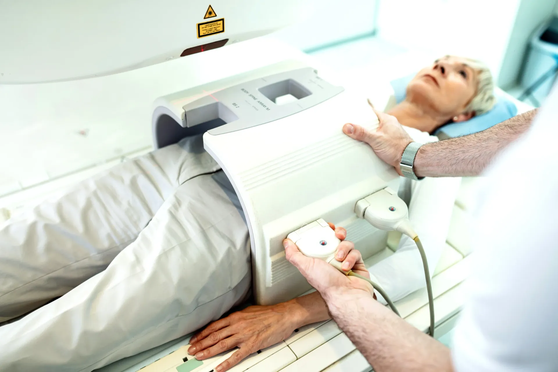
An abdominal MRI scan is the most detailed imaging tool for evaluating the solid and hollow organs of the abdomen, including the liver, kidneys, pancreas, spleen, adrenal glands, and bile ducts. While CT scan is often the first cross-sectional imaging test ordered for abdominal symptoms, MRI provides superior soft tissue contrast that is critical for characterizing liver lesions, detecting early-stage liver cancer, evaluating complex kidney masses, and imaging the biliary system. Whether your gastroenterologist, oncologist, or primary care physician has ordered an abdominal MRI, this comprehensive guide explains what the scan reveals, how much it costs in Dubai, and how to prepare for optimal results.


What Does an Abdominal MRI Show?
An abdominal MRI produces detailed images of all the abdominal organs using magnetic fields and radio waves instead of radiation. Its greatest strength is superior soft tissue contrast — the ability to distinguish between different types of tissue within organs, which is essential for characterizing masses and lesions. MRI is the preferred imaging modality when CT findings are inconclusive or when radiation-free imaging is needed.
The following organs and conditions are commonly evaluated with abdominal MRI:
Liver Imaging
- Liver lesion characterization: This is the most common reason for abdominal MRI. When an ultrasound or CT scan detects a liver lesion, MRI provides definitive characterization. Using specific contrast enhancement patterns across multiple phases (arterial, portal venous, delayed), MRI can distinguish between benign lesions (hemangiomas, focal nodular hyperplasia, adenomas) and malignant tumors (hepatocellular carcinoma, cholangiocarcinoma, metastases). Many liver lesions can be confidently diagnosed on MRI without biopsy.
- Hepatocellular carcinoma (HCC) screening: For patients with liver cirrhosis (regardless of cause — hepatitis B, hepatitis C, alcohol, fatty liver disease), MRI is one of the primary surveillance tools for early detection of hepatocellular carcinoma. The LI-RADS (Liver Imaging Reporting and Data System) classification standardizes reporting of liver observations in at-risk patients, using specific imaging features to categorize the probability of HCC.
- Liver cirrhosis and steatosis: MRI can detect and quantify liver fat content (steatosis) and assess the degree of liver fibrosis using specialized sequences. This is particularly relevant in the growing epidemic of non-alcoholic fatty liver disease (NAFLD) and non-alcoholic steatohepatitis (NASH), where MRI-based fat quantification provides objective measurements for monitoring disease progression and treatment response.
- Iron overload (hemochromatosis): MRI is the definitive non-invasive tool for detecting and quantifying liver iron overload, using specific signal characteristics to estimate iron concentration. This is essential for patients with hereditary hemochromatosis, thalassemia requiring repeated transfusions, and other conditions causing iron deposition.
Kidney Imaging
- Kidney mass characterization: When a kidney mass is found on ultrasound or CT and its nature is uncertain, MRI provides additional tissue characterization. It is particularly useful for distinguishing between solid tumors (renal cell carcinoma), fat-poor angiomyolipomas, complex cysts, and other benign lesions. The Bosniak classification system uses MRI features to categorize kidney cysts and guide management.
- Renal artery evaluation: MR angiography (MRA) can assess the renal arteries for stenosis (narrowing) without radiation, which is relevant for patients with resistant hypertension or declining kidney function suspected to have renal artery disease.
- Kidney infection and obstruction: MRI can evaluate complex kidney infections (abscess formation, pyonephrosis) and obstruction of the urinary tract, particularly when CT is contraindicated due to contrast allergy or pregnancy.
Biliary System (MRCP)
- MRCP (Magnetic Resonance Cholangiopancreatography): MRCP is a specialized MRI technique that produces detailed images of the bile ducts, gallbladder, and pancreatic duct without contrast injection or invasive procedures. It has largely replaced diagnostic ERCP (endoscopic retrograde cholangiopancreatography) for evaluating bile duct stones, strictures, and congenital biliary anomalies. MRCP is also valuable for evaluating primary sclerosing cholangitis (PSC) and bile duct tumors (cholangiocarcinoma).
Pancreas, Spleen & Adrenal Glands
- Pancreatic evaluation: MRI provides excellent visualization of the pancreas and is particularly useful for detecting and characterizing pancreatic cystic lesions (IPMN, mucinous cystic neoplasms, serous cystadenomas), which are increasingly found incidentally and require careful monitoring. MRI combined with MRCP maps the relationship between cystic lesions and the pancreatic duct, which influences management decisions.
- Adrenal gland lesions: Adrenal incidentalomas — adrenal masses found unexpectedly on imaging — are common. MRI using chemical shift imaging can distinguish between benign adrenal adenomas (which contain intracellular lipid) and potentially concerning lesions (metastases, pheochromocytomas) without biopsy in many cases.
- Splenic evaluation: MRI characterizes splenic lesions and assesses splenic size and signal characteristics. It can detect iron deposition (in hemolytic anemias), infarcts, abscesses, and tumors.
"Abdominal MRI is invaluable when we need to characterize a lesion that ultrasound or CT cannot fully define," explains Dr. Osama Elzamzami, Consultant Radiologist at DCDC. "For liver lesions in particular, MRI with liver-specific contrast agents allows us to make a confident diagnosis in the vast majority of cases without subjecting the patient to biopsy. MRCP has also transformed bile duct evaluation, replacing invasive diagnostic procedures with a completely non-invasive alternative."
How Much Does an Abdominal MRI Cost in Dubai?
The cost of an abdominal MRI in Dubai generally ranges from AED 1,200 to AED 2,000. Specialized protocols such as liver-specific contrast studies and MRCP are at the higher end of this range. At Doctors Clinic Diagnostic Center in Dubai Healthcare City, abdominal MRI pricing is all-inclusive — covering the scan, contrast agent if needed, a comprehensive radiologist report, and digital image copies.
| Abdominal MRI Type | Approximate Cost (AED) |
|---|---|
| Abdominal MRI without contrast | 1,200 – 1,600 |
| Abdominal MRI with contrast (gadolinium) | 1,500 – 2,000 |
| Liver MRI with hepatocyte-specific contrast | 1,800 – 2,500 |
| MRCP (bile duct imaging) | 1,200 – 1,800 |
| Abdominal + pelvic MRI combined | 2,200 – 3,000 |
Prices are approximate and include the radiologist report at DCDC. Contact us for exact pricing.
Contrast-enhanced abdominal MRI is used in the majority of cases because the enhancement pattern of lesions across different phases is essential for accurate diagnosis. Hepatocyte-specific contrast agents (such as gadoxetate disodium) are used for detailed liver evaluation and provide an additional hepatobiliary phase that improves the detection and characterization of liver lesions.
Most health insurance plans in the UAE cover abdominal MRI when ordered by a licensed physician with a documented clinical indication. DCDC assists patients with insurance pre-authorization to confirm coverage before the appointment.
Abdominal MRI vs CT Scan vs Ultrasound: Choosing the Right Test
Each imaging modality plays a specific role in abdominal evaluation. Understanding their relative strengths helps explain why your physician has chosen a particular test. For related information, see our guide on Ultrasound Cost Dubai: AED 300-800 by Type [2026 Prices].
| Factor | Ultrasound | CT Scan | MRI |
|---|---|---|---|
| Cost | AED 400 – 800 | AED 800 – 1,500 | AED 1,200 – 2,000 |
| Radiation | None | Moderate to high dose | None |
| Scan time | 15 – 30 minutes | 5 – 10 minutes | 30 – 50 minutes |
| Soft tissue contrast | Moderate | Good | Excellent (superior) |
| Liver lesion characterization | Good for initial detection | Good | Excellent (gold standard) |
| Bile duct imaging (MRCP) | Limited | Moderate | Excellent (replaces diagnostic ERCP) |
| Kidney mass characterization | Good (first-line) | Good | Excellent (problem-solving) |
| Pancreatic cyst evaluation | Limited | Good | Excellent |
| Liver fat/iron quantification | Qualitative only | Limited | Excellent (quantitative) |
| Best for | First-line screening, guided procedures | Emergency, bowel pathology, stones | Definitive lesion characterization, bile duct evaluation |
Ultrasound is the first-line abdominal imaging test. CT scan is fast and broadly available. MRI provides definitive characterization when other tests are inconclusive.
Ultrasound is the appropriate first-line test for most abdominal complaints — it is radiation-free, widely available, and excellent for detecting gallstones, liver lesions, kidney stones, and abdominal aortic aneurysms. CT scan is the primary tool for emergency evaluation (abdominal pain, trauma), bowel pathology, and situations where rapid comprehensive imaging is needed. MRI is the definitive problem-solving tool when CT or ultrasound findings are inconclusive, when superior soft tissue contrast is needed for lesion characterization, and when radiation-free imaging is preferred. In clinical practice, these modalities work together rather than competing.
How to Prepare for an Abdominal MRI
Proper preparation for an abdominal MRI improves image quality and diagnostic accuracy. The following preparation steps are standard:
- Fasting: Do not eat or drink for 4 to 6 hours before the scan. Fasting reduces bowel activity (peristalsis) that can blur images and ensures the gallbladder is distended for optimal visualization. Small sips of water for essential medication are usually permitted.
- Medication: Continue taking your regular medications unless specifically instructed otherwise by your physician. If you take diabetes medication (particularly metformin), inform the radiology team, as special precautions may be needed if contrast dye is used.
- Kidney function: If contrast dye is planned, a recent blood test for kidney function (creatinine/eGFR) is typically required. This is a standard safety measure because gadolinium contrast is processed by the kidneys.
- Metal screening: Complete the MRI safety questionnaire and remove all metal objects before entering the scan room.
- Arrive early: Plan to arrive 15 to 20 minutes before your appointment time to complete paperwork, change into a gown if needed, and allow time for any pre-scan preparation.
Book Your Abdominal MRI at DCDC
At Doctors Clinic Diagnostic Center in Dubai Healthcare City, we offer abdominal MRI scans including MRCP and liver-specific contrast protocols. Comprehensive radiologist reports are delivered within 24 to 48 hours.
Kaugnay na Serbisyo sa DCDC
Dalubhasang pangangalaga at advanced diagnostics sa Dubai Healthcare City
MRI Scan
High-resolution MRI imaging with expert radiologist interpretation
Mag-book ng AppointmentFull Body MRI
Comprehensive whole-body MRI screening for early detection of abnormalities
Mag-book ng AppointmentHealth Checkup
Combine MRI with full-panel blood work for complete health screening
Mag-book ng AppointmentFrequently Asked Questions
Final Thoughts
An abdominal MRI is the most detailed and versatile imaging tool for evaluating the liver, kidneys, pancreas, bile ducts, adrenal glands, and spleen. Its superior soft tissue contrast makes it the definitive test for characterizing organ lesions, staging liver disease, and evaluating the biliary system through MRCP. If your physician has ordered an abdominal MRI, it is because the clinical question requires the level of detail that only MRI can provide.
At Doctors Clinic Diagnostic Center in Dubai Healthcare City, we offer abdominal MRI with all relevant protocols including liver-specific contrast, MRCP, and multiphasic imaging. Our consultant radiologists deliver comprehensive reports using internationally recognized classification systems. Contact us to book your scan.
Mga Sanggunian at Reperensya
Ang artikulong ito ay sinuri ng aming medikal na team at tumutukoy sa mga sumusunod na sanggunian:
- American College of Radiology - LI-RADS (Liver Imaging Reporting)
- Radiological Society of North America - Abdominal MRI
- European Association for the Study of the Liver - HCC Surveillance
- Society of Abdominal Radiology - MRCP Guidelines
- Dubai Health Authority - Diagnostic Imaging Standards
Ang medikal na nilalaman sa site na ito ay sinusuri ng mga DHA-licensed na manggagamot. Tingnan ang aming patakarang editorial para sa higit pang impormasyon.

Isinulat ni
Dr. Osama Elzamzami
Diagnostic Radiology
MD, FRCR
Dr. Osama Elzamzami is a Consultant Radiologist specializing in diagnostic imaging including MRI, CT, and ultrasound at DCDC Dubai Healthcare City.
Related Articles
MRI Scan Dubai: Complete Guide to Types, Cost & Full Body MRI

Pelvic MRI Dubai: What It Detects, Cost & Preparation
Full Body MRI Cost Dubai: AED 5,000-15,000 (2026)
MRI With Contrast: What to Expect, Safety & When It Is Needed
Breast MRI Dubai: When You Need It Beyond Mammogram
More in Diagnostic Imaging
Neck MRI Dubai: What It Shows & Cost (2026)
Basahin Pa
X-Ray Scan Dubai: Complete Guide (2026)
Basahin Pa
MRI vs Ultrasound Dubai: Which Scan? (2026)
Basahin Pa
MRI vs X-Ray Dubai: Which Scan Do You Need? (2026)
Basahin Pa
Kidney Ultrasound vs CT Dubai: Which Test? (2026)
Basahin PaMRI Preparation Dubai: Complete Guide (2026)
Basahin Pa© 2026 Doctors Clinic Diagnostic Center (DCDC), Dubai Healthcare City. Originally published at https://doctorsclinicdubai.ae/blog/abdominal-mri-dubai. All rights reserved. Unauthorized reproduction is prohibited.