نکات کلیدی
- An ankle MRI in Dubai typically costs between AED 900 and AED 1,400 without contrast, making it an accessible diagnostic tool for persistent ankle problems
- Ankle MRI is the gold standard for evaluating ligament tears (ATFL, CFL), Achilles tendon injuries, osteochondral defects, stress fractures, and plantar fasciitis without radiation exposure
- X-ray can miss up to 40 percent of ankle injuries because it only shows bone, while MRI visualizes ligaments, tendons, cartilage, and bone marrow in a single examination
- Your doctor will typically order an ankle MRI after an ankle sprain that has not healed within 6 weeks, chronic ankle instability, persistent pain, or suspected Achilles tendon pathology
- The scan takes approximately 30 to 40 minutes, is completely painless, and requires no special preparation in most cases
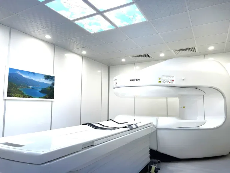
An ankle MRI scan is the most detailed imaging tool for evaluating the complex network of ligaments, tendons, bones, and cartilage that make up the ankle and foot. Ankle injuries are among the most common musculoskeletal problems worldwide, and while many resolve with rest and physiotherapy, persistent pain, instability, or swelling often indicates structural damage that requires precise diagnosis. Whether you are dealing with a stubborn ankle sprain that will not heal, an Achilles tendon injury, or chronic ankle pain that limits your daily activities, an MRI can reveal exactly what is happening beneath the surface. In Dubai, ankle MRI is widely available and provides the detailed information needed to guide treatment decisions, from conservative rehabilitation to surgical intervention.


What Does an Ankle MRI Show?
An ankle MRI produces detailed cross-sectional images of all the structures in and around the ankle joint, including the lateral and medial ligament complexes, Achilles tendon, peroneal tendons, tibialis posterior tendon, articular cartilage (talar dome), bone marrow, and plantar fascia. Unlike X-rays, which only visualize bone, MRI uses magnetic fields and radio waves to generate high-resolution images of soft tissues that are the primary site of most ankle injuries.
The following conditions are commonly identified on an ankle MRI:
- Lateral ligament tears (ATFL and CFL): The anterior talofibular ligament (ATFL) and calcaneofibular ligament (CFL) are the most commonly injured ligaments in the ankle, typically damaged during inversion ankle sprains. MRI can detect partial tears, complete ruptures, chronic ligament laxity, and scar tissue formation. The ATFL is torn in approximately 85 percent of ankle sprains, and MRI helps determine whether the injury will heal with rehabilitation alone or requires surgical reconstruction.
- Achilles tendon injuries: The Achilles tendon is the largest and strongest tendon in the body, yet it is vulnerable to tendinitis, partial tears, and complete ruptures. MRI provides detailed visualization of the tendon's internal structure, identifying areas of degeneration (tendinopathy), partial tears, complete ruptures, and insertional pathology. This information is critical for distinguishing between injuries that can be treated conservatively and those requiring surgical repair.
- Osteochondral defects (OCD): Osteochondral lesions of the talus are areas of damaged cartilage and underlying bone on the dome of the talus, often resulting from an ankle sprain or repetitive trauma. MRI is the definitive test for detecting OCDs, characterizing their size and depth, and determining whether the cartilage is intact, partially detached, or completely loose. This information guides treatment from conservative management to arthroscopic surgery.
- Stress fractures: Stress fractures of the ankle and foot bones (talus, calcaneus, metatarsals, navicular) are common in runners, military personnel, and individuals who have recently increased their activity level. MRI detects stress fractures weeks before they become visible on X-ray by revealing bone marrow edema — a telltale sign of microscopic bone damage.
- Plantar fasciitis: Plantar fasciitis is the most common cause of heel pain, resulting from inflammation and degeneration of the plantar fascia. MRI can confirm the diagnosis by showing fascia thickening (greater than 4 millimeters), increased signal intensity within the fascia, and associated calcaneal bone marrow edema. It also rules out other causes of heel pain such as stress fractures and nerve entrapment.
- Peroneal tendon pathology: The peroneal tendons run along the outer aspect of the ankle and are prone to tendinitis, tears, and subluxation (slipping out of their groove). MRI provides clear visualization of peroneal tendon integrity, tenosynovitis (inflammation of the tendon sheath), and the retinaculum that holds the tendons in place.
- Tibialis posterior tendon dysfunction: Progressive dysfunction of the tibialis posterior tendon is a common cause of acquired flat foot in adults. MRI detects tendinitis, partial tears, and complete ruptures, as well as associated changes in the spring ligament and sinus tarsi that indicate the stage of the condition.
- Ankle arthritis and synovitis: MRI reveals cartilage loss, bone spurs, synovial inflammation, and joint effusion associated with ankle osteoarthritis or inflammatory arthritis (such as rheumatoid arthritis). It provides a more complete assessment than X-ray, particularly for early-stage disease.
"The ankle is a deceptively complex joint, and many significant injuries are invisible on X-ray," explains Dr. Osama Elzamzami, Consultant Radiologist at DCDC. "I frequently see patients who have been told their X-ray is normal after an ankle sprain, yet the MRI reveals ligament tears, osteochondral defects, or bone marrow edema that explain their ongoing symptoms. MRI is the key to understanding why an ankle is not healing as expected."
How Much Does an Ankle MRI Cost in Dubai?
The cost of an ankle MRI in Dubai generally ranges from AED 900 to AED 1,400 for a standard non-contrast examination. The exact price depends on the imaging center, MRI machine specifications, and whether the radiologist report is included. At Doctors Clinic Diagnostic Center in Dubai Healthcare City, ankle MRI pricing is all-inclusive — covering the scan, a comprehensive radiologist report, and digital image copies.
| Ankle/Foot MRI Type | Approximate Cost (AED) |
|---|---|
| Ankle MRI without contrast | 900 – 1,200 |
| Ankle MRI with contrast (gadolinium) | 1,200 – 1,600 |
| Foot MRI without contrast | 900 – 1,200 |
| Both ankles (bilateral) | 1,600 – 2,200 |
| Ankle + foot combined MRI | 1,400 – 1,800 |
Prices are approximate and include the radiologist report at DCDC. Contact us for exact pricing.
Contrast-enhanced ankle MRI is occasionally requested when the referring physician suspects infection, tumors, or inflammatory conditions. For the vast majority of ankle injuries, sprains, and degenerative conditions, a standard non-contrast MRI provides all the diagnostic information needed.
Most health insurance plans in the UAE cover ankle MRI when ordered by a licensed physician with a documented clinical indication. DCDC assists patients with insurance pre-authorization to confirm coverage before the appointment.
When Does a Doctor Order an Ankle MRI?
An ankle MRI is typically ordered when a clinical examination and X-rays do not provide a complete diagnosis, or when specific soft tissue pathology is suspected. The following situations commonly lead to an ankle MRI referral: For related information, see our guide on ACL Tear MRI: How MRI Diagnoses Knee Ligaments.
- Ankle sprain that has not healed: While most ankle sprains heal within 4 to 6 weeks with proper rest and rehabilitation, approximately 20 to 40 percent of patients develop chronic symptoms. An MRI identifies the extent of ligament damage, associated injuries (osteochondral defects, peroneal tendon tears), and helps explain why recovery has stalled.
- Chronic ankle instability: Recurrent ankle sprains or a persistent feeling of the ankle giving way suggests ligament insufficiency. MRI confirms the degree of ligament damage and identifies any associated cartilage or tendon injuries that need to be addressed.
- Suspected Achilles tendon injury: A sudden pop or snap at the back of the ankle during sports, difficulty walking, or a palpable gap in the tendon warrants urgent MRI to determine whether the Achilles is partially or completely torn and to guide treatment decisions.
- Persistent heel pain: When heel pain does not respond to conservative measures for plantar fasciitis (stretching, orthotics, anti-inflammatory medication), MRI can confirm the diagnosis and rule out alternative causes such as stress fracture, nerve entrapment, or calcaneal bone pathology.
- Normal X-ray but ongoing pain: Many significant ankle injuries — ligament tears, osteochondral defects, stress fractures, tendon pathology — are invisible on X-ray. MRI is the essential next step when X-rays are normal but symptoms persist.
- Pre-surgical planning: Before ankle arthroscopy, ligament reconstruction, or Achilles tendon repair, surgeons require detailed MRI to plan the procedure.
- Sports-related foot and ankle pain: Runners, footballers, padel players, and other athletes with persistent foot or ankle pain benefit from MRI to identify stress fractures, tendon injuries, and other pathology that may not be apparent on clinical examination.
Book Your Ankle MRI at DCDC
At Doctors Clinic Diagnostic Center in Dubai Healthcare City, we offer same-week appointments for ankle and foot MRI scans with comprehensive radiologist reports delivered within 24 to 48 hours.
Ankle MRI vs X-Ray vs Ultrasound: Which Imaging Do You Need?
Understanding the strengths and limitations of each imaging modality helps explain why your doctor may recommend one test over another for ankle and foot problems.
| Factor | X-Ray | Ultrasound | MRI |
|---|---|---|---|
| Cost | AED 150 – 300 | AED 400 – 700 | AED 900 – 1,400 |
| Radiation | Low dose | None | None |
| Scan time | 5 minutes | 15 – 20 minutes | 30 – 40 minutes |
| Bone visualization | Excellent | Limited | Good |
| Ligament tears (ATFL, CFL) | Not visible | Moderate (operator-dependent) | Excellent (gold standard) |
| Achilles tendon | Not visible | Good | Excellent |
| Osteochondral defects | May show late changes | Very limited | Excellent |
| Stress fractures | Often missed early | Not visible | Highly sensitive |
| Plantar fascia | Not visible | Good | Excellent |
| Best for | Fractures, dislocations, arthritis | Achilles/tendon assessment, guided injections | Complete ankle/foot evaluation |
X-ray is the first-line test after acute ankle injury. MRI is indicated for persistent symptoms, suspected ligament tears, or when X-ray is normal but pain continues.
X-ray should always be the first imaging test after an acute ankle injury to rule out fractures. The Ottawa Ankle Rules — a well-validated clinical decision tool — help physicians determine which patients need X-rays after an ankle injury. However, X-ray is fundamentally limited to visualizing bone and cannot detect ligament tears, tendon injuries, or cartilage damage. Ultrasound can be helpful for assessing the Achilles tendon and guiding therapeutic injections, but it cannot adequately evaluate the deep ligaments, osteochondral surfaces, or bone marrow. MRI provides the most comprehensive assessment and is the test of choice when soft tissue pathology is suspected.
What to Expect During an Ankle MRI
An ankle MRI is one of the most comfortable MRI examinations because only your lower leg enters the machine. Here is what to expect:
- Before the scan: Complete a safety screening questionnaire. Remove all metal objects from your body. You can wear your normal clothing as long as it does not contain metal components near the ankle area.
- Positioning: You will lie on your back on the MRI table. Your foot and ankle will be placed in a dedicated ankle coil — a device that wraps around the ankle to optimize image quality. Your foot will be positioned in a neutral or slightly flexed position and gently secured.
- During the scan: The table slides into the machine feet-first, with only your lower legs entering the bore. Your head and upper body remain outside the machine, making this one of the least claustrophobia-inducing MRI scans. The machine produces knocking and humming sounds — earplugs or headphones are provided.
- Duration: A standard ankle MRI takes approximately 30 to 40 minutes. Remain as still as possible to ensure clear images.
- After the scan: No recovery period needed. Resume normal activities immediately. Your radiologist report will typically be available within 24 to 48 hours at DCDC.
Get Expert Ankle MRI Interpretation
At DCDC, every ankle MRI is interpreted by a consultant radiologist with musculoskeletal imaging expertise. Our detailed reports assess all ligaments, tendons, cartilage, and bone structures, giving your treating physician the precise information needed for treatment planning. Learn more about our MRI services.
خدمات مرتبط در DCDC
مراقبت تخصصی و تشخیص پیشرفته در شهر بهداشت دبی
Frequently Asked Questions
Final Thoughts
An ankle MRI is the most comprehensive imaging tool for evaluating the ligaments, tendons, cartilage, and bone of the ankle and foot. Whether you are dealing with a sprain that will not heal, an Achilles tendon injury, chronic instability, or unexplained foot pain, MRI reveals the precise diagnosis and guides effective treatment. Do not assume a normal X-ray means your ankle is fine — many significant injuries are only visible on MRI.
At Doctors Clinic Diagnostic Center in Dubai Healthcare City, we combine advanced MRI technology with experienced consultant radiologist interpretation to deliver thorough, reliable ankle and foot MRI reports. Contact us to book your scan or to discuss whether an ankle MRI is the right next step for your symptoms.
منابع و مراجع
این مقاله توسط تیم پزشکی ما بررسی شده و به منابع زیر ارجاع میدهد:
- American College of Radiology - Appropriateness Criteria for Ankle Injury
- Radiological Society of North America - Ankle MRI
- American Academy of Orthopaedic Surgeons - Ankle Sprains and Instability
- European Radiology - MRI Accuracy for Ankle Ligament Injuries
- Dubai Health Authority - Diagnostic Imaging Standards
محتوای پزشکی این سایت توسط پزشکان دارای مجوز DHA بررسی میشود. مشاهده سیاست تحریریه برای اطلاعات بیشتر.

نوشته شده توسط
Dr. Osama Elzamzami
Diagnostic Radiology
MD, FRCR
Dr. Osama Elzamzami is a Consultant Radiologist specializing in diagnostic imaging including MRI, CT, and ultrasound at DCDC Dubai Healthcare City.
Related Articles
MRI Scan Dubai: Complete Guide to Types, Cost & Full Body MRI

Knee MRI in Dubai: What It Shows, Cost & When You Need One
ACL Tear MRI: How MRI Diagnoses Ligament Injuries in Dubai
Hip MRI Dubai: Detect Labral Tears, AVN & Joint Problems

Full Body MRI Cost Dubai: AED 5,000-15,000 (2026)
More in Diagnostic Imaging

Neck MRI Dubai: What It Shows & Cost (2026)
بیشتر بخوانید
X-Ray Scan Dubai: Complete Guide (2026)
بیشتر بخوانید
MRI vs Ultrasound Dubai: Which Scan? (2026)
بیشتر بخوانید
MRI vs X-Ray Dubai: Which Scan Do You Need? (2026)
بیشتر بخوانید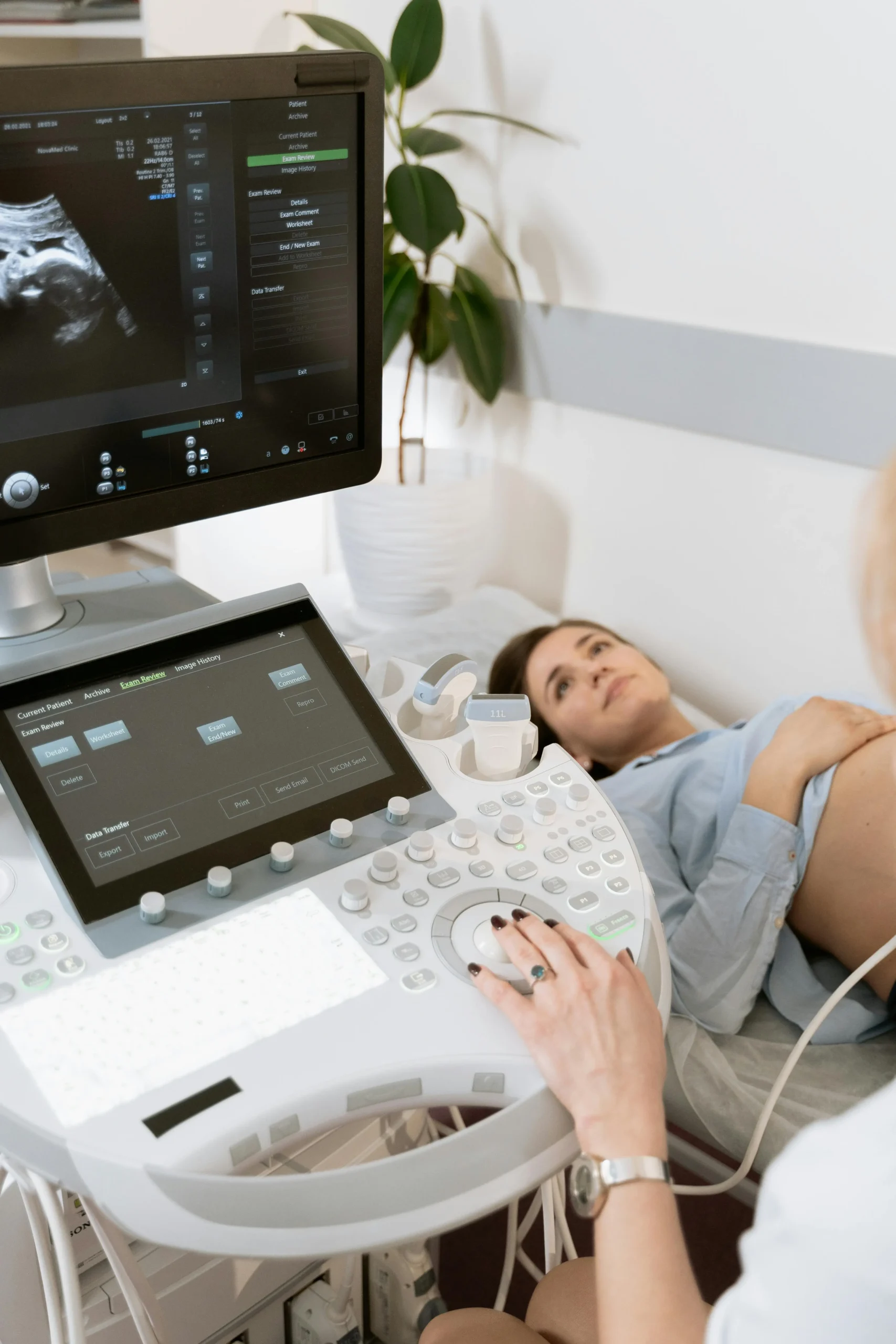
Kidney Ultrasound vs CT Dubai: Which Test? (2026)
بیشتر بخوانیدMRI Preparation Dubai: Complete Guide (2026)
بیشتر بخوانید© 2026 Doctors Clinic Diagnostic Center (DCDC), Dubai Healthcare City. Originally published at https://doctorsclinicdubai.ae/blog/ankle-mri-dubai. All rights reserved. Unauthorized reproduction is prohibited.