نکات کلیدی
- 3D mammography (tomosynthesis) increases breast cancer detection by up to 40% compared to standard 2D mammography.
- 3D mammograms reduce false-positive callbacks by approximately 15%, meaning fewer unnecessary follow-up tests.
- Women with dense breast tissue benefit most from 3D mammography because it separates overlapping tissue layers.
- DCDC offers both 2D and 3D digital mammography from AED 250 with subspecialty breast imaging radiologists.
- Breast cancer accounts for approximately 38% of all cancers in UAE women, making regular screening critical.
- The 5-year survival rate for breast cancer detected at an early stage exceeds 95%, underscoring the value of annual screening from age 40.
- 3D mammograms use only slightly more radiation than 2D and remain well within international safety limits.
- Breast density is assessed and reported with every mammogram at DCDC, helping guide follow-up imaging decisions.
Choosing between a 3D and a 2D mammogram can feel confusing, especially when both options are available at your clinic. The technology behind each type differs significantly, and those differences directly affect how well cancer can be detected and how often patients are called back for false alarms. At DCDC in Dubai Healthcare City, we offer both 2D and 3D digital mammography so that every woman receives the screening approach best suited to her breast tissue and risk profile.
This guide explains how each technology works, compares detection rates, breaks down costs in Dubai, and helps you decide which mammogram type is right for you. Whether you are scheduling your first screening or switching from 2D to 3D, the information below is designed to answer every question you may have.


3D vs 2D Mammogram: What Is the Difference?
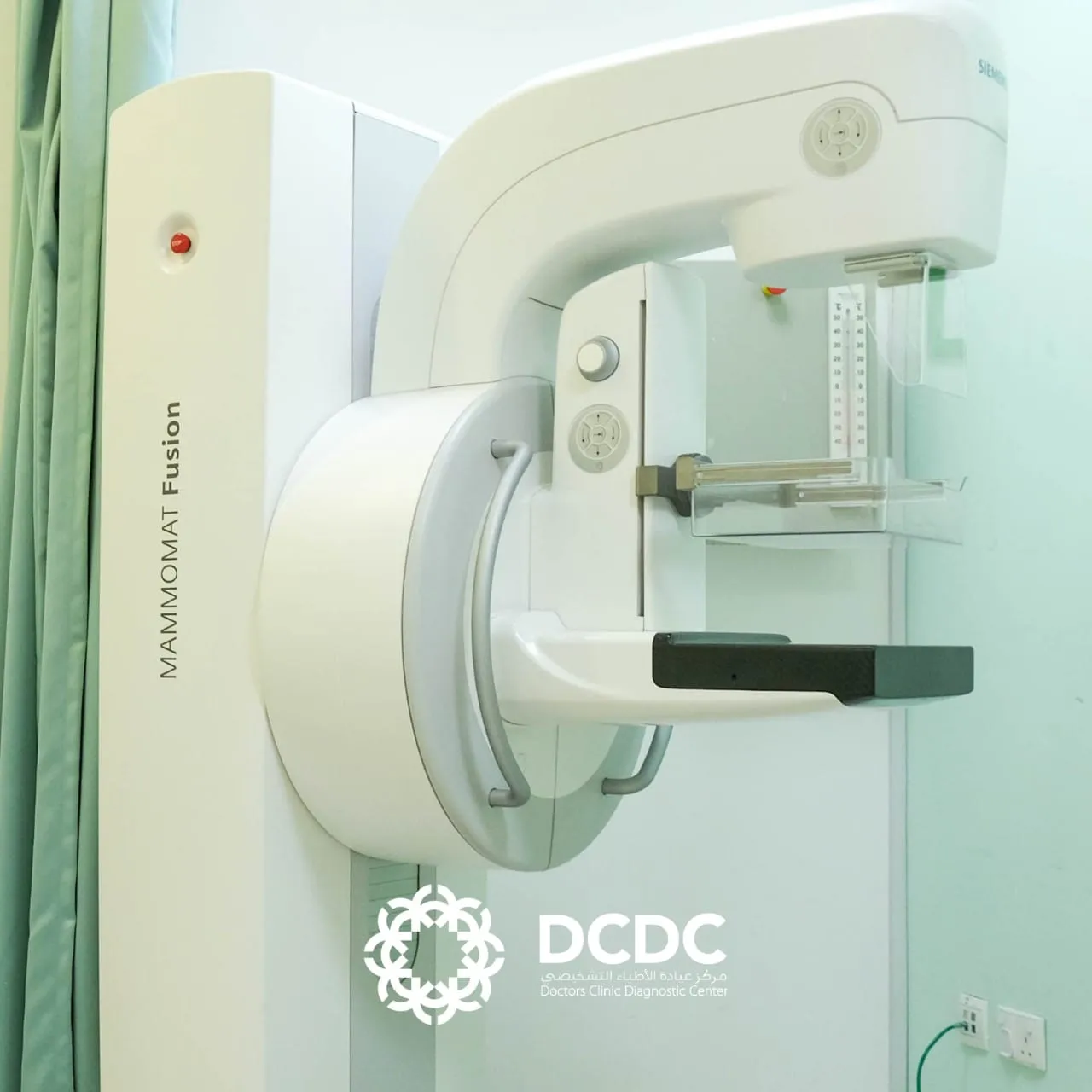
A 2D mammogram (conventional digital mammography) produces a single flat image of the breast from each angle, top-to-bottom and side-to-side. A 3D mammogram, formally called digital breast tomosynthesis (DBT), captures multiple thin-slice images as the X-ray arm sweeps in a short arc around the breast. The computer then reconstructs these slices into a three-dimensional picture that the radiologist can scroll through layer by layer.
The core difference lies in tissue separation. In a 2D image, all breast tissue is compressed into a single plane, which means overlapping structures can either hide a small cancer or mimic one that is not there. In a 3D mammogram, each slice isolates a thin section of tissue, making it easier to spot abnormalities and harder for normal tissue to create false alarms. This distinction has real clinical consequences: studies published in the Journal of the American Medical Association have shown that adding tomosynthesis to standard 2D mammography increases invasive cancer detection rates by 29 to 41 percent while reducing false-positive recall rates by about 15 percent.
How 2D Mammography Works
During a standard 2D mammogram, the breast is positioned on a flat plate and gently compressed by a second plate from above. A single X-ray exposure is taken from two directions for each breast: craniocaudal (top-to-bottom) and mediolateral oblique (angled side view). The entire process for both breasts typically takes about 10 to 15 minutes.
The resulting images are two-dimensional, meaning the radiologist sees all tissue stacked on top of itself. For women with mostly fatty breast tissue, 2D mammography performs well because there is high contrast between fat and any abnormal growth. However, for women with denser breast tissue, overlapping fibroglandular structures can obscure small lesions or create areas of density that require a callback for additional imaging.
Strengths of 2D Mammography
- Widely available — standard equipment in nearly every imaging centre in Dubai.
- Lower cost — generally the most affordable mammography option, starting from AED 250 at DCDC.
- Slightly faster — each compression takes only a few seconds with a single exposure per view.
- Well-established evidence base — decades of clinical trials confirming mortality reduction from screening.
- Lower radiation dose per examination — a single-view exposure delivers a very small dose, typically around 0.4 mSv for a four-view study.
How 3D Mammography (Tomosynthesis) Works
A 3D mammogram uses the same compression technique as a 2D study, so the patient experience feels almost identical. The key difference occurs during the exposure: the X-ray tube moves in a short arc (typically 15 to 50 degrees, depending on the manufacturer) and captures a series of low-dose images at multiple angles. In most systems, between 9 and 25 individual projections are acquired during each sweep, which takes only a few seconds longer than a single 2D exposure.
The raw projections are sent to a computer that reconstructs them into thin slices, each approximately 1 mm thick. The radiologist can then scroll through the breast one slice at a time, much like flipping through the pages of a book. This layer-by-layer view eliminates the tissue overlap problem that limits 2D mammography, revealing small cancers that might otherwise be hidden and confirming that suspicious-looking areas on a flat image are simply normal tissue viewed in cross-section.
For a deeper look at what screening options are available and what they cost in the UAE, see our detailed breakdown of mammogram cost in Dubai.
Strengths of 3D Mammography
- Higher cancer detection rate — up to 40% more cancers found compared to 2D alone, particularly invasive tumours.
- Fewer false positives — approximately 15% reduction in unnecessary callbacks.
- Superior performance in dense breasts — slices separate overlapping fibroglandular tissue.
- Better architectural distortion detection — subtle structural changes are more visible on thin slices.
- Combined 2D + 3D reading — most protocols generate a synthesised 2D image alongside the 3D data set, giving radiologists two complementary views.
Side-by-Side Comparison: 2D vs 3D Mammogram
| Feature | 2D Mammogram | 3D Mammogram (Tomosynthesis) |
|---|---|---|
| Technology | Single flat X-ray image per view | Multiple thin-slice images reconstructed in 3D |
| Cancer detection rate | Baseline standard | Up to 40% higher than 2D alone |
| False-positive callbacks | Higher (overlapping tissue mimics lesions) | ~15% lower than 2D |
| Dense breast performance | Limited; tissue overlap obscures findings | Superior; slices separate dense tissue layers |
| Compression | Standard (same for both) | Standard (same for both) |
| Examination time | ~10–15 minutes total | ~12–17 minutes total (1–2 min longer) |
| Radiation dose | ~0.4 mSv (four views) | ~0.5–0.7 mSv (slightly higher, within safety limits) |
| BI-RADS reporting | Yes | Yes |
| Breast density assessment | Yes | Yes (often more precise) |
| Cost at DCDC (Dubai) | From AED 250 | From AED 450 |
| Best for | Routine screening; fatty breast tissue | Dense breasts; high-risk patients; problem-solving |
| Availability at DCDC | Yes | Yes |
Head-to-head comparison of 2D and 3D mammography features, performance, and cost in Dubai.
Cancer Detection: Which Mammogram Finds More?
The single most important question when comparing mammogram types is whether one finds more cancers than the other. The evidence overwhelmingly favours the combination of 3D and 2D imaging. A landmark study published in JAMA in 2014, covering nearly half a million screening examinations, found that digital breast tomosynthesis combined with 2D mammography detected 29% more invasive cancers compared to 2D mammography alone. Subsequent studies across Europe and the United States have reported detection increases of up to 40%.
These gains are not limited to large, obvious tumours. 3D mammography is especially effective at finding small, node-negative invasive cancers — the ones that carry the best prognosis when caught early. According to the American Cancer Society, the 5-year relative survival rate for breast cancer detected at a localised stage exceeds 99%. Even at the regional stage, survival is around 86%. This means that finding a cancer one or two years earlier through better imaging technology can translate into a significantly higher chance of cure.
The reduction in false positives is equally important. Being called back for additional imaging after a screening mammogram causes anxiety, costs time and money, and may lead to unnecessary biopsies. Studies show that 3D mammography reduces the false-positive recall rate by approximately 15%, which means fewer women experience the stress and inconvenience of a false alarm.
Breast Density and Why It Matters
Breast density is one of the most important factors in mammographic screening, yet many women have never heard the term. The radiologist assigns a breast density category based on the ratio of fibroglandular tissue (which appears white on a mammogram) to fatty tissue (which appears dark). The four standard categories, defined by the American College of Radiology BI-RADS system, are:
- Category A — Almost entirely fatty: Very little fibroglandular tissue. Mammograms are easiest to read.
- Category B — Scattered areas of fibroglandular density: Some dense tissue but mostly fatty. Standard 2D screening usually performs well.
- Category C — Heterogeneously dense: More fibroglandular tissue than fat. Cancers can be obscured. 3D mammography adds meaningful value.
- Category D — Extremely dense: Breast tissue is almost entirely dense. Cancers are very difficult to see on any mammogram; supplemental ultrasound is often recommended.
Approximately 40 to 50 percent of women have heterogeneously or extremely dense breasts (Categories C and D). Dense tissue matters for two reasons. First, it masks cancers on mammography by creating a white background that hides white tumours. Second, dense breast tissue is an independent risk factor for developing breast cancer, meaning women with dense breasts face both a higher likelihood of cancer and a harder time detecting it on screening.
At DCDC, breast density is assessed and reported with every mammogram, whether 2D or 3D. When density falls into Category C or D, our radiologists may recommend supplemental breast ultrasound to further evaluate the tissue. For a broader overview of screening options and risk factors, read our breast cancer screening guide.
Who Should Get a 3D Mammogram?
While 3D mammography benefits all women, certain groups gain the most from the additional imaging detail. Consider requesting a 3D mammogram if any of the following apply to you:
- Dense breast tissue (BI-RADS C or D): The primary indication. 3D slices cut through overlapping dense tissue that can hide cancers on 2D images.
- Family history of breast cancer: First-degree relatives (mother, sister, daughter) with breast cancer increase your lifetime risk.
- Personal history of breast abnormalities: Prior biopsies, atypical ductal hyperplasia, or lobular carcinoma in situ warrant enhanced screening.
- Previous false-positive callbacks: If you have been called back after a 2D mammogram only to learn the finding was benign, 3D may reduce future callbacks.
- BRCA1 or BRCA2 gene mutation carriers: High-risk women often combine 3D mammography with breast MRI for maximum detection.
- First mammogram: Starting with 3D provides a more detailed baseline and establishes a reference for future comparisons.
- Age 40 and above beginning annual screening: The American Cancer Society recommends annual screening mammography from age 40, and 3D offers the highest detection sensitivity.
Breast cancer accounts for approximately 38% of all cancers diagnosed in UAE women, according to the UAE National Cancer Registry. Given this high incidence, choosing the most sensitive screening method available is a practical decision, particularly for women living in Dubai with convenient access to 3D mammography.
Book Your Mammogram at DCDC
DCDC offers both 2D and 3D digital mammography from AED 250 with female technologists and same-day appointments available. View mammogram services or WhatsApp us to book.
Mammogram Cost in Dubai: 2D vs 3D Pricing
Cost is an important consideration when choosing between mammogram types. In Dubai, pricing varies by facility, technology, and whether additional imaging (such as ultrasound) is included. Below is a general pricing overview based on current rates across Dubai clinics and hospitals, including DCDC.
| Mammogram Type | Typical Cost in Dubai | DCDC Price |
|---|---|---|
| Standard 2D mammogram (bilateral) | AED 250–600 | From AED 250 |
| 3D mammogram / tomosynthesis (bilateral) | AED 450–900 | From AED 450 |
| 2D mammogram + breast ultrasound | AED 500–900 | From AED 500 |
| 3D mammogram + breast ultrasound | AED 700–1,200 | From AED 700 |
| Diagnostic mammogram (targeted, with additional views) | AED 400–800 | From AED 400 |
Mammogram pricing comparison in Dubai (2026). Prices are approximate; insurance coverage may reduce out-of-pocket costs.
DCDC provides direct billing with over 20 insurance companies including Daman, AXA, and Bupa. For self-pay patients, no referral is needed — you can book a mammogram directly by calling the clinic or sending a WhatsApp message. Mammograms are also covered under most enhanced insurance plans, and our reception team can verify your coverage before your appointment.
What to Expect During Your Mammogram at DCDC
Understanding what happens during your appointment removes much of the anxiety associated with mammography. Here is a step-by-step overview of the mammogram experience at Doctors Clinic Diagnostic Center in Dubai Healthcare City.
Before Your Appointment
- No special preparation is required. Avoid wearing deodorant, powder, or lotion on the day of your scan, as these products can appear as artefacts on the images.
- Wear a two-piece outfit so you only need to remove your top garment.
- If you have previous mammogram images from another facility, bring them (or have them sent digitally) for comparison.
- Self-referrals are accepted for self-pay patients. If you are using insurance, check whether your plan requires a GP referral.
During the Examination
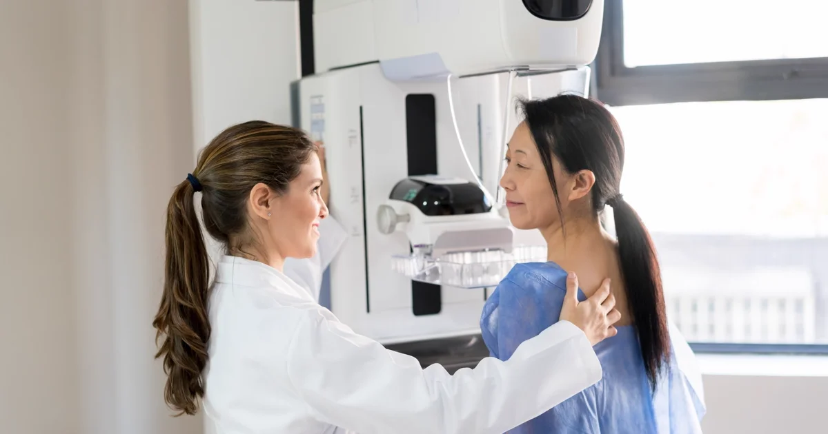
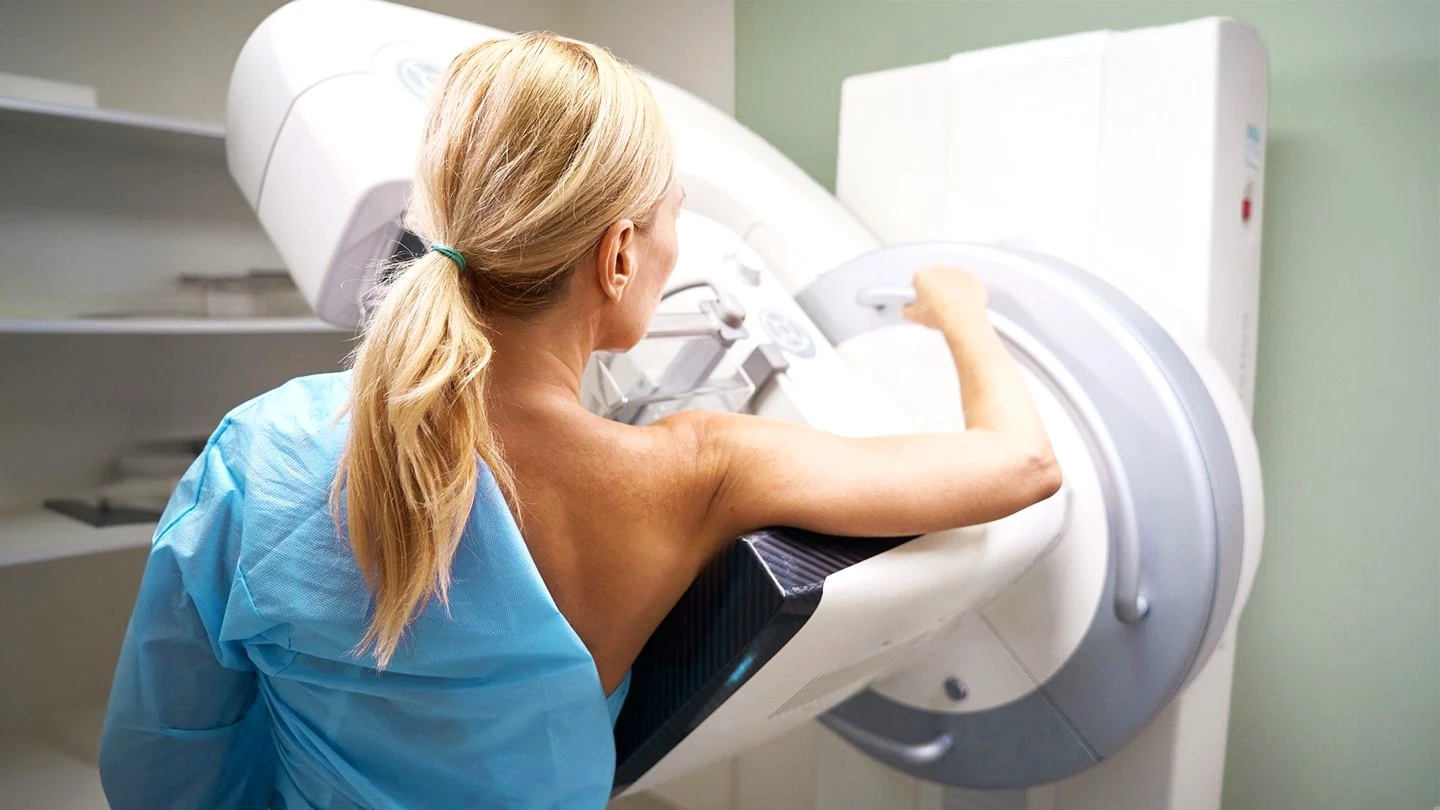
- A female radiographer will position your breast on the mammography platform and apply gentle compression. Compression is essential for image quality and dose reduction.
- For a 2D mammogram, two views of each breast are taken (four images total). Each compression lasts only a few seconds.
- For a 3D mammogram, the X-ray arm sweeps in an arc during each compression. You may feel the machine move slightly, but the compression time is only 1–2 seconds longer.
- The total appointment time is approximately 15–20 minutes, including registration and changing.
- The examination takes place in a private, dedicated women’s imaging area staffed entirely by female technologists for your comfort.
After Your Mammogram
- Your images are reviewed by a subspecialty radiologist experienced in breast imaging who interprets all mammograms at DCDC.
- Results are reported using the standardised BI-RADS (Breast Imaging Reporting and Data System) scoring system, from BI-RADS 0 (needs additional imaging) through BI-RADS 6 (known cancer).
- Breast density is assessed and categorised (A through D) as part of every report.
- Results are typically available within 18–24 hours. For urgent cases, same-day reporting is available.
- If follow-up imaging is recommended (such as breast ultrasound), it can often be arranged the same day or next day at DCDC.
For practical tips on making the most of your screening visit, see our guide on how to prepare for your mammogram.
Radiation Safety: 2D vs 3D Mammogram
Radiation exposure is a common concern among patients considering any X-ray-based examination. Both 2D and 3D mammograms use low-dose ionising radiation, and the doses involved are far below levels associated with any measurable health risk.
A standard four-view 2D mammogram delivers approximately 0.4 mSv of radiation. A 3D mammogram is slightly higher, typically in the range of 0.5 to 0.7 mSv, because multiple exposures are needed during the arc sweep. For context, the average annual background radiation from natural sources (cosmic rays, radon, soil) is about 2.4 mSv. A return flight between Dubai and London exposes passengers to roughly 0.1 mSv.
The Radiological Society of North America (RSNA) and the American College of Radiology (ACR) both confirm that the radiation dose from mammography, whether 2D or 3D, falls well within established safety guidelines and that the benefit of early cancer detection far outweighs the negligible risk from radiation. Modern 3D mammography systems use dose-reduction algorithms and iterative reconstruction to keep exposure as low as reasonably achievable (ALARA principle).
At DCDC, all mammography equipment is regularly quality-tested in accordance with MOHAP (Ministry of Health and Prevention) regulations, and our facility operates under MOHAP License NIMY7VY5-240925. Radiation doses are monitored and optimised for every patient.
Which Mammogram Is Right for You? A Decision Guide
Choosing between a 2D and 3D mammogram depends on several individual factors. Use the following decision framework to determine which option may be most appropriate for your situation.
A 2D Mammogram May Be Sufficient If:
- Your breasts are predominantly fatty (BI-RADS density A or B).
- You have no family history of breast cancer and no known genetic risk factors.
- You have had stable, normal mammograms for several years.
- Cost is a primary concern and insurance does not cover 3D mammography.
A 3D Mammogram Is Recommended If:
- You have dense breast tissue (BI-RADS density C or D), which applies to about 40–50% of women.
- You have a family history of breast cancer, especially in a first-degree relative.
- You carry a known BRCA1 or BRCA2 mutation or have other high-risk genetic factors.
- You have previously been called back after a 2D mammogram for findings that turned out to be benign.
- You have a personal history of breast biopsies, atypical cells, or breast surgery.
- You want the most sensitive screening technology available for your baseline or annual mammogram.
- Your doctor has recommended enhanced screening based on your individual risk assessment.
If you are unsure, our radiologists can advise you at the time of your appointment. In many cases, the initial 2D images are reviewed while you are still in the room, and 3D can be added immediately if the radiologist believes it would provide additional diagnostic value.
Subspecialty Breast Imaging at DCDC: A Radiologist’s Perspective

Diagnostic Radiology
Dr. Osama Elzamzami is a Diagnostic Radiologist at Doctors Clinic Diagnostic Center (DCDC) in Dubai Healthcare City. With specialist training in breast imaging interpretation, Dr. Elzamzami reads every mammogram with a focus on breast density assessment, subtle architectural distortion, and early-stage calcification patterns. He works closely with referring gynaecologists and surgeons to ensure timely, accurate diagnoses that guide patient management decisions.
Not all mammograms are read equally. The accuracy of mammographic interpretation depends as much on the radiologist’s expertise as on the technology itself. At DCDC, every mammogram — whether 2D or 3D — is interpreted by a subspecialty radiologist with dedicated experience in breast imaging. This matters because research published by the Radiological Society of North America has shown that radiologists with higher annual mammography reading volumes and subspecialty breast imaging training achieve significantly higher cancer detection rates and lower false-positive rates.
Our breast imaging workflow includes standardised BI-RADS reporting, density categorisation on every examination, structured comparison with prior studies, and direct communication with referring physicians when findings require follow-up. When a mammogram reveals a BI-RADS 4 or 5 finding (suspicious or highly suggestive of malignancy), our team coordinates with the patient’s gynaecologist or breast surgeon to ensure biopsy and further workup are arranged without delay.
Final Thoughts: Making an Informed Mammogram Decision in Dubai
Both 2D and 3D mammograms are effective screening tools, and having either is far better than having none. However, the evidence is clear that 3D mammography offers meaningful advantages in cancer detection and false-positive reduction, particularly for women with dense breast tissue. In a region where breast cancer accounts for the largest share of female cancers, choosing the most sensitive screening method available is a practical step toward early detection and better outcomes.
Schedule Your Mammogram at DCDC Dubai Healthcare City
Doctors Clinic Diagnostic Center offers 2D and 3D mammography from AED 250, interpreted by subspecialty breast imaging radiologists. Female technologists, private imaging suites, free parking, and same-day appointments available. Located in Building 64, Block A, Dubai Healthcare City. Book your mammogram or contact us on WhatsApp.
خدمات مرتبط در DCDC
مراقبت تخصصی و تشخیص پیشرفته در شهر بهداشت دبی
Frequently Asked Questions
Key Points to Remember
3D mammography detects up to 40% more invasive breast cancers than 2D alone and reduces false-positive callbacks by approximately 15%. For women with dense breast tissue, the advantage is even more pronounced.
Both 2D and 3D mammograms are safe, and the radiation dose from either type is far below any level of concern. Annual screening starting at age 40 remains the most effective way to catch breast cancer early, when the 5-year survival rate exceeds 95%.
At DCDC in Dubai Healthcare City, both technologies are available from AED 250 with subspecialty radiologist interpretation, female-only imaging staff, same-day appointments, and results within 18–24 hours. Whether you choose 2D or 3D, the most important step is to get screened regularly.
منابع و مراجع
این مقاله توسط تیم پزشکی ما بررسی شده و به منابع زیر ارجاع میدهد:
- American College of Radiology — ACR Practice Parameter for the Performance of Digital Breast Tomosynthesis
- Radiological Society of North America (RSNA) — Digital Breast Tomosynthesis: A Better Mammogram
- American Cancer Society — Mammogram Screening Recommendations and Guidelines
- Friedewald SM et al. — Breast Cancer Screening Using Tomosynthesis in Combination With Digital Mammography. JAMA. 2014;311(24):2499–2507
- UAE National Cancer Registry — Cancer Incidence in the United Arab Emirates, Annual Report
محتوای پزشکی این سایت توسط پزشکان دارای مجوز DHA بررسی میشود. مشاهده سیاست تحریریه برای اطلاعات بیشتر.
نوشته شده توسط
Dr. Osama Elzamzami
Diagnostic Radiology
MD, FRCR
Dr. Osama Elzamzami is a Diagnostic Radiologist at Doctors Clinic Diagnostic Center (DCDC) in Dubai Healthcare City.
Related Articles
Mammogram Cost Dubai: AED 250-900
Breast Cancer Screening: Mammogram & BRCA Guide
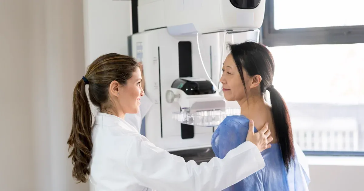
Mammogram Preparation Dubai: What to Expect
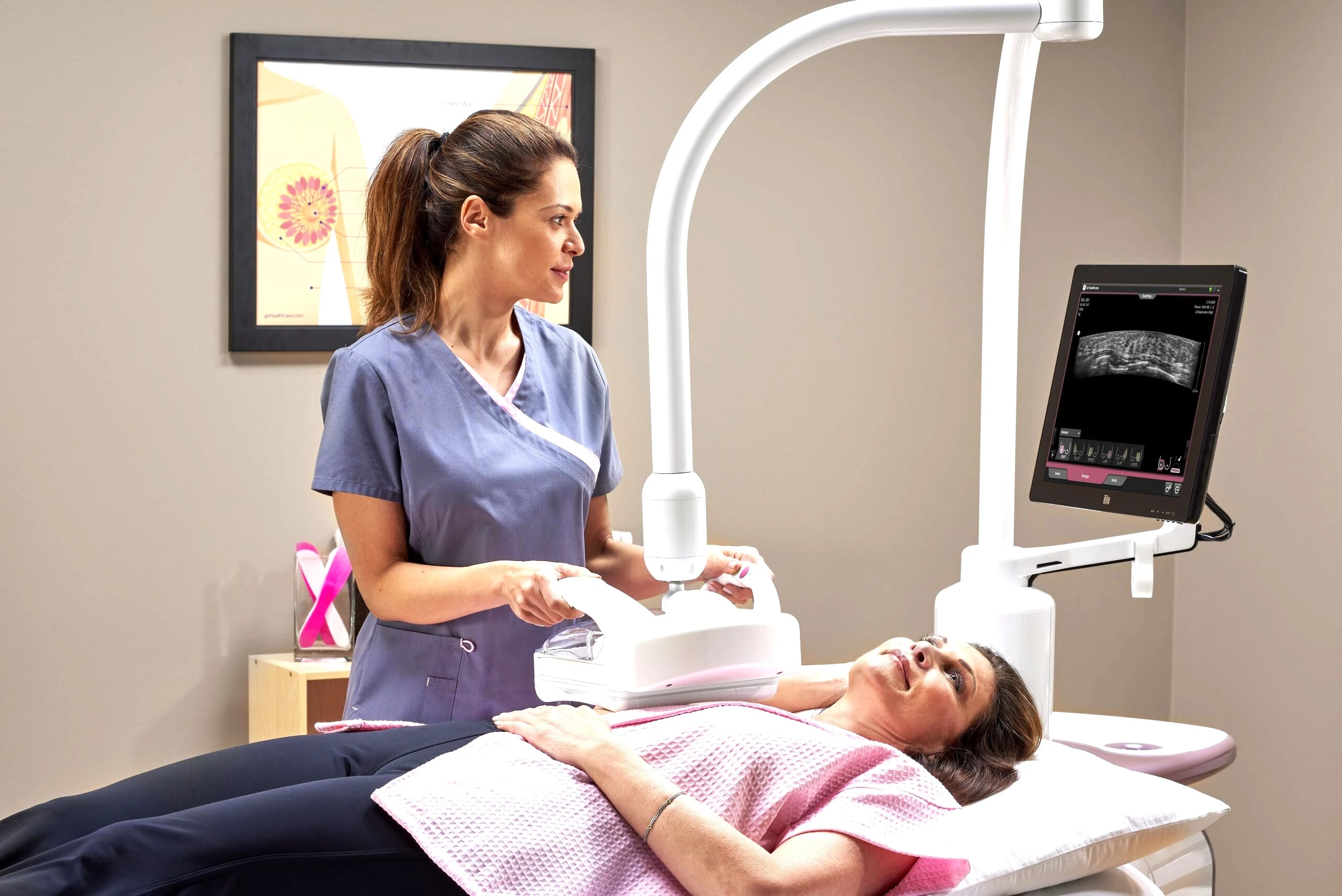
Breast Ultrasound Dubai: Cost & Guide
10 Early Signs of Breast Cancer to Watch For
More in Diagnostic Imaging

X-Ray at Home Dubai: Cost & Guide (2026)
بیشتر بخوانید
Ultrasound at Home Dubai: From AED 599 (2026)
بیشتر بخوانید
Neck MRI Dubai: What It Shows & Cost (2026)
بیشتر بخوانید
X-Ray Scan Dubai: Complete Guide (2026)
بیشتر بخوانید
MRI vs Ultrasound Dubai: Which Scan? (2026)
بیشتر بخوانید
MRI vs X-Ray Dubai: Which Scan Do You Need? (2026)
بیشتر بخوانید© 2026 Doctors Clinic Diagnostic Center (DCDC), Dubai Healthcare City. Originally published at https://doctorsclinicdubai.ae/blog/3d-vs-2d-mammogram-dubai. All rights reserved. Unauthorized reproduction is prohibited.