Wichtigste Erkenntnisse
- A shoulder MRI in Dubai typically costs between AED 900 and AED 1,400 without contrast, with contrast-enhanced scans ranging from AED 1,200 to AED 1,800
- Shoulder MRI is the gold standard for diagnosing rotator cuff tears, labral tears (including SLAP lesions), impingement syndrome, and frozen shoulder without radiation
- MRI provides far superior soft tissue detail compared to X-ray and ultrasound, making it essential for evaluating the rotator cuff tendons, labrum, cartilage, and biceps tendon
- Your doctor will typically order a shoulder MRI after a sports injury, persistent shoulder pain lasting more than 4 to 6 weeks, instability, or limited range of motion that has not responded to conservative treatment
- The scan takes approximately 30 to 45 minutes, is completely painless, and usually requires no special preparation unless contrast dye is needed
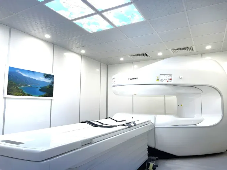
A shoulder MRI scan is the most detailed imaging tool for evaluating the complex anatomy of the shoulder joint. The shoulder has the greatest range of motion of any joint in the body, which also makes it one of the most vulnerable to injury. Whether you are dealing with a rotator cuff tear from a sports injury, a labral tear causing instability, or chronic shoulder pain that has not responded to treatment, an MRI can reveal exactly what is happening inside your shoulder — from tendon tears and cartilage damage to inflammation and structural abnormalities. In Dubai, shoulder MRI is widely available at advanced imaging centers and remains the preferred diagnostic step before planning surgical or conservative treatment for most shoulder conditions.


What Does a Shoulder MRI Show?
A shoulder MRI produces detailed cross-sectional images of all the structures within and around the shoulder joint, including the rotator cuff tendons, labrum, articular cartilage, biceps tendon, joint capsule, bursa, and surrounding muscles. Unlike X-rays, which only visualize bone, MRI uses magnetic fields and radio waves to generate high-resolution images of soft tissues that are invisible on conventional radiographs. This makes MRI indispensable for diagnosing the vast majority of shoulder problems.
The following conditions are commonly identified on a shoulder MRI:
- Rotator cuff tears (partial and full-thickness): The rotator cuff is a group of four tendons (supraspinatus, infraspinatus, teres minor, and subscapularis) that stabilize the shoulder and enable overhead movement. MRI can detect partial-thickness tears, full-thickness tears, and tendinopathy with high accuracy. The supraspinatus tendon is the most commonly torn, and MRI can measure the exact size and retraction of the tear, which directly determines whether surgical repair or conservative management is appropriate.
- Labral tears (SLAP lesions and Bankart lesions): The labrum is a ring of cartilage that deepens the shoulder socket and provides stability. Superior labral anterior-posterior (SLAP) tears affect the top of the labrum where the biceps tendon attaches, while Bankart lesions involve the inferior labrum and typically result from shoulder dislocations. MRI, particularly with intra-articular contrast (MR arthrogram), is the definitive test for detecting labral pathology.
- Shoulder impingement syndrome: Impingement occurs when the rotator cuff tendons become compressed between the humeral head and the acromion during overhead movements. MRI reveals the degree of subacromial space narrowing, any associated bursitis, and early rotator cuff damage that may result from chronic impingement.
- Frozen shoulder (adhesive capsulitis): MRI can demonstrate thickening and inflammation of the joint capsule, particularly at the axillary recess, which is characteristic of frozen shoulder. It also helps rule out other conditions that can mimic frozen shoulder, such as rotator cuff tears or arthritis.
- Biceps tendon pathology: The long head of the biceps tendon is a common source of anterior shoulder pain. MRI can detect biceps tendinitis, partial tears, subluxation out of the bicipital groove, and complete ruptures.
- Shoulder arthritis and cartilage damage: MRI reveals cartilage thinning, bone spurs, synovial inflammation, and joint effusion associated with glenohumeral osteoarthritis or inflammatory arthritis. It provides critical pre-surgical information for patients being evaluated for shoulder replacement.
- Fractures and bone marrow edema: While X-ray detects most acute fractures, MRI is superior for identifying stress fractures, occult fractures (not visible on X-ray), Hill-Sachs lesions (bone dents from dislocations), and bone marrow edema patterns that indicate recent injury.
- Shoulder instability: Recurrent shoulder dislocations or subluxations cause predictable damage patterns visible on MRI, including Bankart lesions, Hill-Sachs deformities, and capsular stretching. MRI maps these injuries precisely, guiding the surgeon's approach to stabilization surgery.
"The shoulder is one of the most complex joints to image because of its remarkable range of motion and the number of structures packed into a relatively small space," explains Dr. Osama Elzamzami, Consultant Radiologist at DCDC. "MRI gives us a complete roadmap of the rotator cuff, labrum, cartilage, and biceps tendon in a single examination, allowing the treating physician to make an informed decision about surgery versus conservative management."
How Much Does a Shoulder MRI Cost in Dubai?
The cost of a shoulder MRI in Dubai generally ranges from AED 900 to AED 1,400 for a standard non-contrast examination. The exact price depends on the imaging center, the strength of the MRI machine (1.5T vs 3T), whether contrast dye is required, and whether the radiologist report is included. At Doctors Clinic Diagnostic Center in Dubai Healthcare City, shoulder MRI pricing is all-inclusive — covering the scan, a comprehensive radiologist report, and digital image copies.
| Shoulder MRI Type | Approximate Cost (AED) |
|---|---|
| Shoulder MRI without contrast | 900 – 1,400 |
| Shoulder MRI with contrast (gadolinium) | 1,200 – 1,800 |
| MR arthrogram (intra-articular contrast) | 1,500 – 2,200 |
| Both shoulders (bilateral) | 1,600 – 2,400 |
| Shoulder MRI + additional joint | 1,800 – 2,500 |
Prices are approximate and include the radiologist report at DCDC. Contact us for exact pricing.
An MR arthrogram, where contrast dye is injected directly into the shoulder joint before the MRI, is sometimes requested by orthopedic surgeons when a labral tear is strongly suspected. The intra-articular contrast distends the joint and outlines the labrum more clearly, increasing diagnostic accuracy for subtle labral pathology. For most rotator cuff evaluations and general shoulder assessments, a standard non-contrast MRI provides excellent diagnostic information.
Most health insurance plans in the UAE cover shoulder MRI when ordered by a licensed physician with a documented clinical indication such as suspected rotator cuff tear, post-dislocation evaluation, or persistent shoulder pain. DCDC assists patients with insurance pre-authorization to confirm coverage before the appointment.
When Does a Doctor Order a Shoulder MRI?
A shoulder MRI is not typically the first imaging test ordered for shoulder pain. Most physicians begin with a clinical examination and standard X-rays to rule out fractures, dislocations, or advanced arthritis. However, when the clinical examination suggests a soft tissue injury, or when initial conservative treatment fails to resolve symptoms, an MRI becomes the essential next step. Your doctor is likely to order a shoulder MRI in the following situations: For related information, see our guide on Hip MRI Dubai: Labral Tears, AVN & Joint Issues.
- Suspected rotator cuff tear: Difficulty raising the arm overhead, weakness with external rotation, night pain that disrupts sleep, or an acute injury involving a fall on an outstretched hand or a forceful pulling motion.
- Shoulder dislocation or instability: After a first-time or recurrent shoulder dislocation, MRI is essential to assess for labral tears (Bankart lesions), Hill-Sachs bone defects, and capsular damage that determine the risk of future dislocations and the need for surgical stabilization.
- Persistent shoulder pain: Shoulder pain lasting more than 4 to 6 weeks despite rest, anti-inflammatory medication, and physiotherapy warrants further investigation with MRI to identify the underlying structural cause.
- Limited range of motion: Progressive stiffness and inability to reach behind the back or overhead, particularly if frozen shoulder (adhesive capsulitis) is suspected or needs to be differentiated from a rotator cuff tear.
- Sports injuries: Throwing athletes (cricket, baseball), overhead sport participants (swimming, tennis, volleyball), and contact sport players (rugby, football) are particularly vulnerable to labral tears, rotator cuff injuries, and impingement that require MRI for accurate diagnosis.
- Pre-surgical planning: Before arthroscopic rotator cuff repair, labral repair, shoulder stabilization, or shoulder replacement, surgeons require detailed MRI to plan the procedure and discuss expected outcomes with the patient.
- Post-surgical evaluation: Following rotator cuff repair or labral surgery, MRI is used to assess healing, graft integrity, and detect re-tears or complications.
Book Your Shoulder MRI at DCDC
At Doctors Clinic Diagnostic Center in Dubai Healthcare City, we offer same-week appointments for shoulder MRI scans with comprehensive radiologist reports delivered within 24 to 48 hours.
Shoulder MRI vs X-Ray vs Ultrasound: Which Imaging Do You Need?
Patients often wonder whether an X-ray or ultrasound is sufficient for diagnosing their shoulder problem or whether an MRI is truly necessary. Each imaging modality has specific strengths and limitations, and the choice depends on what condition is suspected. In many cases, these tests are complementary rather than interchangeable.
| Factor | X-Ray | Ultrasound | MRI |
|---|---|---|---|
| Cost | AED 150 – 300 | AED 400 – 700 | AED 900 – 1,400 |
| Radiation | Low dose | None | None |
| Scan time | 5 minutes | 15 – 20 minutes | 30 – 45 minutes |
| Bone visualization | Excellent | Limited | Good |
| Rotator cuff tears | Not visible | Good (operator-dependent) | Excellent (gold standard) |
| Labral tears | Not visible | Very limited | Excellent (especially MR arthrogram) |
| Cartilage damage | Indirect signs only | Not visible | Direct visualization |
| Biceps tendon | Not visible | Good | Excellent |
| Bone marrow edema | Not visible | Not visible | Excellent |
| Best for | Fractures, arthritis, calcification | Quick rotator cuff assessment | Complete evaluation of all shoulder structures |
X-ray is typically the first-line test. Ultrasound is useful for focused rotator cuff evaluation. MRI provides the most comprehensive assessment.
Ultrasound can be a useful screening tool for rotator cuff tears, particularly in experienced hands, and has the advantage of being dynamic — the radiologist can move the shoulder during the scan. However, ultrasound is highly operator-dependent, cannot adequately visualize the labrum or deep structures, and does not detect bone marrow abnormalities. MRI remains the definitive test when a comprehensive evaluation is needed, when labral pathology is suspected, or when surgical planning requires precise anatomical detail.
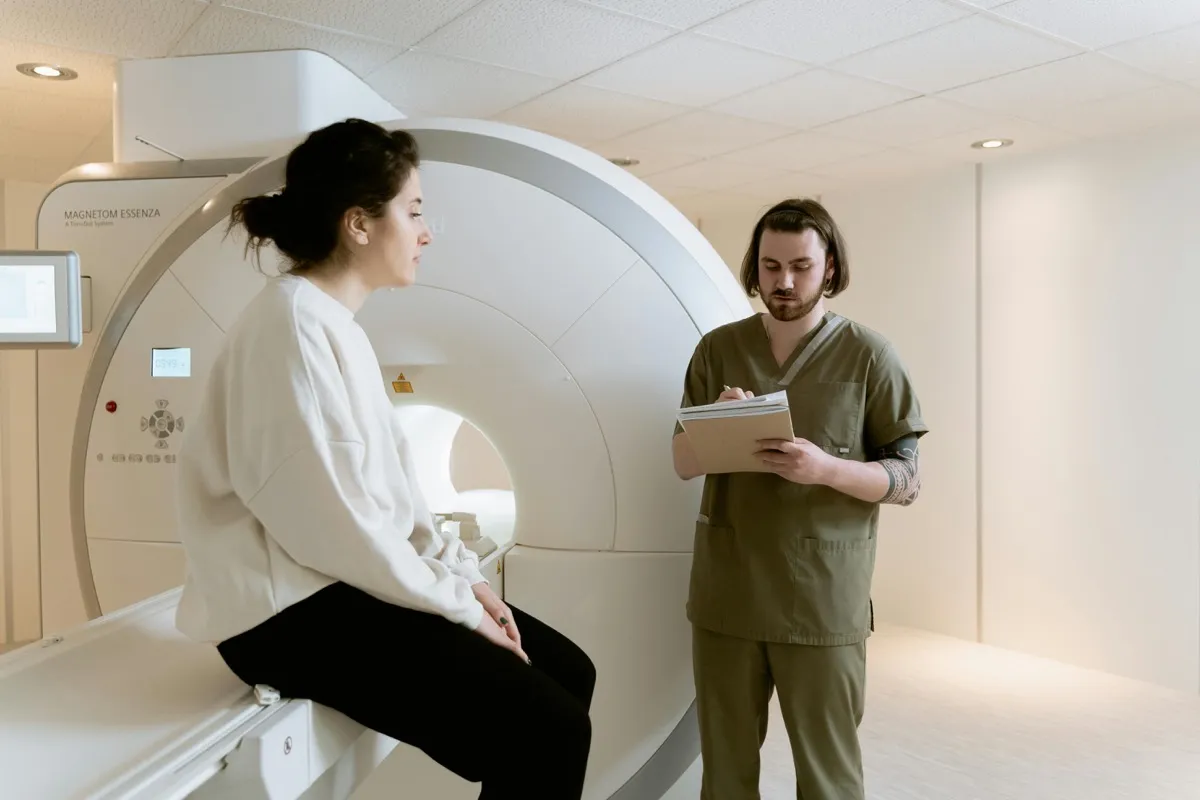
What to Expect During a Shoulder MRI
Understanding what happens during a shoulder MRI helps patients prepare and reduces anxiety. The scan is entirely non-invasive, painless, and does not involve radiation.
- Before the scan: You will complete a safety screening questionnaire to confirm you have no MRI-incompatible implants (pacemakers, certain cochlear implants, metallic foreign bodies). You will remove all metal objects including jewellery, watches, and belts. Wear comfortable clothing without metal zippers or clasps.
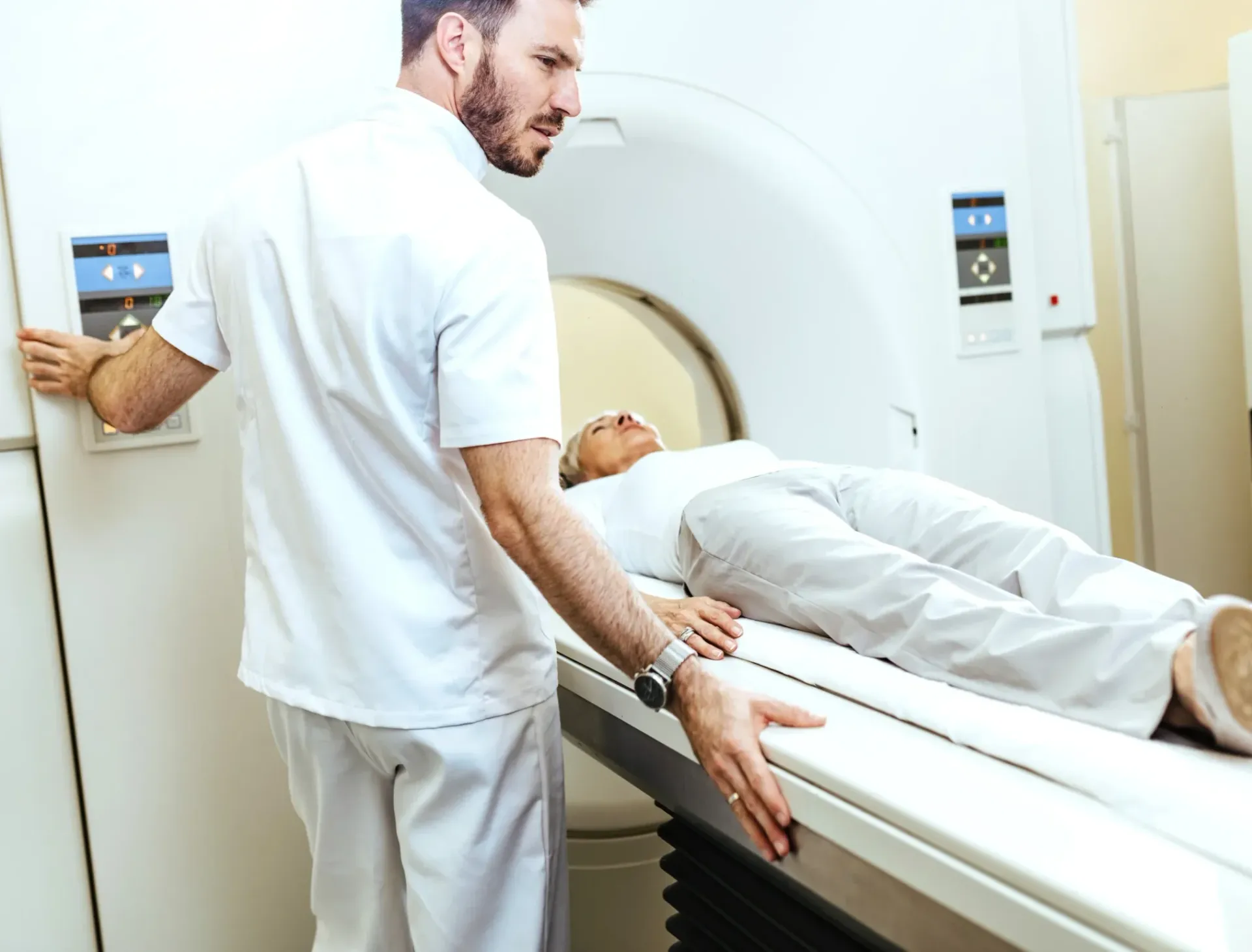
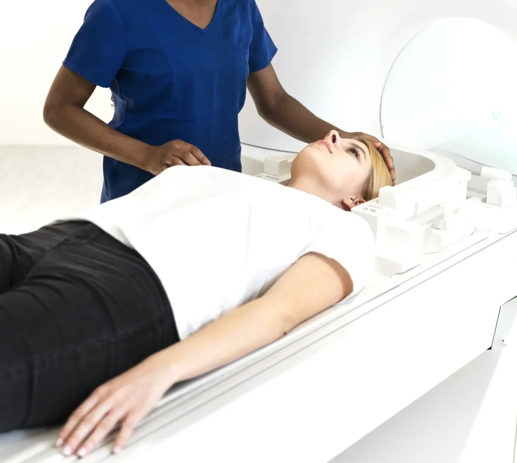
- Positioning: You will lie on your back on the MRI table with the affected shoulder positioned in a dedicated shoulder coil. Your arm will be positioned at your side, typically in a neutral or slightly externally rotated position. The arm will be gently secured to minimize movement during the scan.
- During the scan: The table slides into the MRI machine. The machine produces loud knocking and humming sounds — you will be given earplugs or headphones. For a shoulder MRI, you enter the machine head-first, but your head remains near the opening, which many patients find more comfortable than expected. It is essential to remain as still as possible.
- Duration: A standard shoulder MRI takes approximately 30 to 45 minutes. If an MR arthrogram is performed, an additional 15 to 20 minutes is needed for the intra-articular injection before the scan begins.
- After the scan: There is no recovery period. You can return to normal activities immediately. If contrast was used (IV or intra-articular), drink extra water to help your body eliminate the contrast agent. Your radiologist report will typically be available within 24 to 48 hours at DCDC.
No special preparation is needed for a standard shoulder MRI. You can eat and drink normally before the appointment. If intravenous contrast is required, you may be asked to fast for 2 to 4 hours beforehand. If an MR arthrogram is planned, the radiologist will discuss the intra-articular injection process with you before the procedure.
Common Shoulder Conditions Diagnosed with MRI
The shoulder joint's remarkable mobility comes at the cost of stability, making it susceptible to a wide range of injuries and degenerative conditions. MRI plays a central role in diagnosing and grading the severity of these conditions, which directly impacts treatment decisions. You may also find our Knee MRI Dubai: What It Shows & Cost helpful.
Rotator Cuff Tears
Rotator cuff tears are among the most common shoulder injuries, affecting both athletes and older adults. They can result from acute trauma (a fall, lifting a heavy object) or from chronic degeneration over time. MRI distinguishes between partial-thickness tears (which may respond to physiotherapy and injections) and full-thickness tears (which often require surgical repair). MRI also measures tear size and the degree of tendon retraction and muscle atrophy, which are critical factors in determining whether a tear is repairable and predicting surgical outcomes. Studies show MRI has a sensitivity exceeding 90 percent for detecting full-thickness rotator cuff tears and approximately 80 percent for partial-thickness tears.
Labral Tears and Shoulder Instability
Labral tears are particularly common in young athletes and patients who have experienced shoulder dislocations. SLAP tears affect the superior labrum and are frequently seen in throwing athletes, while Bankart lesions affect the inferior labrum and result from traumatic dislocations. MR arthrography (MRI performed after injecting contrast into the joint) is considered the gold standard for labral evaluation, with sensitivity rates of 82 to 100 percent depending on the tear type and location. Standard MRI without arthrography can also detect many labral tears, particularly larger ones.
Frozen Shoulder (Adhesive Capsulitis)
Frozen shoulder causes progressive pain and stiffness that severely limits shoulder movement. It occurs in approximately 2 to 5 percent of the general population, with higher rates in diabetic patients (up to 20 percent). MRI findings include thickening of the joint capsule (particularly at the axillary recess and rotator interval), synovial enhancement on contrast-enhanced imaging, and reduced joint volume. Importantly, MRI also rules out other conditions that can present similarly, such as rotator cuff tears, calcific tendinitis, and glenohumeral arthritis.
Get Expert Shoulder MRI Interpretation
At DCDC, every shoulder MRI is interpreted by a consultant radiologist with subspecialty musculoskeletal imaging experience. Our detailed, structured reports provide your referring physician with the precise information needed for treatment planning. Learn more about our MRI services.
Verwandte Leistungen im DCDC
Fachkundige Betreuung und moderne Diagnostik in Dubai Healthcare City
Frequently Asked Questions
Final Thoughts
A shoulder MRI is the most reliable and comprehensive imaging tool for evaluating the rotator cuff, labrum, cartilage, tendons, and bone of the shoulder joint. Whether you are dealing with a sports injury, chronic pain, instability, or preparing for surgery, MRI provides the detailed anatomical information your physician needs to make an accurate diagnosis and recommend the most appropriate treatment plan.
At Doctors Clinic Diagnostic Center in Dubai Healthcare City, we combine advanced MRI technology with experienced consultant radiologist interpretation to deliver thorough, reliable shoulder MRI reports. Contact us to book your scan or to discuss whether a shoulder MRI is the right next step for your symptoms.
Quellen und Referenzen
Dieser Artikel wurde von unserem medizinischen Team überprüft und bezieht sich auf folgende Quellen:
- American College of Radiology - Appropriateness Criteria for Shoulder Pain
- Radiological Society of North America - Shoulder MRI
- American Academy of Orthopaedic Surgeons - Rotator Cuff Tears
- European Radiology - MRI Accuracy for Labral Tears
- Dubai Health Authority - Diagnostic Imaging Standards
Medizinische Inhalte auf dieser Website werden von DHA-lizenzierten Ärzten überprüft. Siehe unsere redaktionelle Richtlinien für weitere Informationen.

Verfasst von
Dr. Osama Elzamzami
Diagnostic Radiology
MD, FRCR
Dr. Osama Elzamzami is a Consultant Radiologist specializing in diagnostic imaging including MRI, CT, and ultrasound at DCDC Dubai Healthcare City.
Related Articles
MRI Scan Dubai: Complete Guide to Types, Cost & Full Body MRI
Knee MRI in Dubai: What It Shows, Cost & When You Need One
ACL Tear MRI: How MRI Diagnoses Ligament Injuries in Dubai
Hip MRI Dubai: Detect Labral Tears, AVN & Joint Problems
Full Body MRI Cost Dubai: AED 5,000-15,000 (2026)
More in Diagnostic Imaging
Neck MRI Dubai: What It Shows & Cost (2026)
Weiterlesen
X-Ray Scan Dubai: Complete Guide (2026)
Weiterlesen
MRI vs Ultrasound Dubai: Which Scan? (2026)
Weiterlesen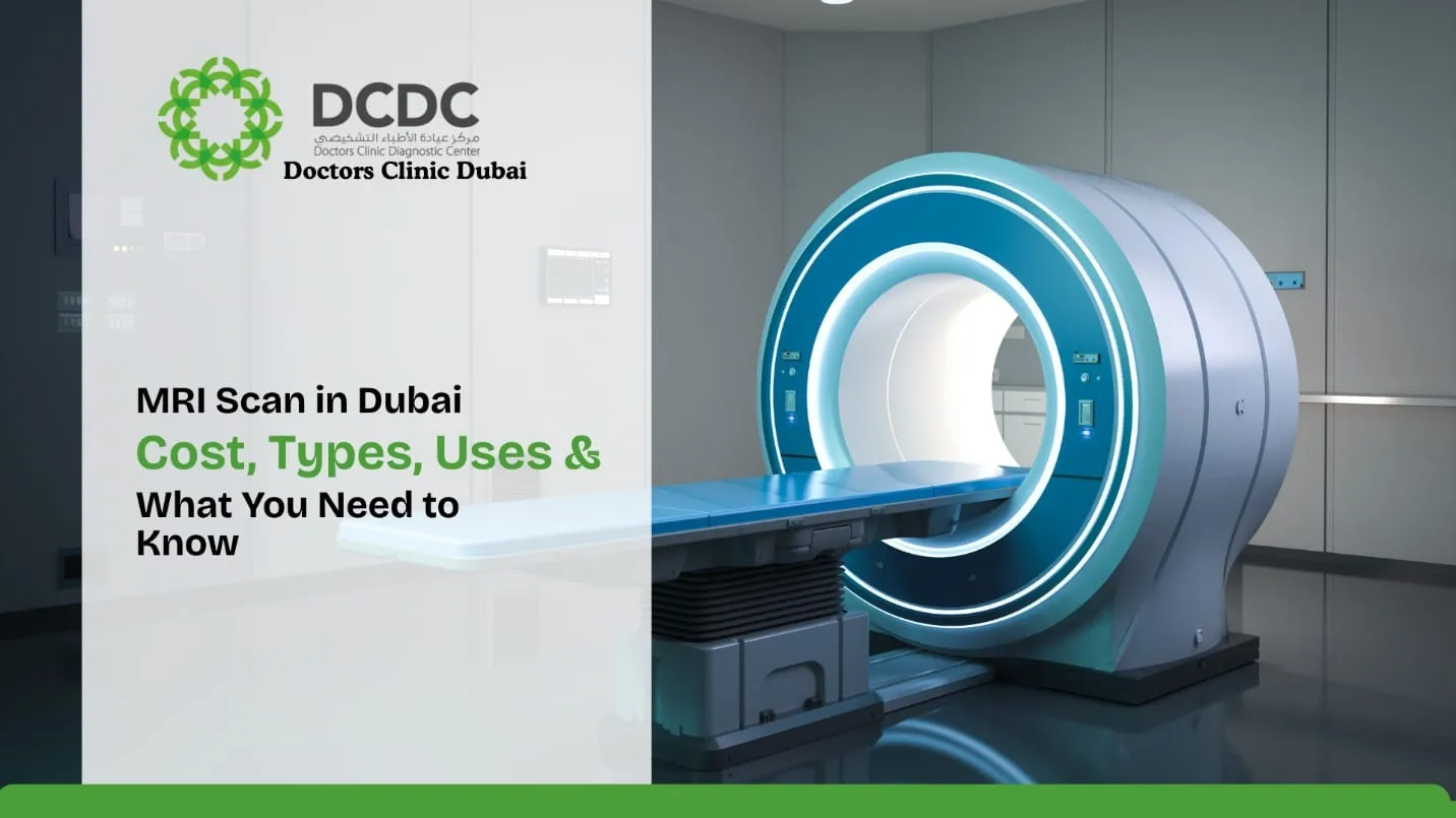
MRI vs X-Ray Dubai: Which Scan Do You Need? (2026)
Weiterlesen
Kidney Ultrasound vs CT Dubai: Which Test? (2026)
WeiterlesenMRI Preparation Dubai: Complete Guide (2026)
Weiterlesen© 2026 Doctors Clinic Diagnostic Center (DCDC), Dubai Healthcare City. Originally published at https://doctorsclinicdubai.ae/blog/shoulder-mri-dubai. All rights reserved. Unauthorized reproduction is prohibited.