
Wichtigste Erkenntnisse
- OPG liefert eine flache 2D-Panoramaansicht des gesamten Kiefers; CBCT erstellt ein detailliertes 3D-Volumen der Zaehne, Knochen und umgebenden Strukturen
- OPG verwendet deutlich weniger Strahlung (5-26 µSv) im Vergleich zu CBCT (30-200 µSv), obwohl beide als niedrig dosiert gelten
- OPG kostet in Dubai 150-350 AED; CBCT kostet 500-1.500 AED je nach Aufnahmebereich
- Waehlen Sie OPG fuer Routine-Screening, kieferorthopaedische Bewertung und allgemeine Zahnuntersuchungen
- Waehlen Sie CBCT fuer Implantatplanung, komplexe Extraktionen, Wurzelkanaldiagnostik und Kieferchirurgieplanung
- Viele Faelle profitieren von einem OPG-Screening zuerst, dann CBCT fuer gezielte 3D-Details bei Bedarf
"Soll ich ein CBCT oder ein OPG machen lassen?" Dies ist eine der haeufigsten Fragen, die ich von Patienten hoere, die zur Dental-Bildgebung ueberwiesen werden. Die ehrliche Antwort ist, dass es ganz davon abhaengt, was Ihr Zahnarzt sehen muss. Ein OPG gibt uns eine zuverlaessige Panoramauebersicht Ihrer Kiefer, Zaehne und Nebenhoehlen in einem einzigen flachen Bild. Ein CBCT gibt uns ein dreidimensionales Modell, das wir drehen, schneiden und mit Submillimeter-Genauigkeit messen koennen. Keines ersetzt das andere. Sie dienen unterschiedlichen Zwecken, und das Verstaendnis dieser Zwecke hilft Ihnen, nicht fuer einen Scan zu bezahlen, den Sie nicht brauchen, und gleichzeitig sicherzustellen, dass Sie den bekommen, den Sie brauchen.
Dieser Leitfaden vergleicht CBCT und OPG Dental-Scans Seite an Seite, einschliesslich Bildgebungstechnologie, Strahlenbelastung, Kosten in Dubai und den spezifischen klinischen Szenarien, in denen jeder Scan die richtige Wahl ist.


CBCT vs OPG auf einen Blick
Bevor wir ins Detail gehen, hier ein kurzer Ueberblick, wie diese beiden Dental-Bildgebungsmethoden im Vergleich stehen. Wenn Sie nur 30 Sekunden haben, sagt Ihnen diese Tabelle das Wesentliche. Die folgenden Abschnitte erklaeren die Gruende hinter jeder Zeile.
| Merkmal | OPG (Panorama-Roentgen) | CBCT (Cone Beam CT) |
|---|---|---|
| Dimensionen | 2D flaches Panoramabild | 3D volumetrischer Datensatz |
| Strahlendosis | Sehr niedrig (5-26 µSv) | Niedrig (30-200 µSv) |
| Scanzeit | 15-20 Sekunden | 10-40 Sekunden |
| Detailgrad | Guter Ueberblick, begrenzte Feindetails | Ausgezeichnete Details, Submillimeter-Genauigkeit |
| Kostenbereich (Dubai) | 150-350 AED | 500-1.500 AED |
| Am besten fuer | Allgemeines Screening, Kieferorthopaedie, Grundbewertung | Implantatplanung, komplexe Chirurgie, Wurzelkanaldiagnostik |
| Versicherungsdeckung | In der Regel mit zahnhaerztlicher Ueberweisung abgedeckt | Erfordert oft Vorabgenehmigung und klinische Begruendung |
OPG vs CBCT Vergleich. Kosten sind Richtwerte und koennen je nach Anbieter und Aufnahmebereich variieren.
Die wichtigste Erkenntnis aus dieser Tabelle ist einfach: OPG ist das Arbeitspferd der routinemaessigen Dental-Bildgebung, waehrend CBCT das Spezialwerkzeug ist, das zum Einsatz kommt, wenn zweidimensionale Informationen nicht ausreichen. Die meisten Patienten beginnen mit einem OPG. Nur ein Bruchteil benoetigt CBCT, aber wenn sie es tun, bietet nichts anderes das gleiche Mass an dreidimensionalem Detail.
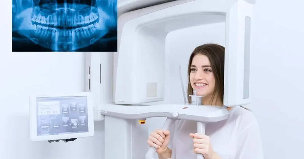
Was ist ein OPG-Roentgen?
Ein OPG, auch Orthopantomogramm oder Panorama-Roentgen genannt, ist ein einzelnes Weitwinkelbild, das beide Kiefer, alle Zaehne, die Kiefergelenke (TMJ) und Teile der Nebenhoehlen und Nasenhoehle in einer Aufnahme erfasst. Das Geraet rotiert um Ihren Kopf, waehrend Sie stillstehen und auf ein kleines Plattchen beissen, und der gesamte Vorgang dauert etwa 15 bis 20 Sekunden.
Das Ergebnis ist ein flacher, zweidimensionaler Panoramastreifen, der Zahnhaerzten einen breiten Ueberblick gibt. Er ist ausgezeichnet zum Erkennen von Karies, zur Bewertung der Knochenniveaus bei Parodontalerkrankungen, zur Ueberpruefung der Position von Weisheitszaehnen, zur Identifizierung von Zysten oder grossen Laesionen und zur Planung kieferorthopaedischer Behandlungen. Da die Strahlendosis extrem niedrig ist, vergleichbar mit etwa ein bis drei Tagen natuerlicher Hintergrundstrahlung, gilt OPG als sicher fuer den routinemaessigen Einsatz bei Erwachsenen, Jugendlichen und sogar Kindern, wenn klinisch gerechtfertigt.
Die Haupteinschraenkung von OPG ist, dass es eine zweidimensionale Darstellung einer dreidimensionalen Struktur ist. Ueberlappende Anatomie, Vergroesserungsverzerrung (typischerweise 20-30%) und die Unfaehigkeit, Knochentiefe zu messen, bedeuten, dass OPG allein nicht jede klinische Frage beantworten kann. Hier kommt CBCT ins Spiel.
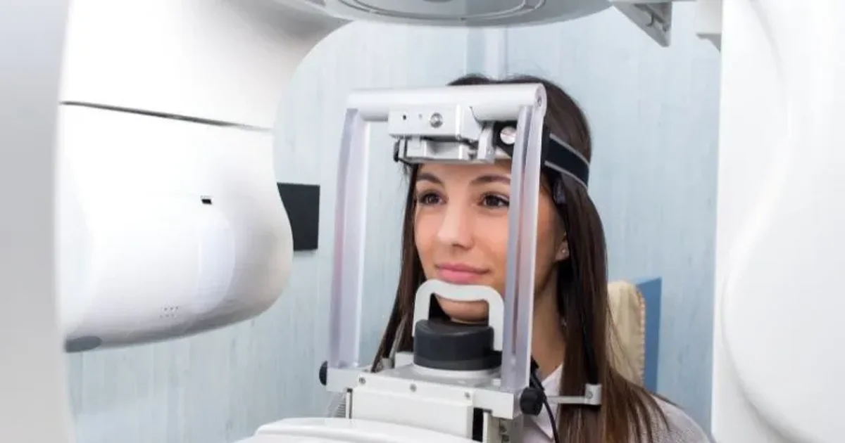
Was ist ein CBCT-Scan?
Cone Beam Computed Tomography (CBCT) ist eine spezielle Form der CT-Bildgebung, die speziell fuer den Kopf- und Halsbereich entwickelt wurde. Anstelle des faecherfoermigen Roentgenstrahls, der bei medizinischem CT verwendet wird, nutzt CBCT einen kegelfoermigen Strahl, der einmal um Ihren Kopf rotiert und Hunderte einzelner Projektionen aufnimmt. Software rekonstruiert dann diese Projektionen zu einem dreidimensionalen Volumen, das in jeder Ebene betrachtet werden kann: axial, koronar, sagittal oder sogar als 3D-Darstellung von Knochenoberflaechen.
Der Aufnahmebereich (Field of View) kann angepasst werden. Ein kleines FOV (4x4 cm oder 5x5 cm) erfasst nur wenige Zaehne in extrem hoher Aufloesung, was ideal fuer die endodontische Diagnostik ist. Ein mittleres FOV deckt einen Kiefer ab. Ein grosses FOV erfasst beide Kiefer, das TMJ, die Nebenhoehlen und Teile der Atemwege. Die Scanzeit reicht von 10 bis 40 Sekunden, abhaengig vom Geraet und dem gewaehlten FOV.
Was CBCT so leistungsfaehig macht, ist seine Faehigkeit, das Ueberlagerungsproblem zu beseitigen, das OPG einschraenkt. Mit CBCT kann jede Struktur isoliert betrachtet werden. Sie koennen Knochenbreite, -hoehe und -dichte an einer geplanten Implantatstelle messen. Sie koennen den genauen Verlauf des Nervus alveolaris inferior verfolgen. Sie koennen die tatsaechliche Anzahl und Kruemmung der Wurzelkanaele sehen. Sie koennen das Ausmass einer Frakturlinie durch eine Wurzel kartieren. Diese dreidimensionale Klarheit ist der Grund, warum CBCT in der Behandlungsplanung fuer Implantologie, Oralchirurgie, Endodontie und Kieferorthopaedie unverzichtbar geworden ist.
Wann OPG statt CBCT waehlen
OPG bleibt die Bildgebung erster Wahl fuer die Mehrheit der zahnhaerztlichen klinischen Situationen. Hier sind die Szenarien, in denen eine Panorama-Roentgenaufnahme der richtige Scan ist und CBCT unnoetig waere:
Allgemeines Dental-Screening
Wenn Sie einen neuen Zahnarzt besuchen oder zu einer Routinekontrolle kommen, bietet ein OPG einen umfassenden Ueberblick. Es zeigt versteckte Karies zwischen den Zaehnen, den Zustand frueherer Fuellungen und Kronen, hebt Knochenverlust durch Zahnfleischerkrankungen hervor und identifiziert sich entwickelnde Probleme wie Zysten oder impaktierte Zaehne. Fuer diesen breiten Screening-Zweck fuegt das zusaetzliche Detail von CBCT Kosten hinzu, ohne klinischen Mehrwert zu bieten.
Kieferorthopaedische Bewertung
Kieferorthopaedn verwenden routinemaessig OPG zusammen mit einer lateralen Kephalometrie-Aufnahme zur Planung von Zahnspangen- oder Aligner-Behandlungen. Die Panoramaansicht zeigt alle sich entwickelnden und durchgebrochenen Zaehne, Wurzellaengen und Kieferbeziehungen. Sofern es keine spezifische Komplikation gibt, wie einen impaktierten Eckzahn in schwieriger Position oder vermutete Wurzelresorption, die eine 3D-Lokalisierung erfordert, ist OPG fuer die kieferorthopaedische Planung ausreichend.
Grundlegende Weisheitszahnbewertung
Die meisten Weisheitszahnbewertungen beginnen und enden mit einem OPG. Das Panoramabild zeigt deutlich die Position des Weisheitszahns relativ zum Kiefer, den Impaktionswinkel und ob eine begleitende Pathologie wie eine follikulaere Zyste vorliegt. CBCT wird nur benoetigt, wenn das OPG zeigt, dass die Wurzeln des Weisheitszahns in sehr enger Naehe zum Kanal des Nervus alveolaris inferior stehen - etwas, das wir im CBCT-Abschnitt ausfuehrlicher besprechen.
Parodontale Knochenbewertung
Zur Bewertung generalisierter Knochenverlustmuster bei Parodontalerkrankungen gibt OPG in Kombination mit Einzelzahnaufnahmen Zahnhaerzten alle notwendigen Informationen. Die zweidimensionale Ansicht ist ausreichend zur Messung der Knochenniveaus um die Zaehne und zur Ueberwachung des Krankheitsverlaufs im Zeitverlauf.
Kostenbewusste und paediatrische Patienten
Mit 150-350 AED kostet OPG einen Bruchteil von CBCT. Fuer Patienten, die aus eigener Tasche zahlen, oder fuer Kinder, die Bildgebung fuer die kieferorthopaedische Planung benoetigen, machen die niedrigeren Kosten und die deutlich geringere Strahlendosis OPG zur offensichtlichen Wahl. In der Kinderzahnheilkunde empfiehlt das ALARA-Prinzip (As Low As Reasonably Achievable - So niedrig wie vernuenftigerweise erreichbar) ausdruecklich die Verwendung der niedrigsten Strahlendosis, die die klinische Frage beantwortet.
"Fuer die ueberwiegende Mehrheit der Patienten, die eine Zahnarztpraxis betreten, ist ein OPG der richtige erste Schritt. Es gibt uns das Gesamtbild. Wenn etwas auf dem OPG eine Frage aufwirft, die nur 3D-Bildgebung beantworten kann, stufen wir auf CBCT fuer diesen spezifischen Bereich auf. Dieser schrittweise Ansatz spart Patienten Geld und haelt die Strahlenbelastung auf dem absoluten Minimum." — Dr. Osama Elzamzami, Facharzt fuer Radiologie
Wann CBCT statt OPG waehlen
CBCT wird zur richtigen Wahl, wenn zweidimensionale Bildgebung nicht die Informationen liefern kann, die fuer eine sichere und praezise Behandlung erforderlich sind. Hier sind die klinischen Szenarien, in denen CBCT bevorzugt oder sogar unverzichtbar ist:
Zahnimplantat-Planung
Dies ist der haeufigste Grund fuer die Anordnung eines CBCT in der zahnhaerztlichen Praxis. Bevor ein Implantat gesetzt wird, muss Ihr Chirurg die genaue Breite, Hoehe und Dichte des verfuegbaren Knochens kennen. Er muss sehen, wo der Nervus alveolaris inferior verlaeuft, wie nah der Kieferhoehlenboden ist und ob verborgene Pathologien im Knochen vorliegen. OPG zeigt die Hoehe, aber nicht die Breite, und seine 20-30% Vergroesserung macht Messungen unzuverlaessig. CBCT liefert echte 1:1-Messungen in allen drei Dimensionen und ermoeglicht es Chirurgen, die richtige Implantatgroesse und -position praezise auszuwaehlen. Viele Implantatsysteme verwenden heute CBCT-Daten zur Erstellung chirurgischer Schablonen, was die Genauigkeit weiter verbessert.
Komplexe Weisheitszahn- und chirurgische Extraktionen
Wenn ein OPG zeigt, dass die Wurzeln eines unteren Weisheitszahns den Kanal des Nervus alveolaris inferior zu ueberlagern oder abzulenken scheinen, wird CBCT empfohlen, um die tatsaechliche raeumliche Beziehung zu bestimmen. Verlaeuft der Nervkanal zwischen den Wurzeln? Liegt er bukkal oder lingual der Wurzeln? Beruehrt die Wurzel tatsaechlich die Kanalwand, oder gibt es einen sicheren Abstand? Diese Informationen beeinflussen direkt den chirurgischen Ansatz und ermoeglichen es dem Chirurgen, den Patienten mit deutlich groesserer Genauigkeit ueber das Nervverletzungsrisiko aufzuklaeren. Ebenso lassen sich tief impaktierte Zaehne, ueberzaehlige Zaehne oder mit dem Knochen verwachsene Zaehne (Ankylose) besser mit CBCT beurteilen.
Wurzelkanaldiagnostik und Revision
Endodontologen verlassen sich zunehmend auf Klein-FOV-CBCT zur Diagnostik der Ursache anhaltender Schmerzen nach Wurzelkanalbehandlung, zur Identifizierung uebersehener Kanaele, zur Erkennung vertikaler Wurzelfrakturen, die auf 2D-Roentgenbildern unsichtbar sind, und zur Bewertung des wahren Ausmasses periapikaler Pathologie. Eine Einzelzahnaufnahme komprimiert dreidimensionale Anatomie in zwei Dimensionen, was bedeutet, dass eine Laesion an der bukkalen Wurzel hinter einer gesunden palatinalen Wurzel verborgen sein kann. CBCT beseitigt diese Ueberlagerung und zeigt jede Wurzel und ihren umgebenden Knochen isoliert.
Detaillierte TMJ-Analyse
Waehrend OPG grobe knoecherne Veraenderungen am Kiefergelenk zeigen kann, bietet CBCT detaillierte dreidimensionale Ansichten des Kondylus, der Fossa und der Eminentia articularis. Dies ist besonders wertvoll zur Beurteilung von Kondyluserosion, Osteophyten, Ankylose und Entwicklungsanomalien. Fuer Patienten mit chronischen Kiefergelenkschmerzen, die auf konservative Behandlung nicht angesprochen haben, kann CBCT knoecherne Pathologie aufdecken, die OPG einfach nicht zeigen kann.
Kieferchirurgie (Orthognathe) Planung
Orthognathe Chirurgie zur Korrektur von Kieferdiskrepanzen erfordert praezise dreidimensionale Messungen des Schaedels, der Kiefer und der Zaehne. Gross-FOV CBCT liefert ein vollstaendiges 3D-Modell, das fuer die virtuelle chirurgische Planung verwendet werden kann, einschliesslich der Simulation von Knochenschnitten und der Vorhersage des endgueltigen Gesichtsprofils. Dieses Planungsniveau ist mit zweidimensionaler Bildgebung allein schlicht nicht moeglich.
Verdacht auf Pathologie und Trauma
Wenn OPG eine verdaechtige Laesion im Kiefer aufdeckt, hilft CBCT, deren genaue Groesse, Ausdehnung, Beziehung zu vitalen Strukturen und ob eine kortikale Perforation vorliegt, zu bestimmen. Bei Zahn- und Gesichtstraumata kann CBCT Wurzelfrakturen, Alveolarknochenfrakturen und Zahnverschiebungen erkennen, die Einzelzahn- und Panoramaaufnahmen moeglicherweise uebersehen.
"Ich hatte eine Patientin, die zur routinemaessigen Implantatkonsultation kam. Ihr OPG sah unkompliziert aus - ausreichend Knochen, keine offensichtlichen Probleme. Aber als wir das CBCT machten, entdeckten wir, dass der Knochen auf der Wangenseite hauchdünn war, nur 3mm breit, wo wir mindestens 6mm brauchten. Ohne das CBCT haetten wir das Implantat gesetzt und mit ziemlicher Sicherheit einen Misserfolg gehabt. Der 3D-Scan veraenderte den gesamten Behandlungsplan. Wir machten zuerst einen Knochenaufbau, warteten vier Monate und setzten dann das Implantat erfolgreich. Das ist die Art von Information, die nur CBCT liefern kann." — Dr. Osama Elzamzami, Facharzt fuer Radiologie
Buchen Sie Ihren CBCT- oder OPG-Scan bei DCDC
DCDC in Dubai Healthcare City bietet sowohl OPG- als auch CBCT-Scans mit fachkundiger Radiologen-Befundung und Ergebnissen am selben Tag. Unser Team hilft Ihnen, den richtigen Scan fuer Ihre klinischen Beduerfnisse zu erhalten.
Koennen Sie sowohl OPG als auch CBCT brauchen?
Ja, und in vielen klinischen Arbeitsablaeufen ist genau das der Fall. Die beiden Scans ergaenzen sich, sie konkurrieren nicht. So arbeiten sie zusammen:
Schritt 1: OPG fuer das Screening. Ihr Zahnarzt ordnet eine Panorama-Roentgenaufnahme als ersten Bildgebungsschritt an. Dies gibt einen vollstaendigen Ueberblick ueber beide Kiefer, alle Zaehne und die umgebenden Strukturen bei minimalen Kosten und minimaler Strahlung. Das OPG kann zeigen, dass alles unkompliziert ist und keine weitere Bildgebung erforderlich ist.
Schritt 2: CBCT fuer gezielte Details. Wenn das OPG eine spezifische Frage aufwirft, wie z.B. ob eine Weisheitszahnwurzel den Nerv beruehrt, wie viel Knochen fuer ein Implantat verfuegbar ist oder wie das wahre Ausmass einer Laesion ist, ordnet Ihr Zahnarzt ein CBCT an, das auf diesen spezifischen Bereich fokussiert ist. Ein Klein-FOV-CBCT der interessierenden Region haelt die Strahlendosis niedrig und liefert gleichzeitig die dreidimensionalen Antworten, die fuer die Behandlungsplanung benoetigt werden.
Dieser schrittweise Ansatz ist die kosteneffektivste Strategie fuer Patienten. Sie vermeiden es, 500-1.500 AED fuer ein CBCT zu bezahlen, wenn ein 150-350 AED teures OPG die Frage beantwortet haette. Und wenn Sie CBCT brauchen, hat das OPG bereits den Fokus eingegrenzt, sodass Sie oft ein kleineres (und guenstigeres) Aufnahmefeld verwenden koennen.
Es gibt Faelle, in denen CBCT direkt ohne vorheriges OPG angeordnet wird, am haeufigsten wenn der klinische Bedarf bereits klar ist. Ein Patient, der speziell zur Implantatplanung kommt, kann direkt zum CBCT gehen, weil der Zahnarzt bereits weiss, dass 3D-Daten erforderlich sein werden. Ebenso geht eine Ueberweisung von einem Endodontologen fuer eine vermutete vertikale Wurzelfraktur direkt zu einem Klein-FOV-CBCT.
Strahlenvergleich: OPG vs CBCT
Die Strahlendosis ist eine der haeufigsten Sorgen der Patienten, und das zu Recht. Lassen Sie uns die Zahlen mit konkreten Vergleichen in Perspektive setzen:
| Bildgebungstyp | Effektive Dosis (µSv) | Aequivalente Hintergrundstrahlung |
|---|---|---|
| Einzelne dentale Einzelzahnaufnahme | 1-8 µSv | < 1 Tag |
| OPG (Panorama) | 5-26 µSv | 1-3 Tage |
| CBCT kleines FOV (einzelne Kieferregion) | 30-80 µSv | 4-10 Tage |
| CBCT mittleres FOV (ein vollstaendiger Kiefer) | 50-150 µSv | 6-19 Tage |
| CBCT grosses FOV (beide Kiefer + Nebenhoehlen) | 100-200 µSv | 12-25 Tage |
| Medizinisches CT des Kopfes | 1.000-2.000 µSv | 4-8 Monate |
| Jaehrliche Hintergrundstrahlung (VAE) | ~2.400 µSv | 365 Tage (Basiswert) |
Strahlendosisvergleich. CBCT liefert 2-10x mehr Strahlung als OPG, aber 5-20x weniger als medizinisches CT. Quellen: SEDENTEXCT, Europaeische Kommission.
Aus diesem Vergleich ergeben sich mehrere wichtige Punkte. Erstens liegen sowohl OPG als auch CBCT deutlich im Niedrigdosisbereich. Keines kommt auch nur annaehernd an die Strahlenbelastung eines medizinischen CT-Scans des Kopfes heran, der einen Faecherstrahl verwendet und erheblich hoehere Dosen abgibt. Zweitens ist der Strahlungsunterschied zwischen OPG und Klein-FOV-CBCT relativ gering, etwa vergleichbar mit einigen zusaetzlichen Tagen natuerlicher Hintergrundbelastung. Drittens liefert Gross-FOV-CBCT spuerbar mehr Strahlung, weshalb es nur verwendet werden sollte, wenn die klinische Fragestellung tatsaechlich eine Bildgebung der gesamten kraniofazialen Region erfordert.
Sicherheit fuer Kinder
Kinder sind strahlenempfindlicher als Erwachsene, da sich ihre Zellen schneller teilen und sie mehr Lebensjahre vor sich haben, in denen sich moegliche Auswirkungen manifestieren koennten. Aus diesem Grund sollte CBCT bei Kindern nur durchgefuehrt werden, wenn die Information nicht aus niedrig dosierten Alternativen wie OPG oder Einzelzahnaufnahmen gewonnen werden kann. Die SEDENTEXCT-Richtlinien und die Europaeische Akademie fuer DentoMaxilloFaziale Radiologie (EADMFR) empfehlen beide, dass CBCT bei paediatrischen Patienten auf das kleinste FOV und die niedrigsten Dosiseinstellungen beschraenkt werden sollte, und nur wenn eine klare klinische Rechtfertigung vorliegt.
Das ALARA-Prinzip
Sowohl OPG als auch CBCT folgen dem ALARA-Prinzip: As Low As Reasonably Achievable (So niedrig wie vernuenftigerweise erreichbar). Das bedeutet, dass die Bildgebungswahl immer die Option mit der niedrigsten Strahlung sein sollte, die die klinische Frage beantworten kann. Wenn ein OPG die Antwort liefern kann, sollte CBCT nicht verwendet werden. Wenn ein Klein-FOV-CBCT die Antwort liefern kann, sollte kein Gross-FOV-CBCT verwendet werden. Bei DCDC ueberpruefen unsere Radiologen jede CBCT-Ueberweisung, um sicherzustellen, dass der Scan klinisch gerechtfertigt ist und das FOV der klinischen Fragestellung angemessen ist.
OPG und CBCT bei DCDC Dubai Healthcare City
Im Diagnostik-Bildgebungszentrum von DCDC in Dubai Healthcare City bieten wir sowohl OPG- als auch CBCT-Scans mit modernster Ausruestung und fachkundiger Radiologen-Interpretation an. Folgendes koennen Sie erwarten:
- Sowohl OPG als auch CBCT vor Ort verfuegbar mit Walk-in- und Terminoptionen
- Board-zertifizierter Radiologe ueberprüft jeden Scan und erstellt einen detaillierten Bericht innerhalb von 24 Stunden
- Radiologische Beratung, welcher Scan fuer Ihre klinische Situation geeignet ist, damit Sie nie fuer unnoetige Bildgebung bezahlen
- Digitale Bilder werden elektronisch an Ihren ueberweisenden Zahnarzt oder Chirurgen fuer nahtlose Behandlungsplanung uebermittelt
- CBCT-Daten im DICOM-Format exportiert, kompatibel mit allen gaengigen Implantatplanungssoftwares
- Paediatrisch optimierte Protokolle mit reduzierten Dosiseinstellungen fuer Kinder
- Versicherungskoordination und Unterstuetzung bei der Vorabgenehmigung fuer abgedeckte Scans
Ob Sie ein schnelles OPG fuer eine zahnhaerztliche Kontrolle oder ein detailliertes CBCT fuer Implantat- oder Chirurgieplanung benoetigen, unser Team stellt sicher, dass Sie den richtigen Scan mit den richtigen Einstellungen erhalten, befundet von einem erfahrenen Radiologen, der den klinischen Kontext versteht.
Brauchen Sie einen Dental-Scan? Wir helfen Ihnen bei der richtigen Wahl
Bringen Sie die Ueberweisung Ihres Zahnarztes mit und unser Radiologe bestaetigt, ob Sie ein OPG, einen CBCT-Scan oder beides benoetigen. Wir akzeptieren Walk-ins und Termine am selben Tag fuer Dental-Bildgebung bei DCDC in Dubai Healthcare City.
Anruf: +971 56 403 3528
Verwandte Leistungen im DCDC
Fachkundige Betreuung und moderne Diagnostik in Dubai Healthcare City
Frequently Asked Questions
Den richtigen Dental-Scan waehlen
OPG und CBCT sind keine Konkurrenten. Sie sind Partner in einem rationalen Bildgebungsablauf. OPG gibt Ihnen den Panoramaeueberblick, den jeder Zahnpatient irgendwann braucht. CBCT gibt Ihnen die dreidimensionale Praezision, die bestimmte klinische Situationen erfordern. Die Wahl zwischen ihnen geht nicht darum, was abstrakt "besser" ist. Es geht darum, welcher Scan die spezifische Frage beantwortet, die Ihr Zahnarzt stellt.
Bei DCDC in Dubai Healthcare City arbeiten unsere Radiologen mit Ihrem ueberweisenden Zahnarzt zusammen, um sicherzustellen, dass Sie genau die Bildgebung erhalten, die Sie brauchen - nicht mehr und nicht weniger. Wenn Sie fuer einen Dental-Scan ueberwiesen wurden und unsicher sind, ob Sie ein OPG, ein CBCT oder beides brauchen, ist unser Team hier, um Sie zur richtigen Wahl zu fuehren.
Quellen und Referenzen
Dieser Artikel wurde von unserem medizinischen Team überprüft und bezieht sich auf folgende Quellen:
- SEDENTEXCT Project - Evidence-Based Guidelines on Cone Beam CT for Dental and Maxillofacial Radiology
- European Commission - Radiation Protection No. 172: Cone Beam CT for Dental and Maxillofacial Radiology
- American Dental Association - Dental Radiographic Examinations: Recommendations for Patient Selection and Limiting Radiation Exposure
- European Academy of DentoMaxilloFacial Radiology (EADMFR) - Position Paper on Use of CBCT in Dentistry
Medizinische Inhalte auf dieser Website werden von DHA-lizenzierten Ärzten überprüft. Siehe unsere redaktionelle Richtlinien für weitere Informationen.

Verfasst von
Dr. Osama Elzamzami
Facharzt fuer Radiologie
FRCR, MD
Dr. Osama Elzamzami ist Facharzt fuer Radiologie bei DCDC mit umfangreicher Erfahrung in der Zahn- und Kieferbildgebung, einschliesslich CBCT, OPG und fortgeschrittener diagnostischer Radiologie. Er setzt sich dafuer ein, dass jeder Patient die geeignetste Bildgebung mit der geringstmoeglichen Strahlendosis fuer eine praezise Diagnose erhaelt.
Verwandte Artikel
OPG-Roentgen: Vollstaendiger Leitfaden zu Panorama-Dental-Bildgebung und Ergebnissen
CBCT-Scan: Vollstaendiger Leitfaden zur 3D-Dental-Bildgebung

CBCT-Scan Kosten in Dubai: Preise und Versicherungsdeckung
More in Diagnostic Imaging

3D vs 2D Mammogram Dubai: Key Differences (2026)
Weiterlesen
X-Ray at Home Dubai: Cost & Guide (2026)
Weiterlesen
Ultrasound at Home Dubai: From AED 599 (2026)
Weiterlesen
Neck MRI Dubai: What It Shows & Cost (2026)
Weiterlesen
X-Ray Scan Dubai: Complete Guide (2026)
Weiterlesen
MRI vs Ultrasound Dubai: Which Scan? (2026)
Weiterlesen© 2026 Doctors Clinic Diagnostic Center (DCDC), Dubai Healthcare City. Originally published at https://doctorsclinicdubai.ae/blog/cbct-vs-opg-xray. All rights reserved. Unauthorized reproduction is prohibited.