Key Takeaways
- A CBCT (Cone Beam Computed Tomography) scan is a specialized 3D X-ray that produces detailed three-dimensional images of teeth, jawbone, nerve pathways, sinuses, airways, and surrounding facial structures in a single 10-to-40-second rotation
- CBCT delivers 50 to 100 times less radiation than a conventional medical CT scan, with typical doses of 30 to 200 microsieverts compared to approximately 2,000 microsieverts for a standard head CT
- CBCT is essential for dental implant planning (changes the surgical plan in approximately 30% of cases), wisdom tooth nerve mapping (reduces nerve injury by 25-30%), root canal diagnosis (detects up to 34% more canals), and orthodontic treatment planning
- CBCT vs OPG: CBCT provides 3D imaging with 0.1-0.3 mm resolution at 30-200 uSv, while OPG provides 2D panoramic imaging at 10-20 uSv. CBCT is used when 3D detail is clinically needed; OPG suffices for routine screening
- The scan requires no special preparation, no fasting, no injection, and no sedation — the entire appointment takes 10 to 15 minutes with no recovery time
- At DCDC in Dubai Healthcare City, CBCT scans cost from AED 350-450 with same-day results, dose-optimized protocols, pediatric settings, and experienced consultant radiologist reporting
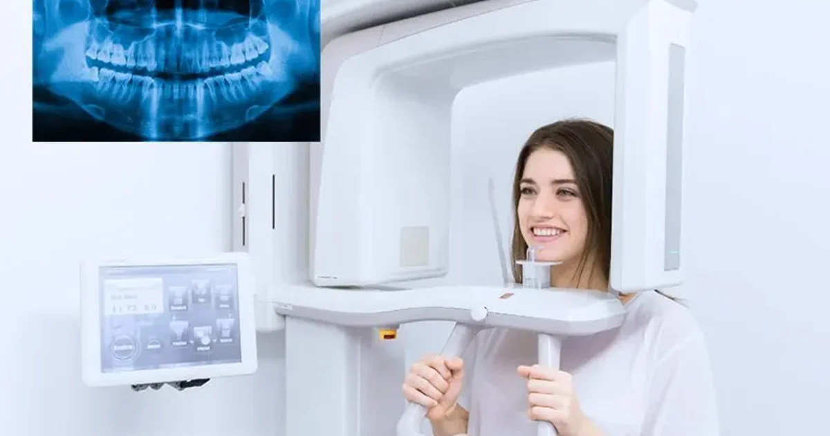
A CBCT scan (Cone Beam Computed Tomography) is a specialized type of X-ray technology that produces detailed, three-dimensional images of the teeth, jawbone, soft tissues, nerve pathways, and surrounding facial structures in a single rotation. Unlike conventional dental X-rays that produce flat, two-dimensional images, CBCT dental imaging captures a complete volumetric dataset that allows dentists, oral surgeons, orthodontists, endodontists, and radiologists to view the anatomy from any angle and in any cross-sectional plane. This technology has transformed modern dentistry by providing the precision needed for complex procedures such as dental implant placement, wisdom tooth extraction planning, root canal treatment, and orthodontic assessment. Our CBCT scan service at DCDC offers same-day appointments in Dubai Healthcare City.
This comprehensive guide covers everything patients and referring clinicians need to know about CBCT scanning in 2026: what CBCT stands for and how the technology works, how it compares to OPG panoramic X-rays and medical CT scans, the different types of CBCT scans by field of view, specific uses in implant planning, wisdom teeth, root canal treatment, orthodontics, TMJ assessment, and airway analysis, how to prepare for a scan, what happens during the procedure step by step, radiation safety and dose comparisons, and where to get a CBCT scan at Doctors Clinic Diagnostic Center (DCDC) in Dubai Healthcare City.


What Is a CBCT Scan?
CBCT stands for Cone Beam Computed Tomography, a diagnostic imaging technique that uses a cone-shaped X-ray beam to capture a cylindrical volume of data in a single 360-degree rotation around the patient's head. The name describes the geometry of the X-ray beam: rather than the narrow, fan-shaped beam used in traditional medical CT scanners, a CBCT machine emits a wide, cone-shaped beam that captures an entire region of interest in one pass. This fundamental difference in beam geometry is what makes CBCT faster, more compact, and lower in radiation dose than conventional CT.
CBCT technology was first introduced for dental and maxillofacial imaging in the late 1990s and has since become a standard diagnostic tool in dental clinics, oral surgery practices, and diagnostic imaging centres worldwide. The technology bridges the gap between limited two-dimensional dental X-rays (such as periapical and panoramic radiographs) and full medical CT scans, providing three-dimensional detail at a fraction of the radiation dose and cost of a hospital CT scanner.
A CBCT scan works by rotating a compact X-ray source and a flat-panel digital detector around the patient's head in a single arc, capturing between 200 and 600 individual projection images over the course of 10 to 40 seconds. These raw projection images are then processed by sophisticated reconstruction algorithms that convert them into a three-dimensional volumetric dataset composed of hundreds of cross-sectional slices, each as thin as 0.1 to 0.3 millimetres.
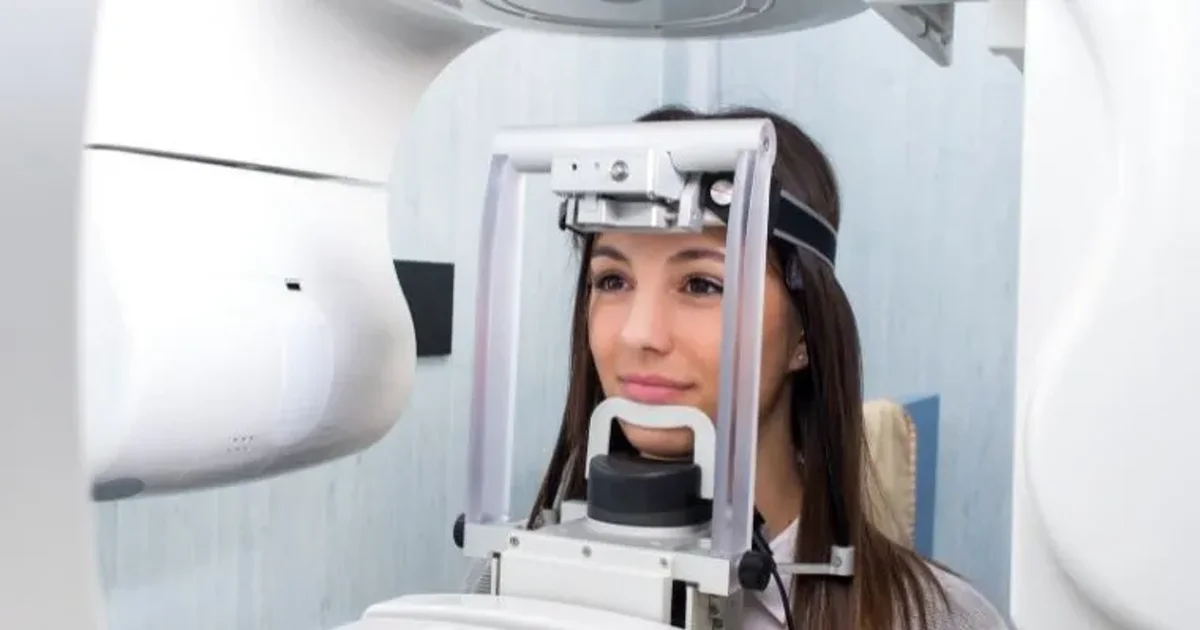
During the scan, the patient stands upright or sits in a chair with their head stabilized by a chin rest and head support. The C-arm of the CBCT machine rotates smoothly around the head, and the patient simply needs to remain still for the duration of the rotation. There is no tunnel or enclosed space, making CBCT significantly more comfortable than a traditional medical CT scanner, especially for patients who experience claustrophobia.
Once the raw data is captured, the computer reconstructs the images into three standard viewing planes: axial (horizontal slices from top to bottom), coronal (front-to-back slices), and sagittal (side-to-side slices). Clinicians can also generate curved panoramic reconstructions, cross-sectional views along the arch of the jaw, and full 3D volume renderings. This versatility is what makes CBCT indispensable for treatment planning across multiple dental specialties.
"At DCDC, we use CBCT technology that captures a complete 3D image of the jaw in under 40 seconds with radiation levels significantly lower than a standard medical CT scan," explains Dr. Osama Elzamzami, Consultant Radiologist at DCDC. "This means patients get the precise imaging their dentist needs without unnecessary exposure."
What Does a CBCT Scan Show?
A CBCT scan reveals a comprehensive range of dental and maxillofacial structures with exceptional clarity:
- Teeth and tooth roots: Position, shape, number, length, curvature, root fractures, and supernumerary or unerupted teeth
- Jawbone (alveolar bone): Bone height, width, density, and volume available for dental implant placement or bone grafting
- Nerve canals: The exact course of the inferior alveolar nerve, mental foramen, and other critical nerve structures
- Maxillary sinuses: Sinus floor position, membrane thickness, mucous retention cysts, and sinusitis
- Airways: Upper airway dimensions relevant to sleep apnoea assessment and orthodontic treatment planning
- Temporomandibular joints (TMJ): Condylar morphology, joint space, degenerative changes, and signs of TMD
- Pathology: Cysts, tumours, periapical lesions, bone infections, root resorption, and other jaw abnormalities
- Impacted teeth: The precise three-dimensional position of impacted wisdom teeth and canines relative to adjacent teeth and nerves
CBCT vs OPG: When Do You Need 3D vs 2D?
One of the most common clinical decisions in dental imaging is whether a patient needs a CBCT scan or whether a standard OPG (orthopantomogram/panoramic X-ray) is sufficient. Understanding the differences helps patients appreciate why their dentist may recommend one over the other.
| Feature | OPG (Panoramic X-Ray) | CBCT Scan |
|---|---|---|
| Imaging type | 2D flat panoramic image | 3D volumetric dataset (hundreds of slices) |
| Dimensions shown | Height and width only (no depth) | Height, width, and depth (all 3 dimensions) |
| Radiation dose | 10-20 uSv | 30-200 uSv (depending on field of view) |
| Bone measurement accuracy | 15-30% magnification error | True 1:1 measurements, accurate to 0.1 mm |
| Nerve canal visibility | Approximation only (2D overlap) | Exact 3D course and dimensions clearly visible |
| Detail level | Limited by overlapping structures | 0.1-0.3 mm voxels (superior bone resolution) |
| Scan time | 10-15 seconds | 10-40 seconds |
| Cost at DCDC | From AED 100-200 | From AED 350-450 |
| Patient position | Standing, biting on tab | Standing or seated, chin rest + head support |
| Best for | Routine screening, general dental check-ups, orthodontic overview, assessing tooth eruption | Implant planning, wisdom tooth nerve mapping, root canal assessment, orthodontic complex cases, TMJ evaluation, surgical planning |
| When to upgrade | N/A — first-line imaging | When OPG shows nerve proximity, insufficient bone detail, or complex anatomy requiring 3D visualization |
CBCT vs OPG comparison. OPG remains the appropriate first-line imaging for most routine dental assessments; CBCT is indicated when 3D detail is clinically required for treatment planning or safety.
When does your dentist upgrade from OPG to CBCT? The decision to perform a CBCT instead of or in addition to an OPG is based on clinical need. A CBCT is typically recommended when the OPG shows signs of nerve proximity to wisdom teeth, when bone dimensions cannot be adequately assessed in 2D for implant planning, when root canal treatment fails and retreatment needs 3D assessment, when orthodontic treatment involves impacted canines or skeletal asymmetry, or when TMJ evaluation requires bony detail. In approximately 30-40 percent of cases where the OPG suggests a problem near the nerve, the CBCT reveals the true relationship is different from what the 2D image suggested.
For a detailed side-by-side comparison, see our dedicated article on CBCT vs OPG X-ray: which do you need?
CBCT vs Regular CT Scan: Key Differences
Patients sometimes confuse CBCT with a regular medical CT scan performed in a hospital radiology department. While both produce 3D images, the technologies are fundamentally different in their design, radiation exposure, image characteristics, and clinical applications.
| Feature | CBCT (Cone Beam CT) | Medical CT Scan |
|---|---|---|
| X-ray beam shape | Cone-shaped (wide beam, single rotation) | Fan-shaped (narrow beam, hundreds of rotations) |
| Radiation dose (head) | 30-200 uSv | 2,000-2,500 uSv (10-60x higher) |
| Bone resolution | 0.1-0.3 mm voxels (superior for dental/jaw) | 0.5-1.0 mm voxels |
| Soft tissue contrast | Limited (not diagnostic for soft tissue) | Excellent (diagnostic quality for brain, organs) |
| Machine size | Compact, office-based | Large, hospital-based |
| Patient position | Standing or seated, open design | Lying down in enclosed tunnel |
| Contrast dye | Not used | Frequently used (IV iodinated contrast) |
| Scan time | 10-40 seconds (single rotation) | 5-30 seconds per body region |
| Best for | Teeth, jawbone, sinuses, TMJ, dental surgery planning | Brain, organs, trauma, cancer staging, vascular imaging |
| Cost (Dubai) | AED 350-1,500 | AED 2,000-5,000+ |
CBCT vs medical CT scan comparison. CBCT is purpose-built for dental and maxillofacial imaging with superior bone detail at a fraction of the radiation dose. Medical CT is required for soft tissue evaluation, trauma, and systemic disease assessment.
The critical takeaway is that CBCT produces sharper images of bony dental anatomy than medical CT despite using far less radiation. A medical CT scan of the head delivers approximately 2,000-2,500 microsieverts, compared to 30-200 microsieverts for a CBCT scan. This means CBCT exposes the patient to 10-60 times less radiation for the specific purpose of dental and jaw imaging. Medical CT becomes necessary only when soft tissue evaluation is required (brain pathology, soft tissue tumours, or vascular assessment).
Radiation Dose Comparison: How Safe Is CBCT?
Patients who are told they need a CBCT scan often ask: is CBCT safe? The short answer is that a CBCT scan is one of the safest advanced imaging technologies in modern dentistry. Putting the radiation dose into perspective with everyday comparisons helps illustrate just how low the exposure is.
| Imaging/Exposure Type | Effective Dose (uSv) | Equivalent to Background Radiation | Comparison to CBCT |
|---|---|---|---|
| Single periapical X-ray | 5 | ~1 day | ~10-20x lower than CBCT |
| Bitewing X-rays (4 films) | 20 | ~3 days | ~5x lower |
| OPG panoramic X-ray | 10-20 | ~2-3 days | ~5-10x lower |
| Chest X-ray | 20 | ~3 days | ~5x lower |
| CBCT scan (small FOV) | 30-80 | ~5-12 days | Baseline (small) |
| CBCT scan (medium FOV) | 50-150 | ~8-23 days | Baseline (medium) |
| CBCT scan (large FOV) | 100-200 | ~15-30 days | Baseline (large) |
| Dubai-London round-trip flight | 60-80 | ~10-12 days | Similar to small CBCT |
| Medical CT scan (head) | 2,000-2,500 | ~10 months | 10-60x higher than CBCT |
| Medical CT scan (abdomen) | 8,000-10,000 | ~3-4 years | 50-300x higher than CBCT |
| Annual background radiation (UAE) | 2,400/year | 365 days | Reference baseline |
Radiation dose comparison across dental, medical, and everyday exposures. CBCT radiation is equivalent to a few days of natural background radiation and comparable to a short-haul flight.
Every CBCT scan follows the ALARA principle: As Low As Reasonably Achievable. This means a scan is recommended only when the clinical benefit of 3D imaging clearly outweighs the minimal radiation exposure. At DCDC, every scan request is reviewed to confirm clinical justification before proceeding, and the smallest field of view and lowest dose settings that produce diagnostically adequate images are always selected.
CBCT Dental Uses: When Is 3D Imaging Needed?
CBCT has specific clinical applications across virtually every dental specialty. Understanding these uses helps patients appreciate why their dentist has recommended a 3D scan rather than a conventional 2D X-ray.
1. Dental Implant Planning
Dental implant placement is the single most common indication for CBCT. Implant surgery involves drilling into bone and placing a titanium post that must integrate with surrounding tissue. A CBCT scan provides precise bone height, width, and density measurements; the exact location and course of the inferior alveolar nerve; maxillary sinus floor height (determining if a sinus lift is needed); neighboring tooth root positions; and detection of existing pathology. Published research shows CBCT imaging changes the surgical plan in approximately 30% of dental implant cases, revealing bone that is thinner than expected or nerves positioned differently than the 2D X-ray suggested.
2. Orthodontic Treatment Planning
CBCT gives orthodontists complete 3D views of teeth, roots, jawbone, TMJ, and upper airway. Research confirms CBCT changes the orthodontic diagnosis or treatment plan in up to 36% of cases. It is particularly valuable for impacted canine localization, skeletal asymmetry assessment, temporary anchorage device (TAD) placement planning, airway analysis for sleep apnoea patients, and detecting pre-existing root resorption before treatment begins.
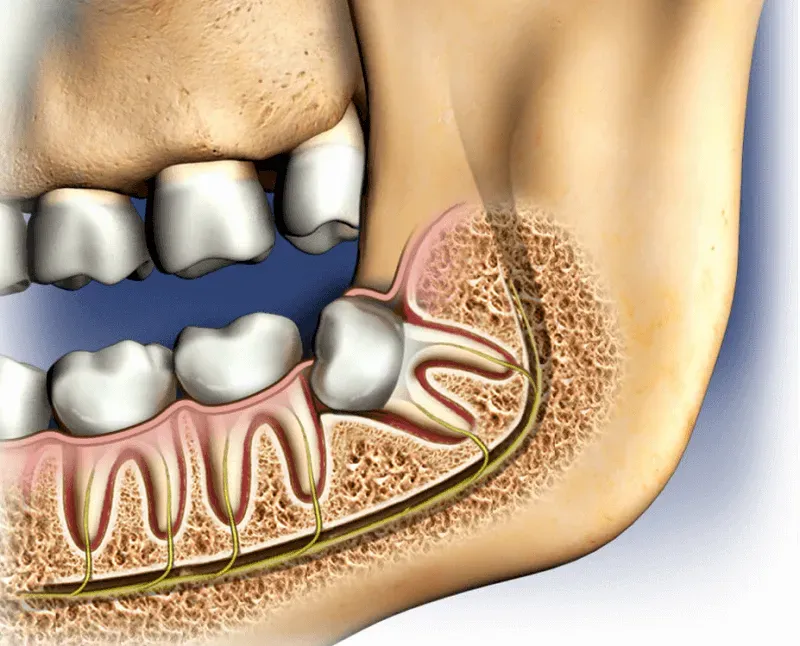
3. Wisdom Tooth Nerve Mapping
CBCT shows the exact three-dimensional relationship between wisdom tooth roots and the inferior alveolar nerve. Research demonstrates CBCT assessment before extraction reduces nerve injury rates by 25-30%. CBCT is recommended when the OPG shows signs of nerve proximity, deep impaction, or unusual root morphology.
4. Root Canal Diagnosis
CBCT detects up to 34% more root canals than conventional X-rays, particularly the MB2 canal of upper molars (present in 90% of teeth but detected on 2D X-rays in fewer than 55%). It is indicated for complex root anatomy, retreatment cases, suspected root fractures, persistent post-treatment symptoms, and pre-surgical planning for apicoectomy.
5. TMJ (Jaw Joint) Assessment
For patients with jaw pain, clicking, locking, or limited mouth opening, CBCT reveals bony changes in the condyle (flattening, erosion, osteophyte formation) that indicate degenerative joint disease. While MRI is the gold standard for soft tissue (disc) evaluation, CBCT provides superior hard tissue visualization at a fraction of the cost.
6. Airway Analysis
Large-field CBCT scans capture the upper airway, allowing volumetric measurement of airway dimensions relevant to obstructive sleep apnoea assessment. This is increasingly used in combined orthodontic-sleep medicine treatment planning.
Types of CBCT Scans: Small, Medium & Large Field of View
Not all CBCT scans are the same. The field of view (FOV) determines how much anatomy is captured and directly affects radiation dose, image resolution, and cost.
| FOV Type | Coverage Area | Typical Size | Radiation Dose | Cost at DCDC (AED) | Common Uses |
|---|---|---|---|---|---|
| Small FOV | A few teeth and surrounding bone | 4x4 to 5x5 cm | 30-80 uSv | From 350 | Single implant site, endodontic assessment, localized pathology, root fracture |
| Medium FOV | One complete jaw or quadrant | 8x8 to 10x10 cm | 50-150 uSv | From 400 | Multiple implants, wisdom teeth, orthodontic assessment of one arch |
| Large FOV | Both jaws and full facial skeleton | 15x15 to 20x17 cm | 100-200 uSv | From 450 | Full-arch implants (All-on-4/6), surgical planning, TMJ bilateral, full orthodontic records, airway analysis |
CBCT field of view options with radiation doses, costs, and clinical indications at DCDC Dubai Healthcare City. Smaller FOV means lower radiation and higher resolution for the target area.
CBCT Scan Procedure: Step by Step
The CBCT scan procedure is straightforward, non-invasive, and remarkably quick. From arrival to departure, the entire appointment typically takes 10 to 15 minutes, with the actual scan lasting only 10 to 40 seconds.
| Step | What Happens | Duration |
|---|---|---|
| 1. Arrival and registration | Check in at reception, provide referral and identification. | 2-3 minutes |
| 2. Preparation | Remove all metal objects from head and neck area. A lead thyroid collar may be placed. | 1-2 minutes |
| 3. Positioning | Stand or sit at the CBCT machine. Chin rests on padded support, forehead stabilizer keeps head in position. | 1-2 minutes |
| 4. The scan | C-arm rotates smoothly around your head. Quiet humming sound. Keep tongue pressed against roof of mouth, lips closed, breathe through nose. | 10-40 seconds |
| 5. Image reconstruction | Computer reconstructs 3D images. Radiographer checks quality. If motion artifact detected, repeat takes only seconds. | 3-5 minutes |
| 6. Reporting and results | Consultant radiologist reviews all slices and reconstructions. Detailed report prepared. | Same day at DCDC |
Complete CBCT scan procedure timeline from arrival to results. Total appointment time is approximately 10-15 minutes.
The procedure is completely painless. There is no injection, no contrast dye, no sedation, and no physical sensation during the scan itself. Unlike MRI (lying in a narrow tube for 20-60 minutes) or contrast-enhanced CT (requiring an IV injection), a CBCT scan requires nothing more than standing or sitting still for seconds.
How to Prepare for a CBCT Scan
CBCT scan preparation is remarkably simple. Unlike many medical imaging procedures that require fasting, contrast injections, or lengthy protocols, CBCT requires almost no advance preparation.
- Eat and drink normally: No fasting requirements. Eating a light meal before your appointment is encouraged.
- Take regular medications: No drug interactions because no contrast or sedatives are used.
- Drive yourself: No sedation involved.
- Return to normal activities immediately: No recovery period.
What to Remove Before the Scan
Remove all metal objects from the head and neck area to prevent image artifacts: earrings, necklaces, eyeglasses, hearing aids, hairpins and clips, removable dentures with metal clasps, removable orthodontic retainers, facial piercings, and scarves with metal fasteners. Fixed dental work (crowns, bridges, implants, fixed braces) does not need to be removed.
Special Considerations
Pregnancy: CBCT scans are generally avoided during pregnancy as a precaution, though the fetal dose is less than 0.01 microsieverts. Elective scans should be postponed until after delivery. Children: CBCT can be safely performed on children when clinically justified using dedicated pediatric protocols that reduce dose by 30-40 percent.
CBCT Scan Cost in Dubai
CBCT scan costs in Dubai vary based on the field of view, facility, and whether a radiologist report is included. At DCDC, pricing is transparent and includes the scan, 3D reconstruction, and a detailed consultant radiologist report.
At DCDC, CBCT scans are priced from AED 350-450 depending on the field of view selected. This compares favourably to the wider Dubai market range of AED 500-1,500 at other facilities. For patients with dental insurance, CBCT scans may be covered when ordered by a dentist for clinical indications. DCDC works with 20+ insurance partners with direct billing.
For detailed pricing information including insurance coverage, see our guide on CBCT scan cost in Dubai.
Book Your CBCT Scan at DCDC
At Doctors Clinic Diagnostic Center in Dubai Healthcare City, the entire CBCT scan appointment takes just 10-15 minutes with same-day results. CBCT from AED 350. Our experienced radiology team ensures a comfortable, efficient experience.
Walk-in or appointment. Sat-Thu 8 AM-10 PM, Fri 9 AM-9 PM. Free parking. 20+ insurance partners.
CBCT for Dental Implants: Detailed Guide
Dental implant placement is the single most common indication for CBCT. The margin for error in implant surgery is extremely small: placing an implant even 1-2 millimetres off course can damage a nerve, perforate the sinus floor, or result in insufficient bone support. CBCT eliminates much of this risk by providing a complete anatomical map before surgery begins.
Traditional 2D dental X-rays compress three-dimensional anatomy into a flat image. A panoramic X-ray shows height but not the buccolingual (cheek-to-tongue) dimension, and magnifies structures by 15-30%, making measurements unreliable. Since an implant occupies space in all three dimensions, planning with only two dimensions is fundamentally incomplete.
Published research in the International Journal of Oral & Maxillofacial Implants reports that implants placed with CBCT-guided planning achieve success rates exceeding 97%, compared to approximately 90-95% with 2D imaging alone. The European Association for Osseointegration (EAO) and the International Team for Implantology (ITI) both recommend CBCT when conventional radiography does not provide sufficient information.
CBCT for Wisdom Teeth: Nerve Mapping
A CBCT scan for wisdom teeth eliminates the guesswork about nerve proximity that plagues 2D imaging. Research published in the British Journal of Oral and Maxillofacial Surgery demonstrates that CBCT assessment before extraction reduces the incidence of inferior alveolar nerve injury by 25-30% compared to surgery planned on panoramic radiographs alone.
"In approximately 30-40% of cases where the panoramic X-ray suggests nerve proximity, the CBCT reveals that the nerve is actually separated from the roots by intact bone," notes Dr. Osama Elzamzami. "In the remaining cases, the CBCT confirms true intimate contact, and the surgeon can modify the approach or perform a coronectomy to protect the nerve."
CBCT for Root Canal Treatment
Root canal treatment relies on locating, cleaning, and sealing every canal inside a tooth. A landmark study in the Journal of Endodontics found that CBCT changed the diagnosis in 62% of endodontic cases compared to periapical radiography alone. CBCT identifies up to 34% more root canals, particularly the MB2 canal of upper first molars (present in 90% of teeth but detected on X-rays in fewer than 55% of cases). It also detects vertical root fractures with 70-80% sensitivity versus only 20-30% on 2D X-rays.
CBCT for Orthodontics
CBCT gives orthodontists the 3D information needed for complex treatment planning. Research in the American Journal of Orthodontics and Dentofacial Orthopedics confirms CBCT changes the orthodontic diagnosis or treatment plan in up to 36% of cases. Key applications include impacted canine localization (CBCT detected root resorption in 48% of cases where it was invisible on panoramic X-rays), alveolar bone thickness assessment, airway volumetric measurement, skeletal asymmetry analysis, and TAD placement planning. CBCT is not needed for every orthodontic case, but is specifically recommended for impacted teeth, skeletal asymmetry, surgical orthodontics, and complex root anatomy.
CBCT Scan at DCDC Dubai Healthcare City
DCDC (Doctors Clinic Diagnostic Center) in Dubai Healthcare City offers advanced CBCT scanning alongside a comprehensive range of dental and medical imaging services. The centre uses a modern CBCT system with adjustable fields of view and optimized dose protocols aligned with international guidelines from the ICRP, EADMFR, and the Dubai Health Authority.
- Clinical justification review: Every scan request is assessed to confirm 3D imaging is necessary
- Optimized dose settings: Lowest kV and mA settings that produce diagnostically acceptable images, with dedicated pediatric protocols
- Smallest field of view: Scan limited to the smallest area needed, reducing radiation by up to 60 percent
- Lead thyroid collar: Placed during every scan to shield the thyroid gland
- Same-day results: Consultant radiologist report and 3D image dataset typically available same day
- Digital sharing: Images shared with referring dentist in DICOM format, compatible with all major planning software
With over 13 years of operation, a 4.8/5 Google rating from over 1,000 patient reviews, and more than 1,000 diagnostic scans performed monthly, DCDC has established a reputation for accurate, timely imaging. Direct billing is available with 20+ insurance providers including Daman, AXA, Bupa, MetLife, and Cigna.
Patient Stories: How CBCT Changed Treatment Plans
Sinus Discovery That Changed an Implant Plan
A 45-year-old Dubai resident needed upper jaw implants. A previous clinic's panoramic X-ray suggested 10 mm of bone below the sinus. CBCT at DCDC revealed only 3 mm of actual bone height. The surgeon planned a sinus lift first, built the necessary bone, and placed implants safely months later.
Nerve Wrapped Around Wisdom Tooth Root
A 28-year-old patient's OPG showed wisdom tooth roots overlapping with the inferior alveolar nerve canal. CBCT revealed the nerve tracked between divergent roots with no cortical bone boundary. A coronectomy was performed instead of complete extraction, preserving nerve function.
Hidden Canal Saves a Tooth From Extraction
A 38-year-old with persistent pain in a previously treated molar had normal-appearing 2D X-rays. CBCT revealed a missed distal canal and a 4 mm periapical lesion hidden within buccal bone. Retreatment targeting the untreated canal resolved symptoms completely.
Get Your 3D Dental Scan at DCDC
DCDC in Dubai Healthcare City offers advanced CBCT scan services from AED 350, with same-day results, experienced consultant radiologists, dose-optimized protocols, and transparent pricing. Walk-in or book ahead.
MOHAP Licensed. Sat-Thu 8 AM-10 PM, Fri 9 AM-9 PM. Free parking.
Related Services at DCDC
Expert care and advanced diagnostics at Dubai Healthcare City
Frequently Asked Questions
Final Thoughts
A CBCT scan represents one of the most significant advancements in dental diagnostic imaging, providing three-dimensional detail that fundamentally changes how dentists, oral surgeons, endodontists, and orthodontists plan and execute treatment. Whether compared to OPG panoramic X-rays or medical CT scans, CBCT occupies a unique position: it delivers superior bone resolution for dental and maxillofacial imaging at radiation doses equivalent to just a few days of natural background radiation, with a scan time of 10-40 seconds and no preparation or recovery required.
At Doctors Clinic Diagnostic Center in Dubai Healthcare City, patients benefit from advanced CBCT technology, strict ALARA safety protocols, pediatric dose settings, experienced radiology specialists, and same-day results. CBCT scans start from AED 350-450, with direct billing available for 20+ insurance partners. If your dentist has recommended a CBCT scan, or if you are planning dental implants, orthodontic treatment, wisdom tooth extraction, or root canal therapy, booking a CBCT scan in Dubai at DCDC is a practical step. For pricing details, visit our guide on CBCT scan cost in Dubai, compare CBCT with panoramic X-rays in our CBCT vs OPG guide, or explore our CBCT imaging service page.
Sources & References
This article was reviewed by our medical team and references the following sources:
- American Dental Association (ADA) - Dental Radiographic Examinations: Recommendations for Patient Selection and Limiting Radiation Exposure
- European Academy of DentoMaxilloFacial Radiology (EADMFR) - Basic Principles for Use of Dental Cone Beam CT
- International Commission on Radiological Protection (ICRP) - Publication 103: The 2007 Recommendations
- Ludlow JB, Timothy R, Walker C, et al. - Effective dose of dental CBCT: a meta-analysis. Dentomaxillofacial Radiology, 2015
- International Journal of Oral & Maxillofacial Implants - CBCT in Implant Dentistry: Systematic Review
- British Journal of Oral and Maxillofacial Surgery - CBCT Assessment of Mandibular Third Molars and the Inferior Alveolar Nerve
- American Association of Endodontists (AAE) - Position Statement on Use of CBCT in Endodontics
Medical content on this site is reviewed by DHA-licensed physicians. See our editorial policy for more information.

Written by
Dr. Osama Elzamzami
Consultant Radiologist
MD, FRCR
Dr. Osama Elzamzami is a Consultant Radiologist specializing in diagnostic imaging including CBCT, CT, MRI, and ultrasound at DCDC Dubai Healthcare City.
Related Articles

CBCT Scan Cost in Dubai: Pricing Guide

CBCT vs OPG X-Ray: Which Do You Need?
OPG X-Ray Results Explained: Complete Guide
Dental Implant Cost Dubai: Complete Guide
Wisdom Teeth Removal Dubai: From AED 399
More in Diagnostic Imaging
CT Scan Cost Dubai: Prices & Types (2026)
Read More
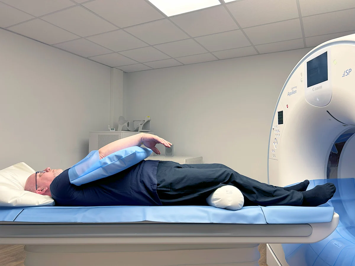
DEXA Scan Dubai: Cost, T-Score & Who Needs It (2026)
Read More![Full Body MRI Cost Dubai: AED 5,000-15,000 [2026]](/wp-media/blog/full-body-mri-scan.webp)
Full Body MRI Cost Dubai: AED 5,000-15,000 [2026]
Read More
MRI Preparation Dubai: Complete Guide (2026)
Read More
Ultrasound Preparation Dubai: What to Expect (2026)
Read More
3D vs 2D Mammogram Dubai: Key Differences (2026)
Read More© 2026 Doctors Clinic Diagnostic Center (DCDC), Dubai Healthcare City. Originally published at https://doctorsclinicdubai.ae/blog/what-is-cbct-scan. All rights reserved. Unauthorized reproduction is prohibited.