اہم نکات
- پیریفرل ویسکولر ڈیزیز (PVD) دل سے باہر کی شریانوں کو متاثر کرتی ہے، خاص طور پر ٹانگوں میں، اور تشخیص نہ ہونے کی صورت میں کٹاؤ سمیت سنگین پیچیدگیوں کا سبب بن سکتی ہے
- ڈاپلر الٹراساؤنڈ PVD کی تشخیص کا بنیادی غیر حملہ آور آلہ ہے، جو نچلے اعضاء کی شریانوں میں خون کے بہاؤ کی رفتار، موج کے پیٹرن اور تنگی کے مقامات کی نقشہ بندی کرتا ہے
- اینکل بریکیئل انڈیکس (ABI)، ایک سادہ پریشر ریشو ٹیسٹ، شریانی بیماری کی پہلی معروضی پیمائش فراہم کرتا ہے اور رہنمائی کرتا ہے کہ مزید ڈاپلر امیجنگ کی ضرورت ہے یا نہیں
- اہم خطرے کے عوامل میں ذیابیطس، تمباکو نوشی، ہائی بلڈ پریشر اور 50 سال سے زائد عمر شامل ہیں، یہ سب UAE کی آبادی میں بہت زیادہ پائے جاتے ہیں
- ڈاپلر سے جلد تشخیص پیچیدگیوں سے پہلے علاج شروع کرنے کی اجازت دیتی ہے، طرز زندگی میں تبدیلیوں اور ادویات سے لے کر اینجیوپلاسٹی یا بائی پاس سرجری تک
پیریفرل ویسکولر ڈیزیز (PVD)، جسے پیریفرل آرٹری ڈیزیز (PAD) بھی کہا جاتا ہے، اس وقت ہوتی ہے جب چربی کے ذخائر ان شریانوں کو تنگ کر دیتے ہیں جو آپ کی ٹانگوں اور پاؤں کو خون فراہم کرتی ہیں۔ خون کے بہاؤ میں کمی سے چلتے وقت ٹانگوں میں درد سے لے کر نہ بھرنے والے زخموں تک اور شدید صورتوں میں بافتوں کی موت کی وجہ سے کٹاؤ کی ضرورت تک علامات پیدا ہوتی ہیں۔ ڈاپلر الٹراساؤنڈ وہ فرنٹ لائن تشخیصی آلہ ہے جو ڈاکٹروں کو سرجری، تابکاری یا کنٹراسٹ ڈائی کے بغیر اس شریانی تنگی کو دیکھنے اور ماپنے کی اجازت دیتا ہے۔
دنیا بھر میں تقریباً 200 ملین لوگوں کو متاثر کرنے کے باوجود، پیریفرل ویسکولر ڈیزیز کی تشخیص کم رہتی ہے کیونکہ اس کی ابتدائی علامات اکثر عام عمر رسیدگی یا پٹھوں کی تھکاوٹ سمجھ کر نظرانداز کر دی جاتی ہیں۔ یہ گائیڈ بتاتی ہے کہ PVD کیا ہے، یہ کیسے پیدا ہوتی ہے، ڈاپلر الٹراساؤنڈ تشخیص کا ترجیحی طریقہ کیوں ہے، ٹیسٹ آپ کی شریانوں کے بارے میں کیا ظاہر کرتا ہے، اور تشخیص کی تصدیق کے بعد کون سے علاج کے راستے دستیاب ہیں۔


پیریفرل ویسکولر ڈیزیز کیا ہے؟
پیریفرل ویسکولر ڈیزیز ایک گردشی حالت ہے جس میں ایتھروسکلروسس، شریان کی دیواروں کے اندر چربی والی تختی کا جمع ہونا، اعضاء تک خون کے بہاؤ کو کم کر دیتا ہے۔ اگرچہ PVD دل اور دماغ سے باہر کسی بھی شریان کو متاثر کر سکتی ہے، یہ عام طور پر ٹانگوں کی شریانوں کو نشانہ بناتی ہے: ایلیاک، فیمورل، پوپلیٹیئل اور ٹبیئل شریانیں۔ بیماری آہستہ آہستہ سالوں میں بڑھتی ہے اور بہت سے مریضوں کو علامات نمایاں ہونے تک احساس نہیں ہوتا کہ انہیں یہ ہے۔
PVD اور PAD اکثر باہم بدل کر استعمال ہوتے ہیں، حالانکہ تکنیکی طور پر PVD ایک وسیع تر اصطلاح ہے جس میں شریانی اور وریدی دونوں حالتیں شامل ہیں۔ طبی عمل میں، جب ڈاکٹر ایتھروسکلروسس کے تناظر میں PVD کا حوالہ دیتے ہیں، تو وہ اسی تنگی کے عمل کو بیان کر رہے ہیں جو دل میں کورونری آرٹری ڈیزیز کا سبب بنتا ہے، سوائے اس کے کہ یہ پیریفرل گردش میں ہو رہا ہے۔ یہ مشترکہ میکانزم کا مطلب ہے کہ PVD کی تشخیص دل کے دورے اور فالج کے بڑھے ہوئے خطرے کی بھی نشاندہی کرتی ہے۔
PVD کی علامات کی پہچان
پیریفرل ویسکولر ڈیزیز کی علامات شریانی تنگی کی شدت اور جسم نے متبادل خون کے بہاؤ کے راستے (کولیٹرل سرکولیشن) کتنے اچھے طریقے سے تیار کیے ہیں اس پر منحصر ہیں۔ بہت سے مریض بیماری کے بگڑنے کے ساتھ قابل شناخت مراحل سے گزرتے ہیں۔
وقفے وقفے سے لنگڑانا
PVD کی نمایاں علامت وقفے وقفے سے لنگڑانا ہے، پنڈلی، ران یا کولہے کے پٹھوں میں اکڑن، درد یا تھکاوٹ کا احساس جو چلنے یا ورزش کے دوران ہوتا ہے اور آرام سے ختم ہو جاتا ہے۔ ایسا اس لیے ہوتا ہے کیونکہ تنگ شریانیں کام کرنے والے پٹھوں کی بڑھی ہوئی آکسیجن کی ضرورت کو پورا کرنے کے لیے کافی خون نہیں پہنچا سکتیں۔ مریض درد شروع ہونے سے پہلے جتنا فاصلہ چل سکتا ہے، جسے لنگڑانے کا فاصلہ کہا جاتا ہے، بیماری کی شدت کی ایک اہم طبی پیمائش ہے۔
آرام کے وقت درد
جیسے جیسے PVD بڑھتی ہے، درد آرام کے وقت بھی ہو سکتا ہے، خاص طور پر رات کو جب ٹانگیں اوپر ہوتی ہیں۔ مریض اکثر پاؤں کی انگلیوں یا اگلے حصے میں جلن یا درد کا احساس بیان کرتے ہیں جو بستر سے پاؤں لٹکانے سے بہتر ہو جاتا ہے، خون کے بہاؤ میں مدد کے لیے کشش ثقل کا استعمال کرتے ہوئے۔ آرام کے وقت درد شدید شریانی خرابی کی نشاندہی کرتا ہے اور فوری تشخیص کی ضرورت ہوتی ہے۔
جلد کی تبدیلیاں اور نہ بھرنے والے زخم
پیشرفتہ PVD متاثرہ عضو میں نظر آنے والی تبدیلیاں پیدا کرتی ہے: جلد پیلی، چمکدار یا بے رنگ نظر آ سکتی ہے؛ ٹانگوں اور پاؤں پر بالوں کا گرنا عام ہے؛ پاؤں کے ناخن موٹے اور بھربھرے ہو جاتے ہیں؛ اور پاؤں چھونے پر ٹھنڈا محسوس ہو سکتا ہے۔ سب سے اہم بات، پاؤں یا انگلیوں پر زخم اور السر نہیں بھرتے کیونکہ ناکافی خون کا بہاؤ بافتوں کی مرمت کے لیے ضروری آکسیجن اور غذائی اجزاء کی فراہمی کو روکتا ہے۔ یہ نہ بھرنے والے زخم انفیکشن اور ممکنہ کٹاؤ کا بڑا خطرہ ہیں۔
سن ہونا اور کمزوری
خون کی فراہمی میں کمی سن ہونا، جھنجھناہٹ یا ٹانگوں میں بھاری پن کا احساس پیدا کر سکتی ہے۔ ذیابیطس کے مریضوں میں، یہ علامت ذیابیطس نیوروپیتھی کے ساتھ اوورلیپ ہو سکتی ہے، جو طبی تفریق کو اہم بناتی ہے۔ کچھ مریض محسوس کرتے ہیں کہ ایک ٹانگ دوسری سے زیادہ کمزور لگتی ہے، یا چلتے وقت ایک طرف زیادہ جلدی تھکاوٹ ہوتی ہے۔
اینکل بریکیئل انڈیکس: تشخیص کا پہلا قدم
پیشرفتہ امیجنگ سے پہلے، مشتبہ PVD کی تشخیص عام طور پر اینکل بریکیئل انڈیکس (ABI) سے شروع ہوتی ہے، ایک سادہ اور انتہائی معلوماتی ٹیسٹ جو ٹخنے میں بلڈ پریشر کا بازو میں بلڈ پریشر سے موازنہ کرتا ہے۔ بلڈ پریشر کف ٹخنے اور بازو پر رکھا جاتا ہے اور ہینڈ ہیلڈ ڈاپلر پروب ہر مقام پر نبض کا پتہ لگاتا ہے تاکہ درست پریشر ریڈنگز حاصل کی جا سکیں۔
ABI ٹخنے میں سسٹولک بلڈ پریشر کو بازو میں سسٹولک بلڈ پریشر سے تقسیم کرکے حساب کیا جاتا ہے۔ نارمل ABI 1.00 سے 1.40 تک ہوتا ہے۔ 0.90 سے کم ABI پیریفرل آرٹیریل ڈیزیز کی موجودگی کی تصدیق کرتا ہے۔ ABI جتنا کم ہوگا، بیماری اتنی زیادہ شدید ہے۔
| ABI قدر | تشریح | طبی اہمیت |
|---|---|---|
| 1.00 – 1.40 | نارمل | کوئی نمایاں شریانی تنگی نہیں پائی گئی |
| 0.91 – 0.99 | بارڈر لائن | ابتدائی بیماری کا امکان؛ ورزش ABI ٹیسٹنگ کی سفارش کی جا سکتی ہے |
| 0.41 – 0.90 | ہلکا سے اعتدال پسند PAD | شریانی بیماری کی تصدیق؛ ڈاپلر میپنگ کی سفارش |
| 0.00 – 0.40 | شدید PAD | اعضاء کی نازک اسکیمیا؛ فوری ویسکولر تشخیص کی ضرورت |
| 1.40 سے زائد | غیر دبنے والی | ذیابیطس کے مریضوں میں کیلسیفائیڈ شریانوں کی وجہ سے عام؛ متبادل ٹیسٹنگ کی ضرورت |
ABI قدریں شریانی بیماری کی شدت کی معروضی پیمائش فراہم کرتی ہیں۔ کیلسیفائیڈ شریانوں والے ذیابیطس کے مریض غلط طور پر بلند ABI ریڈنگز دکھا سکتے ہیں۔
اگرچہ ABI شریانی بیماری کی موجودگی کی تصدیق کرتا ہے، لیکن یہ ظاہر نہیں کرتا کہ تنگی کہاں واقع ہے یا یہ کتنی وسیع ہے۔ یہیں ڈاپلر الٹراساؤنڈ ضروری ہو جاتا ہے، جو ران سے لے کر پاؤں تک پورے شریانی نظام کا تفصیلی نقشہ فراہم کرتا ہے۔
چلتے وقت ٹانگوں میں درد محسوس ہو رہا ہے؟
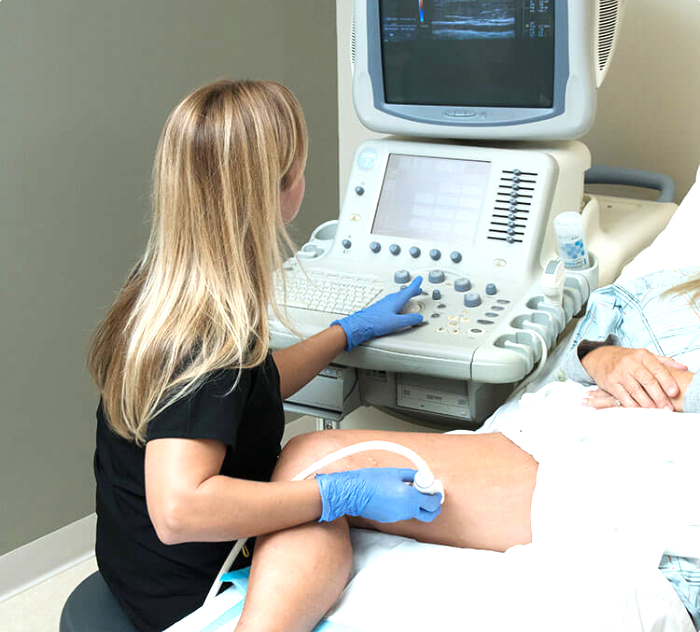
دبئی ہیلتھ کیئر سٹی میں ڈاکٹرز کلینک ڈائیگناسٹک سینٹر میں، ہمارے تجربہ کار ریڈیولوجسٹ شریانی خون کے بہاؤ کی نقشہ بندی اور PAD کی جلد تشخیص کے لیے جامع پیریفرل ویسکولر ڈاپلر تشخیص انجام دیتے ہیں۔
ڈاپلر الٹراساؤنڈ پیریفرل آرٹیریل ڈیزیز کی نقشہ بندی کیسے کرتا ہے
پیریفرل ویسکولر ڈیزیز کے لیے ڈاپلر الٹراساؤنڈ ایک جامع معائنہ ہے جو شریانی خون کی فراہمی کو اورٹک بائفرکیشن (جہاں ایورٹا دو ایلیاک شریانوں میں تقسیم ہوتا ہے) سے لے کر ٹانگ سے ہوتے ہوئے پاؤں تک ٹریس کرتا ہے۔ معائنہ عروق کی ساخت کو دیکھنے کے لیے B-mode (گرے اسکیل) امیجنگ کو خون کے بہاؤ کی سمت دکھانے کے لیے کلر ڈاپلر اور بہاؤ کی رفتار اور موج کے پیٹرن کا تجزیہ کرنے کے لیے سپیکٹرل ڈاپلر کے ساتھ جوڑتا ہے۔
سونوگرافر منظم طریقے سے کامن فیمورل آرٹری، سپرفیشل فیمورل آرٹری (بیماری کا سب سے عام مقام)، گھٹنے کے پیچھے پوپلیٹیئل آرٹری اور گھٹنے کے نیچے ٹبیئل آرٹریز کا معائنہ کرتا ہے۔ ہر سطح پر، معائنہ کرنے والا تختی کی تلاش کرتا ہے، عروق کا قطر ماپتا ہے، خون کے بہاؤ کی رفتار کا جائزہ لیتا ہے اور ڈاپلر ویو فارم پیٹرن ریکارڈ کرتا ہے۔
ویو فارم تجزیہ: شریانی دستخط پڑھنا
پیریفرل ڈاپلر کا سب سے قیمتی پہلو ویو فارم تجزیہ ہے۔ ٹانگ کی صحت مند شریان میں، ڈاپلر ویو فارم ایک خصوصیتی تین مرحلہ پیٹرن دکھاتا ہے: سسٹول (دل کے سکڑنے) کے دوران تیز اوپر کی طرف حرکت، ابتدائی ڈایاسٹول میں بہاؤ کی مختصر واپسی، اور آخری ڈایاسٹول میں آگے کی طرف بہاؤ کا ایک چھوٹا حصہ۔ یہ تین مرحلہ پیٹرن ایک نارمل، لچکدار شریان کی نشاندہی کرتا ہے جس میں کوئی نمایاں اوپر یا نیچے کی بیماری نہیں ہے۔
جیسے جیسے شریانی بیماری بڑھتی ہے، ویو فارم بتدریج بدلتا ہے۔ دو مرحلہ ویو فارم (ابتدائی ڈایاسٹولک واپسی کا نقصان) ہلکی بیماری یا عمر سے متعلق شریانی سختی کی نشاندہی کرتا ہے۔ ایک مرحلہ ویو فارم (مسلسل آگے بہاؤ کے ساتھ سست، گول پیٹرن) نمایاں قریبی تنگی یا رکاوٹ کی نشاندہی کرتا ہے۔ ٹانگ کے ساتھ متعدد مقامات پر ان ویو فارم دستخطوں کو پڑھ کر، ریڈیولوجسٹ تنگی کو براہ راست دیکھنے سے پہلے بھی بیماری کا مقام اور شدت معلوم کر سکتا ہے۔
رفتار کی پیمائش اور تنگی کی درجہ بندی
شریانی تنگی کے مقام پر، خون کی رفتار ڈرامائی طور پر بڑھ جاتی ہے کیونکہ خون کا وہی حجم ایک چھوٹے چینل سے گزرنے پر مجبور ہوتا ہے۔ تنگی پر پیک سسٹولک ویلوسٹی (PSV) کا بالائی نارمل حصے میں PSV سے موازنہ کرکے، ریڈیولوجسٹ ویلوسٹی ریشو حساب کرتا ہے۔ 2.0 سے زائد ریشو عام طور پر کم از کم 50% تنگی کی نشاندہی کرتا ہے، جبکہ 4.0 سے زائد ریشو 75% سے زیادہ تنگی کی نشاندہی کرتا ہے۔ مکمل رکاوٹ اس وقت شناخت کی جاتی ہے جب نظر آنے والی شریان میں کوئی بہاؤ سگنل نہ پایا جائے۔
کلر فلو میپنگ
کلر ڈاپلر بہاؤ کی معلومات کو گرے اسکیل تصویر پر چڑھاتا ہے، جس سے ان جگہوں کی تیزی سے شناخت ممکن ہوتی ہے جہاں خون کا بہاؤ درہم برہم ہو۔ نارمل شریانی بہاؤ ایک یکساں رنگ کے طور پر ظاہر ہوتا ہے جو شریان کی نالی کو بھرتا ہے۔ تنگی پر، رنگ تبدیل ہو کر زیادہ رفتار کی نشاندہی کرتا ہے (عام طور پر چمکدار یا موزیک پیٹرن کے طور پر ظاہر ہوتا ہے)، اور تنگی سے آگے، ہنگامہ خیز بہاؤ مخلوط رنگوں کے طور پر ظاہر ہوتا ہے۔ یہ بصری نقشہ ریڈیولوجسٹ کو لمبے شریانی حصوں کا تیزی سے جائزہ لینے اور تشویش کے علاقوں پر تفصیلی پیمائشوں پر توجہ مرکوز کرنے میں مدد کرتا ہے۔
پیریفرل ویسکولر ڈیزیز کے خطرے کے عوامل
PVD کے خطرے کے عوامل کو سمجھنا اہم ہے کیونکہ ان میں سے زیادہ تر قابل تبدیلی ہیں، یعنی جلد مداخلت بیماری کی پیشرفت کو سست یا روک سکتی ہے۔ اہم خطرے کے عوامل کورونری آرٹری ڈیزیز اور فالج کے عوامل سے نمایاں طور پر مماثل ہیں۔
ذیابیطس
ذیابیطس پیریفرل ویسکولر ڈیزیز کا سب سے مضبوط واحد خطرے کا عامل ہے۔ ذیابیطس والے لوگوں میں PVD ہونے کا امکان دو سے چار گنا زیادہ ہوتا ہے اور وہ کم عمر میں اس سے متاثر ہوتے ہیں۔ زیادہ بلڈ شوگر شریانی اینڈوتھیلیم (اندرونی استر) کو نقصان پہنچاتی ہے، سوزش کو فروغ دیتی ہے اور تختی کی تشکیل کو تیز کرتی ہے۔ اہم بات، ذیابیطس پیریفرل نیوروپیتھی کا بھی سبب بنتی ہے جو لنگڑانے کے درد کو چھپا سکتی ہے، جس سے بیماری کو تشخیص سے پہلے شدید مرحلے تک پہنچنے کا موقع ملتا ہے۔ تیز رفتار بیماری اور کم علامات کا یہ مجموعہ تمام ذیابیطس کے مریضوں کے لیے ڈاپلر اسکریننگ کو خاص طور پر اہم بناتا ہے۔
تمباکو نوشی
سگریٹ نوشی PVD کا سب سے طاقتور قابل تبدیلی خطرے کا عامل ہے۔ تمباکو نوشی کرنے والوں میں غیر تمباکو نوشی کرنے والوں کے مقابلے PAD ہونے کا امکان چار گنا تک زیادہ ہوتا ہے اور انہیں تقریباً ایک دہائی پہلے یہ ہو جاتی ہے۔ تمباکو نوشی اینڈوتھیلیم کو نقصان پہنچاتی ہے، خون کی گاڑھاپن بڑھاتی ہے، جمنے کو فروغ دیتی ہے اور براہ راست شریانوں کی تنگی میں معاون ہوتی ہے۔ خطرہ چھوڑنے کے سالوں بعد بھی بلند رہتا ہے، حالانکہ ترک کرنا پیشرفت کو سست کرتا ہے اور علاج کے نتائج کو نمایاں طور پر بہتر بناتا ہے۔
ہائی بلڈ پریشر
دائمی ہائی بلڈ پریشر شریان کی دیواروں پر میکانیکی دباؤ ڈالتا ہے، ایتھروسکلروٹک عمل کو تیز کرتا ہے۔ ہائی بلڈ پریشر PVD کے خطرے کو تقریباً 50% بڑھا دیتا ہے اور موجودہ بیماری کے بڑھنے کا امکان زیادہ کر دیتا ہے۔ لہذا بلڈ پریشر کا انتظام ایک روک تھام کا اقدام اور PVD کے علاج کا بنیادی جزو دونوں ہے۔
عمر اور دیگر عوامل
PVD کا پھیلاؤ 50 سال کی عمر کے بعد تیزی سے بڑھتا ہے اور 60 سال سے زائد تقریباً 12% سے 20% بالغوں کو متاثر کرتا ہے۔ زیادہ کولیسٹرول، موٹاپا، جسمانی غیر فعالیت، دائمی گردے کی بیماری اور قلبی بیماری کی خاندانی تاریخ سب بڑھے ہوئے خطرے میں حصہ ڈالتے ہیں۔ مردوں میں خواتین کی نسبت قدرے زیادہ شرح سے پایا جاتا ہے، حالانکہ عمر بڑھنے کے ساتھ یہ فرق کم ہو جاتا ہے۔
ڈاپلر تشخیص کے بعد علاج کے راستے
پیریفرل ڈاپلر معائنے کے نتائج براہ راست علاج کے فیصلوں کی رہنمائی کرتے ہیں۔ انتظام بیماری کی شدت اور مقام، مریض کی علامات اور ان کے مجموعی قلبی خطرے کے پروفائل پر منحصر ہوتا ہے۔
طرز زندگی میں تبدیلیاں
PVD کے تمام مراحل کے لیے، طرز زندگی میں تبدیلیاں علاج کی بنیاد بناتی ہیں۔ تمباکو نوشی ترک کرنا سب سے اہم واحد مداخلت ہے جو پیشرفت کی شرح کو نصف کر سکتی ہے۔ نگرانی میں ورزش پروگرام، خاص طور پر پیدل چلنے کے پروگرام جو مریضوں کو لنگڑانے کے شروع ہونے سے آگے چلنے کی ترغیب دیتے ہیں، تین سے چھ ماہ میں چلنے کی دوری کو 50% سے 200% تک بڑھانے کا ثبوت رکھتے ہیں۔ کم سیچوریٹڈ چکنائی والی دل کے لیے صحت مند غذا، ذیابیطس کے مریضوں کے لیے باقاعدہ بلڈ شوگر مانیٹرنگ اور وزن کا انتظام سب بیماری کی پیشرفت کو سست کرنے میں مدد کرتے ہیں۔
طبی علاج
دوائی سے علاج بنیادی خطرے کے عوامل اور خود بیماری کو نشانہ بناتا ہے۔ اینٹی پلیٹلیٹ ایجنٹس (ایسپرین یا کلوپیڈوگریل) قلبی واقعات کا خطرہ کم کرتے ہیں۔ سٹیٹنز کولیسٹرول کم کرتے ہیں اور موجودہ تختی کو مستحکم کرتے ہیں۔ بلڈ پریشر کی دوائیں، خاص طور پر ACE انہیبیٹرز، شریانی تحفظ فراہم کرتی ہیں۔ سیلوسٹازول خاص طور پر لنگڑانے کے لیے تجویز کیا جا سکتا ہے کیونکہ یہ پلیٹلیٹ جمع ہونے کو کم کرکے اور عروق کو کشادہ کرکے چلنے کی دوری بہتر کرتا ہے۔
اینڈوویسکولر اور سرجیکل مداخلتیں
جب طرز زندگی میں تبدیلیاں اور دوائیں ناکافی ہوں، یا جب نازک اعضاء کی اسکیمیا بافتوں کی بقا کو خطرے میں ڈالے، تو مداخلتی طریقہ کار پر غور کیا جاتا ہے۔ اینجیوپلاسٹی، جس میں ایک بیلون کیتھیٹر تنگ شریان کو کھولتا ہے اور اکثر اسٹینٹ لگانے کے ساتھ ملایا جاتا ہے، سب سے عام کم سے کم حملہ آور طریقہ ہے۔ لمبی یا زیادہ پیچیدہ رکاوٹوں کے لیے، جراحی بائی پاس گرافٹنگ، جو ورید یا مصنوعی گرافٹ کا استعمال کرتے ہوئے خون کے بہاؤ کو بیمار حصے سے آگے منتقل کرتی ہے، ضروری ہو سکتی ہے۔ ڈاپلر مطالعے پر شناخت شدہ بیماری کا مقام اور حد ان مداخلتوں کی منصوبہ بندی کے لیے ضروری ہے۔
دبئی میں PVD: ایک بڑھتی ہوئی تشویش
دبئی اور وسیع تر UAE میں پیریفرل ویسکولر ڈیزیز کا پھیلاؤ ایک اہم صحت عامہ کی تشویش ہے جو کئی اکٹھے ہونے والے عوامل سے پیدا ہوتی ہے۔ UAE میں ٹائپ 2 ذیابیطس کی دنیا میں سب سے زیادہ شرحوں میں سے ایک ہے، تقریباً 17% بالغ آبادی متاثر ہے۔ چونکہ ذیابیطس PVD کا سب سے مضبوط واحد خطرے کا عامل ہے، اس لیے یہ براہ راست بیماری کی بلند شرحوں میں تبدیل ہوتا ہے۔
خطے میں غیر فعال طرز زندگی عام ہے، جزوی طور پر شدید گرمیوں کی وجہ سے جو سال کے کئی مہینوں تک باہر جسمانی سرگرمی کو محدود کرتی ہے۔ زیادہ کیلوریز والی غذاؤں کے پھیلاؤ اور موٹاپے اور ہائی بلڈ پریشر کی نمایاں شرحوں کے ساتھ مل کر، ایتھروسکلروٹک بیماری کے لیے حالات مکمل طور پر فراہم ہیں۔ متنوع غیرملکی آبادی میں جنوبی ایشیا کی بڑی برادریاں بھی شامل ہیں، جہاں قلبی بیماری کم عمر میں ظاہر ہونے کا رجحان رکھتی ہے۔
ان خطرے کے عوامل کے باوجود، PVD خطے میں وسیع پیمانے پر کم تشخیص شدہ رہتی ہے۔ بہت سے مریض ٹانگوں کے درد یا تھکاوٹ کو عمر رسیدگی، جسمانی بے ضابطگی یا جوڑوں کے مسائل سے منسوب کرتے ہیں بجائے اس کے کہ ویسکولر تشخیص کرائیں۔ PVD کی علامات کے بارے میں بیداری بڑھانا اور غیر حملہ آور ڈاپلر ٹیسٹنگ کی دستیابی جلد تشخیص اور بہتر نتائج کے لیے ضروری ہے۔
DCDC دبئی ہیلتھ کیئر سٹی میں پیریفرل ویسکولر ڈاپلر
دبئی ہیلتھ کیئر سٹی میں ڈاکٹرز کلینک ڈائیگناسٹک سینٹر میں، پیریفرل ویسکولر ڈاپلر تجربہ کار ریڈیولوجسٹ اعلیٰ ریزولوشن امیجنگ اور درست رفتار کی پیمائش کے قابل جدید ڈاپلر الٹراساؤنڈ آلات کا استعمال کرتے ہوئے انجام دیتے ہیں۔ معائنہ نچلے اعضاء کے مکمل شریانی درخت کا احاطہ کرتا ہے، بیماری کے مقام، شدت اور حد کا مکمل جائزہ فراہم کرتا ہے۔
سینٹر ویسکولر تشخیص کے حصے کے طور پر ABI پیمائش بھی فراہم کرتا ہے، جو مجموعی شریانی صحت کا ایک تکمیلی معروضی پیمانہ فراہم کرتا ہے۔ جامع رپورٹس میں ویو فارم تجزیہ، رفتار کے تناسب، تنگی کی درجہ بندی اور طبی ہم آہنگی شامل ہوتی ہے، سب کچھ مریض کی طبی تاریخ اور خطرے کے عوامل کے تناظر میں تشریح کیا جاتا ہے۔ حوالہ دینے والے ڈاکٹروں، بشمول داخلی طب کے ماہرین، اینڈوکرائنولوجسٹ اور ویسکولر سرجنز کے ساتھ قریبی ہم آہنگی اس بات کو یقینی بناتی ہے کہ تشخیصی نتائج بروقت اور مناسب علاج کے فیصلوں تک پہنچیں۔
ٹانگوں میں درد یا سن ہونا محسوس ہو رہا ہے؟
دبئی ہیلتھ کیئر سٹی میں ڈاکٹرز کلینک ڈائیگناسٹک سینٹر میں پیریفرل ویسکولر ڈاپلر آپ کی ٹانگ کی شریانوں کی تنگی اور رکاوٹوں کے لیے نقشہ بندی کر سکتا ہے۔ ہمارے تجربہ کار ریڈیولوجسٹ آپ کے علاج کے منصوبے کی رہنمائی کے لیے تفصیلی رپورٹس فراہم کرتے ہیں۔
DCDC میں متعلقہ خدمات
دبئی ہیلتھ کیئر سٹی میں ماہرانہ دیکھ بھال اور جدید تشخیص
Doppler Ultrasound
Non-invasive arterial and venous imaging for peripheral vascular disease
اپائنٹمنٹ بک کریںCardiology Consultation
Expert cardiovascular evaluation and vascular risk assessment
اپائنٹمنٹ بک کریںPreventive Cardiology
Proactive heart and vascular disease prevention programs
اپائنٹمنٹ بک کریںFrequently Asked Questions
حتمی خیالات
پیریفرل ویسکولر ڈیزیز ایک عام، سنگین اور کم تشخیص شدہ حالت ہے جو دل کے دوروں اور فالج کے ساتھ اپنی بنیادی وجہ، ایتھروسکلروسس شیئر کرتی ہے۔ ٹانگوں کے درد، اکڑن، سن ہونے اور نہ بھرنے والے زخموں کی علامات کو صرف عمر رسیدگی کا حصہ سمجھ کر کبھی نظرانداز نہیں کرنا چاہیے۔ ڈاپلر الٹراساؤنڈ پوری ٹانگوں میں شریانی بیماری کا پتہ لگانے اور نقشہ بندی کرنے کا بے درد، تابکاری سے پاک اور انتہائی درست طریقہ فراہم کرتا ہے، جو ڈاکٹروں کو مؤثر علاج کی منصوبہ بندی کے لیے درکار تفصیلی معلومات دیتا ہے۔
اگر آپ کو ذیابیطس ہے، تمباکو نوشی کرتے ہیں یا تمباکو نوشی کی تاریخ ہے، ہائی بلڈ پریشر ہے یا 50 سال سے زائد عمر کے ساتھ قلبی خطرے کے عوامل ہیں، تو پیریفرل ویسکولر ڈاپلر تشخیص سب سے قیمتی احتیاطی اقدامات میں سے ایک ہے جو آپ اٹھا سکتے ہیں۔ جلد تشخیص کا مطلب ہے پہلے علاج، بہتر علامات پر قابو اور پیشرفتہ بیماری کی تباہ کن پیچیدگیوں کا کم خطرہ۔ قیمتوں کی تفصیلات کے لیے، دبئی میں ڈاپلر الٹراساؤنڈ کی قیمت پر ہماری گائیڈ دیکھیں۔ دبئی ہیلتھ کیئر سٹی میں ڈاکٹرز کلینک ڈائیگناسٹک سینٹر میں، ہماری ویسکولر امیجنگ ٹیم آپ کی ٹانگوں کی صحت کے لیے مکمل تشخیص فراہم کرنے کے لیے تیار ہے۔
ذرائع اور حوالہ جات
یہ مضمون ہماری طبی ٹیم نے جائزہ لیا ہے اور درج ذیل ذرائع کا حوالہ دیتا ہے:
- امریکن ہارٹ ایسوسی ایشن - پیریفرل آرٹری ڈیزیز
- سوسائٹی فار ویسکولر سرجری - PAD گائیڈ لائنز
- یوروپین سوسائٹی آف کارڈیالوجی - PAD تشخیص اور انتظام
- ریڈیولوجیکل سوسائٹی آف نارتھ امریکا - ویسکولر الٹراساؤنڈ
- انٹرنیشنل ذیابیطس فیڈریشن - ذیابیطس اور قلبی بیماری
اس سائٹ پر طبی مواد کا جائزہ DHA لائسنس یافتہ ڈاکٹرز نے لیا ہے۔ ہماری دیکھیں تحریری پالیسی مزید معلومات کے لیے۔

تحریر
ڈاکٹر اسامہ الزمزمی
کنسلٹنٹ ریڈیولوجسٹ
ایم ڈی، ریڈیولوجی
ڈاکٹر اسامہ الزمزمی ایک کنسلٹنٹ ریڈیولوجسٹ ہیں جو DCDC دبئی ہیلتھ کیئر سٹی میں MRI، CT، الٹراساؤنڈ اور ڈاپلر مطالعات سمیت تشخیصی امیجنگ میں مہارت رکھتے ہیں۔
متعلقہ مضامین

ڈاپلر الٹراساؤنڈ کیا ہے؟ یہ کیسے کام کرتا ہے آسان الفاظ میں
ٹانگ ڈاپلر الٹراساؤنڈ: مکمل گائیڈ
دبئی میں ڈاپلر الٹراساؤنڈ کی قیمت
الٹراساؤنڈ اسکین کیا ہے؟ مکمل گائیڈ
More in Diagnostic Imaging

X-Ray at Home Dubai: Cost & Guide (2026)
مزید پڑھیں
Ultrasound at Home Dubai: From AED 599 (2026)
مزید پڑھیں
Neck MRI Dubai: What It Shows & Cost (2026)
مزید پڑھیں
X-Ray Scan Dubai: Complete Guide (2026)
مزید پڑھیں
MRI vs Ultrasound Dubai: Which Scan? (2026)
مزید پڑھیں
MRI vs X-Ray Dubai: Which Scan Do You Need? (2026)
مزید پڑھیں© 2026 Doctors Clinic Diagnostic Center (DCDC), Dubai Healthcare City. Originally published at https://doctorsclinicdubai.ae/blog/peripheral-vascular-disease-doppler. All rights reserved. Unauthorized reproduction is prohibited.