اہم نکات
- OPG (آرتھوپینٹوموگرام) ایک پینورامک ایکس رے ہے جو دونوں جبڑوں، تمام دانتوں، TMJ جوڑوں، سائنسز اور اعصابی نہروں کو ایک 15 سیکنڈ کے اسکین میں بغیر درد اور بغیر منہ میں سینسر کے پکڑتا ہے
- اسکین کے لیے تقریباً کوئی تیاری نہیں چاہیے - صرف سر اور گردن کے علاقے سے دھاتی زیورات اتاریں؛ فاقہ، انجکشن یا کنٹراسٹ ڈائی کی ضرورت نہیں
- OPG صرف 10-20 مائیکروسیورٹ ریڈی ایشن دیتا ہے، جو 1-2 دن کی قدرتی پس منظر ریڈی ایشن کے برابر ہے اور ایک لمبی پرواز سے کم ہے
- ڈینٹسٹ OPG نتائج کو منظم طریقے سے پڑھتے ہیں، دانت، ہڈی کی سطح، جبڑے کے جوڑ، سائنسز اور اعصابی نہروں کا جائزہ لیتے ہیں
- OPG ڈینٹل امپلانٹ پلاننگ، آرتھوڈونٹک تشخیص، عقل داڑھ کے جائزے، جبڑے کے درد کی تشخیص اور معمول کی ڈینٹل اسکریننگ کے لیے ضروری فرسٹ لائن امیجنگ ہے
- جب OPG ایسی دریافت کی نشاندہی کرتا ہے جس میں تین جہتی تفصیل چاہیے تو CBCT اسکین سرجیکل پلاننگ کے لیے ضروری درستگی فراہم کرتا ہے
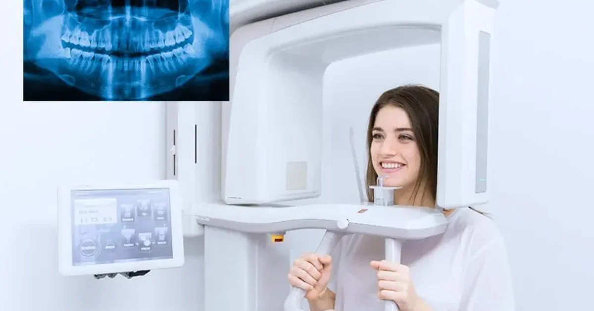
OPG ایکس رے (آرتھوپینٹوموگرام) دنیا میں سب سے زیادہ استعمال ہونے والی ایکسٹراورل ڈینٹل امیجنگ تکنیک ہے۔ 15 سیکنڈ سے کم کے ایک بے درد اسکین میں، یہ دونوں جبڑوں، تمام دانتوں، ٹمپورومینڈیبولر جوڑوں، میکسلری سائنسز اور ارد گرد کی ہڈی کی ایک فلیٹ، دو جہتی پینورامک تصویر کان سے کان تک پکڑتی ہے۔
یہ 2026 میگا گائیڈ ڈاکٹر اسامہ الزمزمی، کنسلٹنٹ ریڈیولوجسٹ (MD, FRCR) ڈاکٹرز کلینک ڈائیگناسٹک سینٹر (DCDC)، دبئی ہیلتھ کیئر سٹی میں جائزہ لیا گیا ہے۔ ہر سیکشن طبی طور پر تصدیق شدہ ہے۔


OPG ایکس رے کیا ہے؟
OPG کا مطلب آرتھوپینٹوموگرام ہے، یونانی الفاظ "آرتھو" (سیدھا)، "پین" (سب) اور "ٹوموگرام" (مقطعی تصویر) سے بنا اصطلاح۔ طبی عمل میں اسکین کو پینورامک ریڈیوگراف یا صرف "پینو" بھی کہا جاتا ہے۔
یہ تصور فنش ریڈیولوجسٹ یورجو ویلی پاٹیرو نے 1940 کی دہائی میں تیار کیا تھا۔ آج ڈیجیٹل OPG سسٹمز نے فلم پر مبنی آلات کی جگہ لے لی ہے۔
OPG ایکس رے کیسے کام کرتی ہے؟
OPG ایکس رے روٹیشنل ٹوموگرافی نامی تکنیک استعمال کرتی ہے جس میں ایکس رے ٹیوب اور ڈیجیٹل ڈیٹیکٹر بیک وقت مریض کے سر کے گرد نیم دائرے میں گھومتے ہیں۔ سافٹ ویئر ان ٹکڑوں کو ایک پینورامک تصویر میں دوبارہ بناتا ہے۔
کلیدی اصول نیرو بیم ٹوموگرافی کہلاتا ہے۔ پورے سر کو ایک ساتھ ریڈی ایشن دینے کی بجائے، OPG مشین ایک پتلی، مرکوز شعاع استعمال کرتی ہے۔
چونکہ ایکس رے ذریعہ پوری کارروائی کے دوران منہ کے باہر رہتا ہے، OPG کو ایکسٹراورل امیجنگ تکنیک کے طور پر درجہ بندی کیا جاتا ہے۔ جن مریضوں کو شدید گیگ ریفلیکس، محدود منہ کھلنا یا ڈینٹل اضطراب ہے وہ OPG کو انٹراورل متبادلات سے کافی آرام دہ پاتے ہیں۔
"OPG ڈینٹل دنیا کا سب سے ورسٹائل اسکریننگ ٹول ہے،" ڈاکٹر اسامہ الزمزمی بتاتے ہیں۔ "ایک 15 سیکنڈ کے اسکین میں ہم دونوں جبڑے، تمام دانت، ٹمپورومینڈیبولر جوڑ اور ارد گرد کی ہڈی پکڑ لیتے ہیں۔"
OPG بمقابلہ انٹراورل ایکس رے بمقابلہ CBCT
فرق سمجھنے کا سب سے آسان طریقہ انہیں وائیڈ اینگل لینز بمقابلہ میکرو لینز سمجھنا ہے۔ OPG آپ کے سر کے باہر سے گھومتا ہے اور ایک فلیٹ تصویر بناتا ہے۔ انٹراورل ایکس رے منہ میں ایک چھوٹا سینسر رکھتا ہے اور صرف 2-4 دانتوں کی تصویر لیتا ہے۔ CBCT تین جہتی والیومیٹرک تصاویر بناتا ہے۔
| خصوصیت | OPG (پینورامک) | پیری ایپیکل | بائٹ ونگ | CBCT |
|---|---|---|---|---|
| کوریج | دونوں جبڑے کان سے کان تک، TMJ، سائنسز | 2-3 دانت + جڑ کی نوک | اوپر/نیچے پری مولرز اور مولرز کے تاج | منتخب علاقے یا دونوں جبڑوں کا 3D والیوم |
| سینسر مقام | منہ کے باہر (ایکسٹراورل) | منہ کے اندر | منہ کے اندر | منہ کے باہر (ایکسٹراورل) |
| تفصیل کی سطح | درمیانی (وسیع جائزہ) | بہت زیادہ (دانت مخصوص) | بہت زیادہ (تاج اور اوپری جڑ) | بہت زیادہ (3D کراس سیکشنز) |
| ریڈی ایشن ڈوز | 10-20 μSv | 1-8 μSv فی تصویر | 1-5 μSv فی تصویر | 30-200 μSv |
| پورے منہ کے لیے تصاویر | 1 تصویر | 14-20 تصاویر (مکمل سیریز) | 4 تصاویر (معیاری سیریز) | 1 اسکین |
| اسکین کا وقت | 15-20 سیکنڈ | < 1 سیکنڈ فی ایکسپوژر | < 1 سیکنڈ فی ایکسپوژر | 15-30 سیکنڈ |
| مریض کا آرام | بہت آرام دہ؛ منہ میں کوئی سینسر نہیں | تکلیف دہ ہو سکتا ہے؛ گیگ ریفلیکس کا خطرہ | درمیانی؛ بائٹ ٹیب مددگار | آرام دہ؛ منہ میں کوئی سینسر نہیں |
| بہترین استعمال | اسکریننگ، امپیکٹڈ دانت، فریکچر، آرتھوڈونٹکس، امپلانٹ جائزہ، TMJ | روٹ کینال، پیری ایپیکل انفیکشنز، جڑ کے فریکچر | ابتدائی کیویٹی کی تشخیص، ریسٹوریشن کی جانچ | امپلانٹ پلاننگ، پیچیدہ سرجری، 3D اناٹومی |
| دبئی میں تخمینی لاگت (AED) | 150-300 | 50-150 فی تصویر | 50-100 فی تصویر | 400-1,200 |
OPG پینورامک ایکس رے، انٹراورل ایکس رے ذیلی اقسام اور CBCT کا تفصیلی موازنہ۔ CBCT موازنے کے لیے ہماری <a href="/blog/cbct-vs-opg-xray" class="text-primary-600 hover:underline">CBCT بمقابلہ OPG</a> گائیڈ دیکھیں۔
لاگت فی کوریج کے لحاظ سے OPG کافی زیادہ کفایتی ہے۔ تفصیلی قیمتوں کے لیے ہماری دبئی میں OPG ایکس رے کی لاگت گائیڈ دیکھیں۔
OPG کیا دکھاتا ہے؟
OPG ایکس رے مکمل ڈینٹل اور میکسلوفیشل اناٹومی ایک تصویر میں دکھاتا ہے۔
- تمام نکلے ہوئے دانت دونوں جبڑوں میں بشمول جڑیں، تاج اور ارد گرد کی ہڈی
- نہ نکلے اور امپیکٹڈ دانت، خاص طور پر مسوڑوں کے نیچے پھنسی عقل داڑھ
- نشوونما پانے والے دانت بچوں اور نوجوانوں میں
- دانت کا سڑنا (کیریز)، خاص طور پر دانتوں کے درمیان بڑے سوراخ
- پیری ایپیکل پیتھولوجی جیسے ڈینٹل ایبسیسز اور گرینولوماز
- ہڈی کا نقصان پیریوڈونٹل (مسوڑے) بیماری سے
- جبڑے کی ہڈی کی غیر معمولیات بشمول سسٹ، ٹیومر اور نیک ورم
- جبڑے کے فریکچر صدمے سے
- ٹمپورومینڈیبولر جوڑ (TMJ) آرتھرائٹس، کٹاؤ یا بے جگہ ہونے کی علامات کے ساتھ
- میکسلری سائنسز، سائنوسائٹس، پولپس یا ریٹینشن سسٹ دکھاتے ہوئے
- ناک کی گہا اور ناک کا پردہ انحراف یا رکاوٹ کے لیے
- ڈینٹل ریسٹوریشنز جیسے فلنگز، کراؤنز، برجز اور امپلانٹس
- انفیریئر الویولر اعصابی نہر، امپلانٹ پلاننگ اور عقل داڑھ نکالنے کے لیے اہم
جبکہ OPG ایک بہترین جائزہ فراہم کرتا ہے، اس کی دو جہتی نوعیت کا مطلب ہے کہ کچھ باریک تفصیلات کے لیے اضافی انٹراورل ایکس ریز یا CBCT امیجنگ کی ضرورت ہو سکتی ہے۔
OPG کی تیاری کیسے کریں
OPG ایکس رے کی تیاری آسان ہے۔ کوئی فاقہ، خون کا ٹیسٹ، انجکشن، کنٹراسٹ ڈائی یا صحت یابی کا وقت نہیں۔ سب سے اہم قدم سر اور گردن سے تمام دھاتی اشیاء اتارنا ہے۔
"مریضوں کا سب سے عام سوال ہے کہ کیا انہیں کچھ تیاری کرنی چاہیے،" ڈاکٹر الزمزمی کہتے ہیں۔ "جواب بہت سادہ ہے: دھات اتاریں، 15 سیکنڈ ساکن کھڑے رہیں، اور اسکین ہو گیا۔"
| اتارنے کی چیز | کیوں اتارنی چاہیے |
|---|---|
| بالیاں (سٹڈز، ہوپس، ایئر کفس) | دھاتی بالیاں اسکین کے راستے میں ہوتی ہیں اور روشن سفید دھبے بناتی ہیں |
| ہار اور زنجیریں | ہار لکیری آرٹیفیکٹس بناتے ہیں جو فریکچر جیسے لگ سکتے ہیں |
| چہرے اور منہ کی پیئرسنگ | پیئرسنگ فوکل سفید آرٹیفیکٹس بناتی ہیں |
| عینکیں اور دھوپ کی عینکیں | دھاتی فریم اوپری جبڑے اور ناک کی گہا کو چھپاتے ہیں |
| سماعت کے آلات | TMJ علاقے میں گھنے آرٹیفیکٹس بناتے ہیں |
| اتارنے والے مصنوعی دانت اور ریٹینرز | دھاتی کلیمپ قدرتی دانتوں پر چڑھ جاتے ہیں |
| ہیئر پن، دھاتی کلپس | پوری تصویر میں بکھرے آرٹیفیکٹس بناتے ہیں |
| دھاتی پن والے سکارف | دھاتی پن نقطہ دار آرٹیفیکٹس بناتے ہیں؛ کپڑا رہ سکتا ہے اگر ساری دھات اتار لی جائے |
سر، گردن اور اوپری سینے سے تمام دھاتی اشیاء اتاریں۔ مستقل ڈینٹل ورک (کراؤنز، برجز، بریسز، امپلانٹس) اتارنے کی ضرورت نہیں۔
کھانا پینا اور ادویات
OPG ایکس رے سے پہلے آپ عام طور پر کھا پی سکتے ہیں۔ کوئی فاقہ ضروری نہیں۔ تمام معمول کی ادویات لیں۔
حمل اور OPG تیاری
اگر آپ حاملہ ہیں یا حاملہ ہونے کا امکان ہے تو اسکین سے پہلے ریڈیوگرافر کو بتائیں۔ اختیاری ڈینٹل امیجنگ حمل کے دوران معمول کے مطابق ملتوی کی جاتی ہے۔ تاہم طبی ایمرجنسی میں لیڈ ایپرن کے ساتھ OPG محفوظ طریقے سے کیا جا سکتا ہے۔
ریڈیوگرافر کو کیا بتائیں
- حمل یا ممکنہ حمل
- اسکین کی وجہ - ریڈیولوجسٹ کو رپورٹ فوکس کرنے میں مدد کرتا ہے
- پچھلی جبڑے کی سرجری یا صدمہ
- ساکن کھڑے رہنے میں مشکل
- پچھلی ڈینٹل امیجنگ - موازنے کے لیے
عملی مشورہ: زیورات گھر چھوڑیں اور بغیر اونچی دھاتی زپ والے سادہ کپڑے پہنیں۔
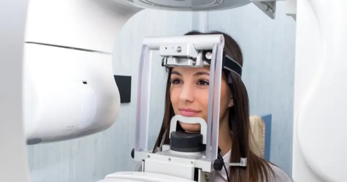
OPG کے دوران کیا ہوتا ہے
مکمل OPG ایکس رے طریقہ کار 5 منٹ سے کم لیتا ہے، اصل اسکین صرف 15 سے 20 سیکنڈ ہے۔ کوئی تیاری، صحت یابی یا ضمنی اثرات نہیں۔
- مرحلہ 1 - آمد اور رجسٹریشن: ایمریٹس آئی ڈی یا پاسپورٹ کے ساتھ چیک ان کریں۔ 2-3 منٹ۔
- مرحلہ 2 - دھاتی اشیاء اتاریں: ریڈیوگرافر تمام دھاتی اشیاء اتارنے کو کہے گا۔ تقریباً 30 سیکنڈ۔
- مرحلہ 3 - حفاظتی شیلڈنگ پہنیں: لیڈ تھائرائیڈ کالر اور/یا لیڈ ایپرن لگایا جاتا ہے۔
- مرحلہ 4 - مشین میں پوزیشن: اپنی ٹھوڑی چن ریسٹ پر رکھیں۔ صحیح پوزیشننگ اعلیٰ معیار کی تصویر کا سب سے اہم عنصر ہے۔
- مرحلہ 5 - پوزیشننگ گائیڈ کاٹیں: ایک جراثیم سے پاک پلاسٹک بائٹ بلاک آہستہ سے کاٹیں۔ ہونٹ بند کریں اور زبان تالو پر سیدھی دبائیں۔
- مرحلہ 6 - اسکین: C شکل کا بازو آہستہ آہستہ آپ کے سر کے گرد گھومتا ہے۔ آپ کو مکمل طور پر ساکن رہنا ہوگا۔ 15 سے 20 سیکنڈ۔
- مرحلہ 7 - اسکین مکمل: ڈیجیٹل تصویر چند سیکنڈوں میں ورک سٹیشن پر ظاہر ہوتی ہے۔ کمرے میں کل وقت: 3 سے 5 منٹ۔
کوئی صحت یابی کا وقت نہیں۔ اسکین مکمل طور پر بے درد ہے - کوئی سوئی، انجکشن، کنٹراسٹ ڈائی یا منہ میں سینسر نہیں۔
بچوں کے لیے OPG
5 سال کی عمر سے بچے OPG کروا سکتے ہیں۔ جدید ڈیجیٹل مشینوں میں بچوں کے لیے خودکار ڈوز کمی پروٹوکول شامل ہیں۔ بچوں کا OPG تقریباً 5 سے 14 μSv دیتا ہے۔
7 سالہ یوسف اپنی ماں کے ساتھ DCDC آیا۔ ریڈیوگرافر نے اسے مشین دکھانے میں وقت لگایا۔ یوسف 16 سیکنڈ بالکل ساکن کھڑا رہا اور اسکین ہو گیا۔ OPG نے دو مستقل دانت غیر معمولی زاویوں پر نشوونما پاتے دکھائے۔
DCDC دبئی ہیلتھ کیئر سٹی میں ڈیجیٹل OPG ایکس رے
ڈاکٹرز کلینک ڈائیگناسٹک سینٹر میں اسی دن ریڈیولوجسٹ رپورٹنگ کے ساتھ ہائی ریزولیوشن ڈیجیٹل OPG اسکین حاصل کریں۔ واک اِن خوش آمدید۔
سیلف پے مریضوں کے لیے ریفرل ضروری نہیں
اپنے OPG نتائج پڑھنا
ڈینٹسٹ اور ریڈیولوجسٹ ایک منظم، سسٹیمیٹک اپروچ کی پیروی کرتے ہیں۔
منظم پڑھنے کا طریقہ
- مجموعی تصویر کے معیار کی جانچ: ریڈیولوجسٹ درست ایکسپوژر اور پوزیشننگ کی تصدیق کرتا ہے۔
- ہم آہنگی کا جائزہ: بائیں اور دائیں طرف تقریباً ہم آہنگ نظر آنی چاہیے۔
- دانتوں کا جائزہ (کواڈرنٹ بہ کواڈرنٹ): ہر دانت کی انفرادی جانچ ہوتی ہے۔
- ہڈی کی سطح کا جائزہ: ہر دانت کے ارد گرد الویولر ہڈی کی اونچائی کا جائزہ لیا جاتا ہے۔
- TMJ جائزہ: دونوں ٹمپورومینڈیبولر جوڑوں کی شکل، سائز اور ہم آہنگی کی جانچ۔
- میکسلری سائنس ریویو: سائنسز کی اوپیسیفکیشن اور پولپس کی جانچ۔
- اعصابی نہر ٹریسنگ: انفیریئر الویولر اعصابی نہر کو ٹریس کیا جاتا ہے۔
- پیتھولوجی کی شناخت: غیر معمولی سیاہ یا روشن علاقے دستاویز کیے جاتے ہیں۔
DCDC میں ہر OPG ایک کنسلٹنٹ ریڈیولوجسٹ پڑھتا ہے۔
OPG ایکس ریز پر عام دریافتیں
| دریافت | OPG پر شکل | اس کا کیا مطلب ہے |
|---|---|---|
| دانتوں کا سڑنا | دانت کے تاج میں سیاہ علاقے | دانت کا سڑنا؛ فلنگ سے لے کر روٹ کینال تک علاج |
| ہڈی کا نقصان (پیریوڈونٹل بیماری) | جڑوں کے ارد گرد ہڈی کی اونچائی میں کمی | مسوڑوں کی بیماری سے ہڈی کا پیچھے ہٹنا |
| امپیکٹڈ دانت | دانت جزوی یا مکمل طور پر مسوڑوں یا ہڈی کے نیچے | عقل داڑھ میں عام؛ درد یا سسٹ کا سبب بن سکتا ہے |
| پیری ایپیکل پیتھولوجی | جڑ کی نوک پر سیاہ علاقہ | انفیکشن جس میں روٹ کینال یا دانت نکالنا ضروری ہے |
| سسٹ | جبڑے کی ہڈی میں واضح طور پر بیان شدہ گول سیاہ علاقہ | سیال سے بھرا تھیلا؛ سرجیکل ہٹانا ضروری |
| TMJ غیر معمولیات | کونڈائل کا چپٹا ہونا، کٹاؤ یا عدم توازن | جبڑے کے جوڑ میں تنزلی کی تبدیلیاں |
| جڑ کا ریزورپشن | چھوٹی یا بے قاعدہ شکل کی جڑ | مختلف وجوہات سے جڑ کا گھلنا |
| سائنس غیر معمولیات | میکسلری سائنس میں اوپیسیفکیشن | سائنوسائٹس یا ریٹینشن سسٹ |
عام OPG دریافتیں اور ان کی طبی اہمیت۔
عام بمقابلہ غیر معمولی OPG نتائج
ایک عام OPG دکھاتا ہے: بغیر سیاہ دھبوں کے صحت مند دانت، صحت مند جڑیں، یکساں ہڈی کی سطح، متوازن کونڈائل، صاف ہوا سے بھرے سائنسز اور نظر آنے والی اعصابی نہریں۔
غیر معمولی دریافتیں شامل ہیں: جبڑے کی ہڈی میں سیاہ علاقے، روشن سفید ماس، نمایاں ہڈی کا نقصان، جڑ کا چھوٹا ہونا اور دھندلے سائنسز۔
OPG رپورٹ کی اصطلاحات سمجھنا
| رپورٹ کی اصطلاح | سادہ زبان میں مطلب |
|---|---|
| ریڈیولوسنٹ / ریڈیولوسنسی | سیاہ علاقہ - ہڈی کی تباہی، سسٹ، ایبسیس یا ٹیومر کی نشاندہی |
| ریڈیو اوپیک / ریڈیو اوپیسٹی | روشن سفید علاقہ - دھاتی فلنگز، امپلانٹس یا گھنی ہڈی |
| پیری ایپیکل پیتھولوجی | دانت کی جڑ کی نوک پر بیماری |
| ریزورپشن | جڑ کا گھلنا - چھوٹی یا بے قاعدہ شکل میں ظاہر |
| افقی ہڈی کا نقصان | دائمی مسوڑوں کی بیماری سے ہڈی کی اونچائی میں یکساں کمی |
| عمودی ہڈی کا نقصان | جڑ کے ایک طرف مقامی V شکل کا ہڈی کا نقصان |
| امپیکشن | دانت جو اپنی عام پوزیشن میں نہیں نکلا |
| واضح بمقابلہ غیر واضح حدود | زخم کی سرحدیں: واضح = عام طور پر سلیم؛ غیر واضح = جارحانہ عمل کا شبہ |
| آسٹیواسکلیروسس | ہڈی کی بڑھی ہوئی کثافت کا علاقہ؛ عام طور پر سلیم |
| پیریکورونائٹس | جزوی طور پر نکلے دانت کے ارد گرد انفیکشن |
| فولیکولر سسٹ | نہ نکلے دانت کے تاج کے ارد گرد سیال سے بھرا تھیلا |
عام OPG رپورٹ اصطلاحات کی وضاحت۔
ڈینٹل امپلانٹس کے لیے OPG
OPG ڈینٹل امپلانٹ امیدواروں کے لیے معیاری فرسٹ لائن اسکریننگ ایکس رے ہے۔
امپلانٹ تشخیص کے لیے OPG کیا دکھاتا ہے
- عمودی ہڈی کی اونچائی: OPG مکمل الویولر رج اونچائی دکھاتا ہے۔
- انفیریئر الویولر اعصابی نہر کی پوزیشن: OPG اعصابی نہر کو ریڈیولوسنٹ بینڈ کے طور پر دکھاتا ہے۔
- میکسلری سائنس فلور پوزیشن: دکھاتا ہے کہ سائنس لفٹ ضروری ہوگا یا نہیں۔
- ملحقہ دانتوں کی حالت: کیریز، پیری ایپیکل پیتھولوجی اور ہڈی کا نقصان۔
- موجودہ پیتھولوجی: سسٹ، ٹیومر، بچے ہوئے جڑ کے ٹکڑے۔
- مجموعی جبڑے کا فن تعمیر: متعدد امپلانٹس والے مریضوں کے لیے۔
OPG کی حدود اور CBCT کب ضروری ہے
OPG ہڈی کی چوڑائی، ہڈی کی کثافت یا عصب کی درست 3D پوزیشن نہیں دکھا سکتا۔ جن صورتوں میں تقریباً ہمیشہ فالو اپ CBCT اسکین چاہیے: اعصابی نہر کے قریب پچھلے نچلے جبڑے کے امپلانٹس، سائنس کے قریب اوپری پچھلے امپلانٹس اور متعدد امپلانٹس۔
آرتھوڈونٹکس کے لیے OPG
OPG پہلی امیجنگ اسٹڈی ہے جو ہر آرتھوڈونٹسٹ بریسز کی سفارش سے پہلے مانگتا ہے۔
| ساخت / حالت | OPG کیا دکھاتا ہے | آرتھوڈونٹکس کے لیے کیوں اہم ہے |
|---|---|---|
| نکلے ہوئے دانت | پوزیشن، صف بندی اور فاصلے | طبی دریافتوں کی تصدیق؛ بریکٹ لگانے میں مدد |
| نہ نکلے / امپیکٹڈ دانت | مقام، زاویہ اور گہرائی | امپیکٹڈ کینائنز کو سرجیکل نمائش کی ضرورت ہو سکتی ہے |
| پیدائشی طور پر غائب دانت | دانت کے جراثیم کی غیر موجودگی | علاج کا مقصد بدل دیتا ہے - خلا بند کرنا یا کھلا رکھنا |
| زائد دانت | عام نکلنے کو روکنے والے اضافی دانت | صف بندی سے پہلے نکالنے ضروری |
| جڑ کی لمبائی اور شکل | ہر جڑ کی لمبائی، شکل اور سالمیت | چھوٹی جڑوں میں ریزورپشن کا زیادہ خطرہ |
| ہڈی کی سطح | ہڈی کی سپورٹ کی اونچائی | کم ہڈی محفوظ دانت کی حرکت کو محدود کرتی ہے |
| جبڑے کی ہم آہنگی | بائیں اور دائیں رامس کا نسبتی سائز | عدم توازن ہڈی کے مسئلے کی نشاندہی کر سکتا ہے |
| TMJ جوڑ | کونڈائل کی شکل اور پوزیشن | موجودہ تنزل آرتھوڈونٹکس کے دوران خراب ہو سکتی ہے |
| پچھلا ڈینٹل علاج | کراؤنز، برجز، روٹ کینال، امپلانٹس | بریکٹ امپلانٹس پر نہیں لگائے جا سکتے |
OPG آرتھوڈونٹسٹ کو جامع تشخیصی جائزہ فراہم کرتا ہے۔
عقل داڑھ کے لیے OPG
OPG عقل داڑھ (تیسرے مولرز) کے جائزے کے لیے معیاری امیجنگ اسٹڈی ہے۔
| امپیکشن کی قسم | OPG پر شکل | تعدد | نکالنے کی مشکل | اہم دریافت |
|---|---|---|---|---|
| میزیو اینگولر | دوسرے مولر کی طرف آگے جھکا ہوا | 40-45% | درمیانی | تاج دوسرے مولر پر دبا رہا ہے |
| عمودی | سیدھا لیکن نہیں نکلا | 25-30% | درمیانی سے مشکل | درست محور لیکن مسدود |
| افقی | اپنی طرف لیٹا ہوا | 10-15% | مشکل | تاج دوسرے مولر کی جڑوں کی طرف |
| ڈسٹو اینگولر | رامس کی طرف پیچھے جھکا ہوا | 5-10% | سب سے مشکل (نچلا جبڑا) | نکالنے کے راستے تک محدود رسائی |
| بکل/لنگوئل (عرضی) | گال یا زبان کی طرف ہٹا ہوا | نایاب | متغیر | OPG ہٹاؤ کو کم اندازہ لگا سکتا ہے؛ اکثر CBCT ضروری |
OPG کی بنیاد پر عقل داڑھ کی امپیکشن کی درجہ بندی۔
ڈینٹل ایکس رے سیفٹی
ہاں، ڈینٹل ایکس ریز محفوظ ہیں۔ معیاری OPG کی ریڈی ایشن ڈوز تقریباً 10 سے 20 مائیکروسیورٹ (μSv) ہے - تقریباً 1 سے 2 دن کی قدرتی پس منظر ریڈی ایشن کے برابر۔
| ریڈی ایشن ذریعہ | تخمینی ڈوز (μSv) | OPG اسکینز میں مساوی |
|---|---|---|
| 1 کیلا کھانا | 0.1 | ایک OPG کا 1/140واں |
| واحد پیری ایپیکل ایکس رے | 1-8 | < 1 OPG |
| ایک دن کی قدرتی پس منظر ریڈی ایشن | 6.5 | تقریباً آدھا OPG |
| OPG (پینورامک ڈینٹل ایکس رے) | 10-20 | 1 OPG |
| سینے کا ایکس رے | 20 | ~1 OPG |
| دبئی سے لندن پرواز | 40-80 | 3-6 OPG |
| CBCT | 30-200 | 2-14 OPG |
| سر کا CT اسکین | 2,000 | ~140 OPG |
| کل سالانہ پس منظر ریڈی ایشن | 2,400 | ~170 OPG |
ڈینٹل ایکس ریز طبی ریڈی ایشن سپیکٹرم کے بالکل نچلے حصے میں ہیں۔
حمل میں OPG سیفٹی
ACOG اور ADA دونوں تصدیق کرتے ہیں کہ طبی اشارے ہونے پر حمل کے دوران ڈینٹل ایکس ریز محفوظ طریقے سے کیے جا سکتے ہیں۔ فوری تشخیصی امیجنگ میں تاخیر نہیں ہونی چاہیے۔
بچوں کے لیے OPG سیفٹی
مناسب تکنیک کے ساتھ بچوں کے لیے ڈینٹل ایکس ریز محفوظ ہیں۔ جدید مشینوں میں بچوں کے لیے خودکار ڈوز کمی پروٹوکول ہیں۔
DCDC میں محفوظ، کم ڈوز ڈینٹل ایکس ریز
DCDC جدید ترین ڈیجیٹل OPG اور ڈیجیٹل ایکس رے سسٹمز استعمال کرتا ہے۔ DCDC دبئی ہیلتھ کیئر سٹی میں واک اِن خوش آمدید۔
سیلف پے مریضوں کے لیے ریفرل ضروری نہیں
DCDC دبئی ہیلتھ کیئر سٹی میں OPG ایکس رے
ڈاکٹرز کلینک ڈائیگناسٹک سینٹر (DCDC) دبئی ہیلتھ کیئر سٹی میں جامع ڈیجیٹل OPG ایکس رے خدمات فراہم کرتا ہے۔
- ایک چھت کے نیچے مکمل امیجنگ: OPG، CBCT، پیری ایپیکل ایکس ریز، سیفالومیٹرک امیجنگ، CT، MRI اور الٹراساؤنڈ آن سائٹ۔
- جدید ترین ڈیجیٹل OPG ٹیکنالوجی: کم ریڈی ایشن ڈوز پر واضح تصاویر۔
- بچوں کے ڈوز پروٹوکول: بچوں کے لیے خودکار ڈوز کمی۔
- اسی دن کنسلٹنٹ ریڈیولوجسٹ رپورٹنگ: ہر OPG ماہر ریڈیولوجسٹ جائزہ لیتا ہے۔
- واک اِن دستیابی: سیلف پے مریضوں کے لیے ریفرل ضروری نہیں۔
- انشورنس قبول: DCDC دبئی کی بڑی انشورنس کمپنیوں کے ساتھ کام کرتا ہے۔
- آسان DHCC مقام: عود میتھا، ام حریر 2، کرامہ سے آسانی سے قابل رسائی۔
قیمت کی تفصیلات کے لیے ہماری دبئی میں OPG ایکس رے کی لاگت گائیڈ دیکھیں۔
آج ہی اپنا OPG اسکین بک کریں
DCDC دبئی ہیلتھ کیئر سٹی میں ڈیجیٹل OPG ایکس رے کے لیے واک اِن کریں یا ایڈوانس بکنگ کریں۔ کنسلٹنٹ ریڈیولوجسٹ سے اسی دن نتائج۔
DCDC میں متعلقہ خدمات
دبئی ہیلتھ کیئر سٹی میں ماہرانہ دیکھ بھال اور جدید تشخیص
Frequently Asked Questions
حتمی خیالات
OPG ایکس رے ڈینٹل اور میکسلوفیشل امیجنگ کے سب سے قیمتی اور ورسٹائل آلات میں سے ایک ہے۔ 15 سیکنڈ سے کم میں جامع پینورامک منظر فراہم کرتا ہے۔ بہت کم ریڈی ایشن (10-20 μSv)، صفر درد اور بغیر خاص تیاری کے، یہ سب سے مریض دوست امیجنگ اسٹڈیز میں سے ایک ہے۔
چاہے آپ کے ڈینٹسٹ نے OPG کی سفارش کی ہو یا آپ کو جامع ڈینٹل اسکریننگ چاہیے، ڈاکٹرز کلینک ڈائیگناسٹک سینٹر دبئی ہیلتھ کیئر سٹی میں اسی دن کنسلٹنٹ ریڈیولوجسٹ رپورٹنگ اور واک اِن دستیابی کے ساتھ ڈیجیٹل OPG اسکین فراہم کرتا ہے۔ تمام فالو اپ امیجنگ بشمول CBCT، CT اور MRI آن سائٹ دستیاب ہے۔ تفصیلی قیمتوں کے لیے ہماری دبئی میں OPG ایکس رے کی لاگت گائیڈ دیکھیں۔
ذرائع اور حوالہ جات
یہ مضمون ہماری طبی ٹیم نے جائزہ لیا ہے اور درج ذیل ذرائع کا حوالہ دیتا ہے:
- American Dental Association - Dental Radiographic Examinations: Recommendations for Patient Selection and Limiting Radiation Exposure
- European Commission - Radiation Protection 136: European Guidelines on Radiation Protection in Dental Radiology
- NCRP Report No. 177: Radiation Protection in Dentistry and Oral & Maxillofacial Imaging
- AAOMS - White Paper on Third Molar Data
- AAO - Clinical Practice Guidelines for Orthodontic Radiography
- EAO - Consensus Statements on Radiographic Examination for Implant Patients
- RadiologyInfo.org - Panoramic Dental X-ray
- ACOG - Guidelines for Diagnostic Imaging During Pregnancy and Lactation
اس سائٹ پر طبی مواد کا جائزہ DHA لائسنس یافتہ ڈاکٹرز نے لیا ہے۔ ہماری دیکھیں تحریری پالیسی مزید معلومات کے لیے۔

تحریر
Dr. Osama Elzamzami
تشخیصی ریڈیولوجی
MD, FRCR
ڈاکٹر اسامہ الزمزمی DCDC دبئی ہیلتھ کیئر سٹی میں ریڈیولوجی شعبے کے سربراہ ہیں، ایکس رے، CT، MRI، OPG، CBCT اور الٹراساؤنڈ سمیت تشخیصی امیجنگ میں ماہر ہیں۔ ان کے پاس MD اور FRCR (فیلوشپ آف دی رائل کالج آف ریڈیولوجسٹس) ہے اور وہ ڈینٹل، میکسلوفیشل اور عام تشخیصی امیجنگ اسٹڈیز کے لیے ماہرانہ رپورٹنگ فراہم کرتے ہیں۔
متعلقہ مضامین

دبئی میں OPG ایکس رے کی لاگت: تازہ ترین قیمتیں اور انشورنس گائیڈ
CBCT بمقابلہ OPG ایکس رے: آپ کو کون سی ڈینٹل امیجنگ چاہیے؟
© 2026 Doctors Clinic Diagnostic Center (DCDC), Dubai Healthcare City. Originally published at https://doctorsclinicdubai.ae/blog/opg-xray-results-explained. All rights reserved. Unauthorized reproduction is prohibited.