اہم نکات
- 3D/4D الٹراساؤنڈ کا بہترین وقت حمل کے 26 سے 32 ہفتوں کے درمیان ہے، جب بچے میں چہرے کی تفصیلی خصوصیات کے لیے کافی چربی ہوتی ہے لیکن ابھی بھی حرکت کے لیے جگہ ہوتی ہے
- دبئی میں 3D/4D الٹراساؤنڈ کی لاگت AED 500 سے AED 1,200 کے درمیان ہے، جو پیکج کی قسم، تصاویر کی تعداد، اور ویڈیو ریکارڈنگ شامل ہے یا نہیں اس پر منحصر ہے
- 4D الٹراساؤنڈ معیاری 2D الٹراساؤنڈ کی طرح آواز کی لہروں کی ٹیکنالوجی استعمال کرتا ہے اور ماں اور بچے دونوں کے لیے یکساں طور پر محفوظ سمجھا جاتا ہے، اس میں کوئی آئنائزنگ ریڈی ایشن شامل نہیں ہے
- 3D الٹراساؤنڈ بچے کی سہ جہتی ساکن تصاویر لیتا ہے، جبکہ 4D الٹراساؤنڈ ریئل ٹائم حرکت کا جہت شامل کرتا ہے، جو والدین کو بچے کو جمائی لیتے، تانتے اور مسکراتے ہوئے دیکھنے کی اجازت دیتا ہے
- دبئی ہیلتھ کیئر سٹی میں DCDC جدید امیجنگ آلات کا استعمال کرتے ہوئے تجربہ کار ریڈیولوجسٹس کے ذریعے 3D/4D الٹراساؤنڈ پیش کرتا ہے، جس میں اسی دن نتائج اور پرنٹ شدہ یادگاری تصاویر شامل ہیں
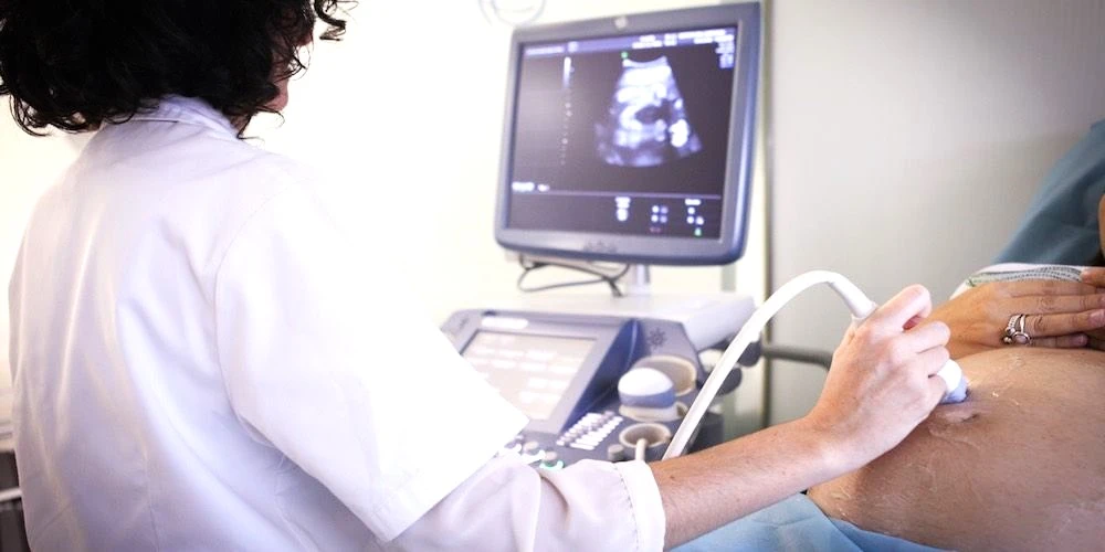
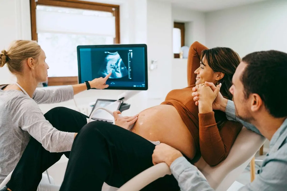
3D/4D الٹراساؤنڈ ایک جدید قبل از پیدائش امیجنگ ٹیکنیک ہے جو رحم کے اندر پیدا نہ ہونے والے بچے کی تفصیلی سہ جہتی تصاویر اور ریئل ٹائم ویڈیو تیار کرتی ہے۔ معیاری 2D الٹراساؤنڈ کے برعکس جو ہموار، کراس سیکشنل تصاویر بناتا ہے، دبئی میں 4D الٹراساؤنڈ بچے کے چہرے کی خصوصیات، تاثرات اور حرکات کو حقیقت پسندانہ تفصیل میں ظاہر کرتا ہے، جس سے حاملہ والدین کو پیدائش سے پہلے اپنے بچے کی پہلی حقیقی جھلک ملتی ہے۔ یہ ٹیکنالوجی دبئی میں سب سے زیادہ طلب کی جانے والی قبل از پیدائش خدمات میں سے ایک بن گئی ہے۔
یہ گائیڈ دبئی میں 3D/4D الٹراساؤنڈ حمل امیجنگ کے بارے میں وہ سب کچھ شامل کرتی ہے جو حاملہ والدین کو جاننے کی ضرورت ہے: 2D، 3D، اور 4D اسکینز کے درمیان فرق، بہترین تصاویر کے لیے بہترین حملاتی عمر، اسکین کیا دکھا سکتا ہے اور کیا نہیں، حفاظت کے ثبوت، دبئی کے کلینکس میں لاگت، اور دبئی ہیلتھ کیئر سٹی میں ڈاکٹرز کلینک ڈائیگناسٹک سینٹر (DCDC) میں دبئی میں 4D بیبی اسکین کیسے بک کریں۔
Health Screening Packages
Save with our bundled screening packages — specialist consultation included


3D/4D الٹراساؤنڈ کیا ہے؟
3D الٹراساؤنڈ ایک امیجنگ تکنیک ہے جو مختلف زاویوں سے متعدد دو جہتی الٹراساؤنڈ تصاویر لیتی ہے اور انہیں بچے کی ایک سہ جہتی تصویر میں دوبارہ تشکیل دینے کے لیے کمپیوٹر پروسیسنگ کا استعمال کرتی ہے۔ نتیجہ ایک ساکن تصویر ہے جو بچے کی جلد کی سطح دکھاتی ہے، چہرے کی خصوصیات جیسے ناک، ہونٹ، گال اور ٹھوڑی کو قابل ذکر تفصیل میں ظاہر کرتی ہے۔ والدین دیکھ سکتے ہیں کہ بچے کا چہرہ گول ہے یا نوکیلی ٹھوڑی ہے، اور بہت سے معاملات میں خاندانی مشابہتیں پیدائش سے پہلے ہی نظر آتی ہیں۔
4D الٹراساؤنڈ وقت کا جہت شامل کر کے اس ٹیکنالوجی کو ایک قدم آگے لے جاتا ہے، جس سے ریئل ٹائم سہ جہتی ویڈیو سٹریم بنتی ہے۔ "چوتھا جہت" حرکت ہے۔ ایک منجمد 3D تصویر کے بجائے، 4D بیبی اسکین بچے کے اعمال کو ان کے ہوتے ہوئے ریکارڈ کرتا ہے: جمائی لینا، انگوٹھا چوسنا، تاننا، لات مارنا، منہ کھولنا اور بند کرنا، اور یہاں تک کہ مسکرانا۔ یہ زندہ حرکت کی صلاحیت 4D اسکین کو والدین کے لیے ایک گہرا جذباتی تجربہ بناتی ہے۔
3D اور 4D دونوں الٹراساؤنڈ روایتی 2D الٹراساؤنڈ کی طرح بالکل اسی قسم کی آواز کی لہروں کی توانائی استعمال کرتے ہیں۔ ٹرانسڈیوسر اعلی تعدد کی آواز کی لہریں خارج کرتا ہے جو بچے کے جسم سے ٹکراتی ہیں، اور واپس آنے والی بازگشتیں کمپیوٹر کے ذریعے پروسیس ہوتی ہیں۔ فرق صرف اس بات میں ہے کہ کمپیوٹر ڈیٹا کو کیسے جمع اور ظاہر کرتا ہے۔ 2D اسکین ایک ہموار ٹکڑا دکھاتا ہے۔ 3D اسکین متعدد ٹکڑوں کو سطحی حجم میں مرتب کرتا ہے۔ 4D اسکین وہی 3D رینڈرنگ کرتا ہے لیکن ریئل ٹائم میں حرکت دکھانے کے لیے تصویر کو مسلسل اپ ڈیٹ کرتا ہے۔
"بہت سے والدین یہ جان کر حیران ہوتے ہیں کہ 3D اور 4D الٹراساؤنڈ وہی محفوظ آواز کی لہروں کی ٹیکنالوجی استعمال کرتے ہیں جو معمول کے اسکینز میں استعمال ہوتی ہے جو انہوں نے حمل کے دوران پہلے ہی کروائے ہیں،" DCDC کے شعبہ ریڈیولوجی کے سربراہ ڈاکٹر اسامہ الزمزمی کہتے ہیں۔ "فرق خالصتاً سافٹ ویئر پروسیسنگ میں ہے، بچے کو دی جانے والی توانائی میں نہیں۔"
2D بمقابلہ 3D بمقابلہ 4D الٹراساؤنڈ: اہم فرق
2D، 3D، اور 4D الٹراساؤنڈ کے درمیان فرق کو سمجھنا حاملہ والدین کو یہ جاننے میں مدد کرتا ہے کہ ہر قسم کے اسکین سے کیا توقع رکھنی ہے اور حمل کے دوران ہر ایک کب سب سے مناسب ہے۔ تینوں اقسام ایک ہی بنیادی الٹراساؤنڈ ٹیکنالوجی استعمال کرتی ہیں لیکن تصاویر کی پروسیسنگ اور ڈسپلے میں مختلف ہیں۔
| خصوصیت | 2D الٹراساؤنڈ | 3D الٹراساؤنڈ | 4D الٹراساؤنڈ |
|---|---|---|---|
| تصویر کی قسم | ہموار، کراس سیکشنل (گرے اسکیل) | ساکن سہ جہتی سطحی تصویر | ریئل ٹائم 3D ویڈیو (براہ راست حرکت) |
| آپ کیا دیکھتے ہیں | کراس سیکشن میں اندرونی ڈھانچے | 3D میں بچے کا چہرہ اور جسم کی سطح | ریئل ٹائم میں بچہ حرکت کرتا، جمائی لیتا، تانتا ہوا |
| بنیادی استعمال | معمول کی طبی جانچ، نشوونما کی پیمائش، اعضاء کی جانچ | چہرے کی خصوصیات کا مشاہدہ، سطحی اناٹومی کا جائزہ | تعلق کا تجربہ، رویے کا مشاہدہ، چہرے کی جانچ |
| طبی قدر | تمام معیاری قبل از پیدائش جانچوں کے لیے ضروری | پھٹے ہونٹ کا پتہ لگانے اور چہرے کی جانچ کے لیے مددگار | 3D کی طرح، نیز جنین کی حرکت کے نمونوں کی جانچ |
| بہترین وقت | پوری حمل میں (سہ ماہی کے مطابق مختلف) | بہترین چہرے کی تصاویر کے لیے 26-32 ہفتے | بہترین چہرے کی تصاویر اور حرکت کے لیے 26-32 ہفتے |
| تصویر کی وضاحت | تشریح کے لیے تربیت یافتہ آنکھ درکار | بدیہی، حقیقت پسندانہ سطحی تصاویر | بدیہی، حقیقت پسندانہ متحرک تصاویر |
| قیمت کی حد (دبئی) | AED 200-500 | AED 400-900 | AED 500-1,200 |
| نتیجہ | پرنٹ شدہ یا ڈیجیٹل گرے اسکیل تصاویر | پرنٹ شدہ یا ڈیجیٹل 3D تصاویر | 3D تصاویر اور ویڈیو ریکارڈنگ |
حمل کے دوران 2D، 3D، اور 4D الٹراساؤنڈ امیجنگ کا موازنہ۔ ہر قسم قبل از پیدائش دیکھ بھال میں مختلف مقصد پورا کرتی ہے۔
یہ سمجھنا اہم ہے کہ 2D الٹراساؤنڈ معمول کی قبل از پیدائش دیکھ بھال کے لیے طبی معیار رہتا ہے۔ 18 سے 22 ہفتوں میں کی جانے والی اناٹومی اسکین، جو بچے کے اعضاء، دماغ، ریڑھ کی ہڈی، دل اور اعضاء کی جانچ کرتی ہے، 2D الٹراساؤنڈ کا استعمال کرتے ہوئے کی جاتی ہے۔ 3D/4D اسکین ایک اضافی معائنہ ہے جو سطحی مشاہدے اور جذباتی تعلق کو شامل کرتا ہے، لیکن یہ معیاری 2D اناٹومی اسکین کی جگہ نہیں لیتا۔
تاہم، 3D/4D امیجنگ کے تعلق سے آگے حقیقی طبی استعمال ہیں۔ یہ چہرے کی غیر معمولیات جیسے پھٹے ہونٹ اور تالو کا پتہ لگانے، کانوں کی پوزیشن اور شکل کی جانچ، اور ہڈیوں کی غیر معمولیات کا جائزہ لینے کے لیے خاص طور پر قیمتی ہے۔
4D الٹراساؤنڈ کا بہترین وقت کب ہے؟
4D الٹراساؤنڈ کا بہترین وقت حمل کے 26 سے 32 ہفتوں کے درمیان ہے۔ یہ مدت دو مسابقتی عوامل کے درمیان مثالی توازن کی نمائندگی کرتی ہے: بچے میں چہرے کی خصوصیات بھرنے کے لیے کافی زیر جلد چربی ہونی چاہیے، لیکن صوتی تضاد فراہم کرنے اور بچے کو حرکت کرنے کے لیے کافی ایمنیوٹک سیال بھی ہونا چاہیے۔
26-32 ہفتے مثالی کیوں ہیں
26 ہفتوں سے پہلے، بچے میں بہت کم زیر جلد چربی ہوتی ہے، جس کا مطلب ہے کہ 3D/4D اسکین پر چہرے کی خصوصیات ہڈی دار دکھائی دیتی ہیں۔ 32 ہفتوں کے بعد، بچہ عام طور پر اتنا بڑا ہو جاتا ہے کہ رحم کی دیوار کے خلاف دبتا ہے، جو حرکت کو محدود کرتا ہے۔ 28 سے 30 ہفتوں کا وقت وہ ہے جب زیادہ تر سونوگرافرز سب سے واضح اور فوٹو جینک تصاویر حاصل کرتے ہیں۔
اس حملاتی عمر میں، بچے کے گال بھر رہے ہوتے ہیں، ناک اور ہونٹ اچھی طرح واضح ہوتے ہیں، اور چہرے کے سامنے عام طور پر تیز، اعلی تضاد والی 3D سطحی تصاویر بنانے کے لیے کافی سیال ہوتا ہے۔
تصویر کے معیار کو متاثر کرنے والے عوامل
- بچے کی پوزیشن: واضح چہرے کی تصویر کے لیے چہرے کا ٹرانسڈیوسر کی طرف ہونا ضروری ہے۔ اگر بچے کا منہ ماں کی پیٹھ کی طرف ہے، تو سونوگرافر ماں سے چلنے، ٹھنڈا پانی پینے، یا کروٹ لیٹنے کو کہہ سکتا ہے
- نال کی پوزیشن: سامنے والی نال الٹراساؤنڈ شعاع کو جزوی طور پر روک سکتی ہے، جس سے تصویر کی وضاحت کم ہو جاتی ہے۔ اسکین ابھی بھی ممکن ہیں لیکن بہترین مناظر حاصل کرنے کے لیے زیادہ وقت اور صبر درکار ہو سکتا ہے
- ایمنیوٹک سیال کی سطح: بچے کے چہرے کے ارد گرد کافی سیال صوتی کھڑکی کا کام کرتا ہے۔ کم سیال تصویر کے معیار کو کم کر سکتا ہے
- ماں کا جسم: زیادہ باڈی ماس انڈیکس (BMI) تصویر کی ریزولیوشن کو کم کر سکتا ہے۔ اپائنٹمنٹ سے پہلے پانی پینا تصویر کے معیار کو بہتر بنا سکتا ہے
- بچے کے ہاتھ اور پاؤں: بچے اکثر اپنے ہاتھ یا پاؤں اپنے چہرے پر رکھتے ہیں، جو منظر کو روک سکتا ہے۔ سونوگرافر بچے کے حرکت کرنے کا انتظار کرے گا
"ہم حاملہ والدین کو ہمیشہ بتاتے ہیں کہ بچہ شو کا ہدایت کار ہے،" DCDC کے شعبہ ریڈیولوجی کے سربراہ ڈاکٹر اسامہ الزمزمی کہتے ہیں۔ "ہم وقت اور تکنیک کو بہتر بنا سکتے ہیں، لیکن آخر کار، تصاویر کا معیار اس بات پر منحصر ہے کہ بچہ آگے کی طرف منہ کر کے اور اپنے ہاتھ چہرے سے دور رکھ کر تعاون کرتا ہے یا نہیں۔ 26 سے 30 ہفتوں میں زیادہ تر معاملات میں، ہم خوبصورت تصاویر حاصل کرتے ہیں۔"
3D/4D الٹراساؤنڈ کیا دکھا سکتا ہے؟
3D/4D الٹراساؤنڈ بچے کی بیرونی اناٹومی اور ریئل ٹائم رویے کا منفرد تفصیلی نظارہ فراہم کرتا ہے۔ جبکہ 2D الٹراساؤنڈ اندرونی اعضاء کی جانچ میں بہترین ہے، 3D/4D اسکین خاص طور پر سطحی ڈھانچے اور حرکت کو دیکھنے کے لیے بنایا گیا ہے۔
والدین کیا دیکھنے کی توقع کر سکتے ہیں
- چہرے کی خصوصیات: ناک، ہونٹ، ٹھوڑی، گال اور پیشانی کی شکل واضح طور پر نظر آتی ہے
- چہرے کے تاثرات: 4D موڈ میں، بچوں کو جمائی لیتے، منہ کھولتے اور بند کرتے، زبان باہر نکالتے، مسکراتے، بھنویں سکیڑتے اور پلکیں جھپکاتے دیکھا جا سکتا ہے
- ہاتھ اور پاؤں: انفرادی انگلیاں اور پاؤں کی انگلیاں نظر آتی ہیں
- جسم کی حرکات: تاننا، لات مارنا، لڑھکنا اور ہچکی لینا سب ریئل ٹائم 4D ویڈیو میں قابل مشاہدہ ہیں
- جنس کی تصدیق: جبکہ جنس عام طور پر 18-22 ہفتوں میں 2D اسکین پر طے ہوتی ہے، 3D/4D اسکین اضافی تصدیق فراہم کر سکتا ہے
- نافی کی ہڈی: بچے کی گردن اور جسم کے سلسلے میں نافی کی ہڈی کی پوزیشن تین جہتوں میں دیکھی جا سکتی ہے
طبی استعمال
تعلق کے تجربے سے آگے، 3D/4D الٹراساؤنڈ کے مخصوص طبی استعمال ہیں:
- پھٹے ہونٹ اور تالو کا پتہ لگانا: 3D امیجنگ پھٹے ہونٹ کو دیکھنے کے لیے 2D سے بہتر ہے کیونکہ یہ چہرے کی سطح کو براہ راست دکھاتی ہے
- چہرے کی ڈسمورفولوجی کی جانچ: بعض جینیاتی حالات مخصوص چہرے کی خصوصیات سے منسلک ہیں۔ 3D امیجنگ ڈاؤن سنڈروم جیسی حالات سے منسلک چہرے کی خصوصیات کی شناخت میں مدد کر سکتی ہے
- ہڈیوں کی غیر معمولیات: اعضاء، ہاتھوں اور پاؤں کی سطحی غیر معمولیات جیسے کلب فٹ تین جہتوں میں آسانی سے جانچی جا سکتی ہیں
- نیورل ٹیوب کے نقائص: سپائنا بائفڈا اور دیگر نقائص جو ریڑھ کی ہڈی کی سطح کو متاثر کرتے ہیں 3D رینڈرنگ سے زیادہ واضح طور پر جانچے جا سکتے ہیں
پہلی جھلک: ایک جوڑے کا تجربہ
سارہ اور احمد، دبئی میں حاملہ والدین، نے حمل کے 29ویں ہفتے میں DCDC میں 4D الٹراساؤنڈ بک کیا۔ سارہ متجسس تھی کہ کیا ان کے بچے کو اس کے شوہر کی نمایاں ناک ملے گی یا اس کی اپنی چھوٹی ناک۔ اسکین شروع ہونے کے چند منٹوں میں، بچے کے چہرے کی کرسٹل واضح 3D تصویر حاصل ہو گئی۔ بچے نے واضح طور پر احمد کی ناک اور سارہ کے گول گال وراثت میں لیے تھے۔ پھر 4D ویڈیو شروع ہوئی، بچے نے جمائی لی، دونوں بازو سر کے اوپر پھیلائے، اور مسکرایا۔
"مجھے توقع نہیں تھی کہ میں اتنی جذباتی ہو جاؤں گی،" سارہ نے یاد کیا۔ "ہمارے بچے کا اصل چہرہ دیکھنا، اسے ریئل ٹائم میں جمائی لیتے اور حرکت کرتے دیکھنا - اس نے سب کچھ اتنا حقیقی بنا دیا۔" احمد نے حمل کے بقیہ ہفتوں کے لیے پرنٹ شدہ 3D تصویر اپنی دفتر کی میز پر رکھی۔ جوڑے نے بعد میں تصدیق کی کہ ان کی بیٹی پیدائش کے وقت بالکل 4D تصاویر جیسی نظر آتی تھی۔
3D/4D الٹراساؤنڈ کی حدود
حقیقت پسندانہ توقعات رکھنا اہم ہے۔ 3D/4D الٹراساؤنڈ معیاری اناٹومی اسکین کی جگہ نہیں لیتا اور اس کی کچھ حدود ہیں:
- یہ 2D الٹراساؤنڈ کا تشخیصی متبادل نہیں ہے
- تصویر کا معیار بچے کی پوزیشن پر بہت زیادہ منحصر ہے
- سامنے والی نال، کم سیال کی سطح، یا زیادہ BMI 3D/4D تصاویر کی وضاحت کو کم کر سکتے ہیں
- اسکین صرف سطحی اناٹومی دکھاتا ہے اور اندرونی اعضاء کو اس طرح نہیں دکھا سکتا جیسے 2D اسکین کرتا ہے
DCDC میں 3D/4D الٹراساؤنڈ بک کریں
پیدائش سے پہلے اپنے بچے کا چہرہ دیکھیں۔ دبئی ہیلتھ کیئر سٹی میں ڈاکٹرز کلینک ڈائیگناسٹک سینٹر میں، ہماری ریڈیولوجی ٹیم جدید امیجنگ آلات کا استعمال کرتے ہوئے 3D/4D الٹراساؤنڈ اسکینز کرتی ہے۔ یادگاری تصاویر، ویڈیو ریکارڈنگز، اور اسی دن نتائج حاصل کریں۔
کیا 4D الٹراساؤنڈ بچے کے لیے محفوظ ہے؟
جی ہاں، 4D الٹراساؤنڈ ماں اور بچے دونوں کے لیے محفوظ سمجھا جاتا ہے۔ یہ ہر بڑی طبی اور ریگولیٹری تنظیم کا متفقہ موقف ہے جس نے اس ٹیکنالوجی کا جائزہ لیا ہے، بشمول ACOG، AIUM، BMUS، اور WHO۔
الٹراساؤنڈ کے محفوظ ہونے کی بنیادی وجہ یہ ہے کہ یہ غیر آئنائزنگ آواز کی لہریں استعمال کرتا ہے، ریڈی ایشن نہیں۔ ایکس رے، CT اسکین، یا نیوکلیئر میڈیسن کے برعکس، الٹراساؤنڈ ماں یا بچے کو کسی آئنائزنگ ریڈی ایشن سے بے نقاب نہیں کرتا۔
3D/4D الٹراساؤنڈ معیاری 2D تشخیصی الٹراساؤنڈ کی طرح ایک ہی تعدد، پاور آؤٹ پٹ، اور صوتی توانائی استعمال کرتا ہے۔ "3D" اور "4D" کی اصطلاحات صرف اس بات کا حوالہ دیتی ہیں کہ کمپیوٹر تصویری ڈیٹا کو کیسے پروسیس اور ظاہر کرتا ہے۔ حفاظتی پروفائل یکساں ہے۔
محفوظ استعمال کے رہنما خطوط
جبکہ 3D/4D الٹراساؤنڈ محفوظ ہے، طبی تنظیمیں تجویز کرتی ہیں کہ تمام قبل از پیدائش الٹراساؤنڈ معائنے مناسب طور پر دیکھ بھال کیے گئے طبی درجے کے آلات کا استعمال کرتے ہوئے اہل طبی پیشہ ور افراد کے ذریعے کیے جائیں:
- ALARA اصول: الٹراساؤنڈ کی نمائش کو "جتنا کم معقول طور پر حاصل کیا جا سکے" اصول کی پیروی کرنی چاہیے
- طبی نگرانی: اسکینز اہل طبی پیشہ ور افراد جیسے ریڈیولوجسٹ، سونوگرافرز، یا ماہرین امراض نسواں کے ذریعے یا ان کی نگرانی میں کیے جانے چاہییں
- طبی درجے کے آلات: صرف درست طریقے سے کیلیبریٹ کیے گئے طبی درجے کے الٹراساؤنڈ مشینیں استعمال ہونی چاہییں
- غیر طبی یادگاری اسٹوڈیوز سے بچیں: FDA اور ACOG غیر طبی تجارتی "یادگاری الٹراساؤنڈ" اسٹوڈیوز کے استعمال کے خلاف مشورہ دیتے ہیں۔ DCDC میں، تمام اسکینز طبی ماحول میں اہل طبی عملے کے ذریعے کیے جاتے ہیں
"حمل میں الٹراساؤنڈ کی حفاظت 50 سال سے زیادہ طبی استعمال اور وسیع تحقیق سے ثابت ہے،" ڈاکٹر اسامہ الزمزمی کہتے ہیں۔ "DCDC میں، ہم تمام بین الاقوامی حفاظتی رہنما خطوط کی پیروی کرتے ہیں اور تربیت یافتہ پیشہ ور افراد کے ذریعے چلائے جانے والے طبی درجے کے آلات استعمال کرتے ہیں۔"
دبئی میں 3D/4D الٹراساؤنڈ کی لاگت
دبئی میں 3D/4D الٹراساؤنڈ کی لاگت پیکج کی قسم، سہولت، ڈاکٹر کے تجربے کی سطح، اور فیس میں کیا شامل ہے اس کے مطابق مختلف ہوتی ہے۔ دبئی کے کلینکس اور تشخیصی مراکز میں، قیمتیں عام طور پر 3D/4D بیبی اسکین کے لیے AED 500 سے AED 1,200 ہوتی ہیں۔
قیمت کو کیا متاثر کرتا ہے
- اسکین کی قسم: بنیادی 3D اسکین (صرف ساکن تصاویر) 4D اسکین (براہ راست ویڈیو اور ساکن تصاویر) سے کم لاگت آتا ہے
- شامل اضافی چیزیں: کچھ پیکجز میں پرنٹ شدہ تصاویر، USB یا ڈیجیٹل ڈاؤن لوڈ، جنس ظاہر کرنا، اور تحریری طبی رپورٹ شامل ہیں
- ڈاکٹر بمقابلہ ٹیکنیشن: مشاور ریڈیولوجسٹ کے ذریعے کیے گئے یا تشریح کیے گئے اسکینز کی فیس زیادہ ہو سکتی ہے لیکن وہ طبی جانچ کی اضافی قدر پیش کرتے ہیں
- سہولت کی قسم: ہسپتال پر مبنی امیجنگ شعبے عام طور پر آزاد تشخیصی مراکز سے زیادہ چارج کرتے ہیں
- انشورنس کوریج: امارات میں زیادہ تر انشورنس پلانز معیاری قبل از پیدائش الٹراساؤنڈ (2D اناٹومی اسکینز) کا احاطہ کرتے ہیں لیکن اختیاری 3D/4D امیجنگ کا احاطہ نہیں کرتے
اپنے 4D اسکین سے بہترین قیمت حاصل کرنا
قیمتوں کا موازنہ کرتے وقت، صرف سب سے کم قیمت پر توجہ دینے کی بجائے فیس میں کیا شامل ہے اس پر غور کرنا قابل قدر ہے۔ DCDC میں، 3D/4D الٹراساؤنڈ میں اسکین، پرنٹ شدہ یادگاری تصاویر، ڈیجیٹل کاپی، اور ریڈیولوجسٹ کی تشخیص شامل ہے، سب ایک ہی اپائنٹمنٹ میں۔
دبئی میں تمام اقسام کے اسکینز کی الٹراساؤنڈ قیمتوں کے جامع جائزے کے لیے، دبئی میں الٹراساؤنڈ لاگت پر ہماری تفصیلی گائیڈ دیکھیں۔
DCDC دبئی میں 4D بیبی امیجنگ
دبئی ہیلتھ کیئر سٹی میں DCDC (ڈاکٹرز کلینک ڈائیگناسٹک سینٹر) حاملہ والدین کے لیے جامع 3D/4D الٹراساؤنڈ خدمات فراہم کرتا ہے، جو تجربہ کار ریڈیولوجی ماہرین کی طبی مہارت کے ساتھ جدید امیجنگ ٹیکنالوجی کو ملاتا ہے۔
اپنی اپائنٹمنٹ میں کیا توقع کریں
- مرحلہ 1 - آمد اور تیاری: استقبالیہ ٹیم آپ کا خیرمقدم کرے گی۔ آپ تجربہ بانٹنے کے لیے اپنے ساتھی یا ایک خاندان کے فرد کو لا سکتے ہیں۔ کوئی خاص تیاری درکار نہیں، اگرچہ پہلے سے پانی پینا تصویر کی وضاحت کو بہتر بنا سکتا ہے
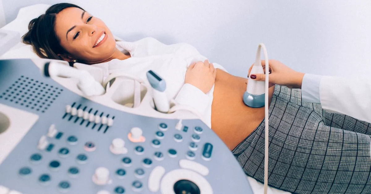
- مرحلہ 2 - اسکین: پیٹ پر گرم الٹراساؤنڈ جیل لگایا جاتا ہے، اور ریڈیولوجسٹ تصاویر لینے کے لیے ٹرانسڈیوسر استعمال کرتا ہے۔ اسکین عام طور پر 20 سے 30 منٹ لیتا ہے
- مرحلہ 3 - تصاویر اور ویڈیو: بہترین 3D ساکن تصاویر منتخب اور یادگار کے طور پر پرنٹ کی جاتی ہیں۔ 4D ویڈیو پیکج میں بچے کی حرکات کی ریکارڈنگ ڈیجیٹل طور پر محفوظ کی جاتی ہے
- مرحلہ 4 - طبی فیڈبیک: ریڈیولوجسٹ بچے کی پوزیشن، تخمینی وزن، اور متعلقہ طبی مشاہدات کا مختصر جائزہ فراہم کرتا ہے۔ باقاعدہ رپورٹ اسی دن دستیاب ہوتی ہے
3D/4D الٹراساؤنڈ کے لیے DCDC کیوں چنیں
- طبی درجے کی امیجنگ: تمام اسکینز لائسنس یافتہ طبی سہولت میں جدید، کیلیبریٹ شدہ الٹراساؤنڈ آلات کا استعمال کرتے ہوئے کیے جاتے ہیں
- ریڈیولوجسٹ کی زیر قیادت خدمت: ہر اسکین مشاور ریڈیولوجسٹ کے ذریعے انجام دیا یا نگرانی کیا جاتا ہے
- اسی دن نتائج: پرنٹ شدہ تصاویر، ڈیجیٹل کاپیاں، اور ریڈیولوجسٹ کی رپورٹ اسکین کے دن ہی دستیاب ہیں
- آسان مقام: DCDC دبئی ہیلتھ کیئر سٹی میں واقع ہے، ڈاؤن ٹاؤن دبئی، بزنس بے، عود میتھا، کرامہ، اور وسیع تر امارات سے آسانی سے قابل رسائی
- جامع امیجنگ سینٹر: DCDC ابتدائی حمل ڈیٹنگ اسکینز سے لے کر اناٹومی اسکینز سے لے کر 3D/4D امیجنگ تک، قبل از پیدائش الٹراساؤنڈ خدمات کی مکمل رینج پیش کرتا ہے
الٹراساؤنڈ ٹیکنالوجی کیسے کام کرتی ہے اور کسی بھی قسم کے الٹراساؤنڈ کی تیاری کیسے کریں اس کے بارے میں عمومی معلومات کے لیے، الٹراساؤنڈ اسکین کیا ہے اور الٹراساؤنڈ کی تیاری کیسے کریں پر ہماری گائیڈز دیکھیں۔
DCDC میں اپنے بچے کو 3D/4D میں دیکھیں
دبئی ہیلتھ کیئر سٹی میں ڈاکٹرز کلینک ڈائیگناسٹک سینٹر میں 3D/4D الٹراساؤنڈ بک کریں۔ ہماری تجربہ کار ریڈیولوجی ٹیم آپ کو اپنے بچے کا بہترین نظارہ دینے کے لیے جدید امیجنگ آلات استعمال کرتی ہے۔ یادگاری تصاویر، ویڈیو ریکارڈنگز، اور طبی اطمینان شامل ہے۔
یا اپنی اپائنٹمنٹ شیڈول کرنے کے لیے ہمیں براہ راست کال کریں۔
DCDC میں متعلقہ خدمات
دبئی ہیلتھ کیئر سٹی میں ماہرانہ دیکھ بھال اور جدید تشخیص
Frequently Asked Questions
حتمی خیالات
3D/4D الٹراساؤنڈ حمل کے سب سے یادگار لمحات میں سے ایک ہے، جو حاملہ والدین کو پیدائش سے ہفتے پہلے اپنے بچے کے چہرے، تاثرات اور شخصیت کی حقیقی پہلی جھلک پیش کرتا ہے۔ ٹیکنالوجی محفوظ، غیر حملہ آور ہے اور ہر معیاری قبل از پیدائش الٹراساؤنڈ کی طرح آواز کی لہروں کا اصول استعمال کرتی ہے۔ جب 26 سے 32 ہفتوں کی مثالی مدت میں ایک اہل طبی پیشہ ور کے ذریعے انجام دیا جاتا ہے، تو 4D بیبی اسکین قابل ذکر طور پر واضح اور جذباتی طور پر بامعنی تصاویر بناتا ہے جو خاندان زندگی بھر سنبھال کر رکھتے ہیں۔
دبئی ہیلتھ کیئر سٹی میں ڈاکٹرز کلینک ڈائیگناسٹک سینٹر میں، حاملہ والدین کو جذباتی یادگاری تجربہ اور طبی اطمینان دونوں ملتے ہیں کہ ان کا اسکین تجربہ کار ریڈیولوجی ماہرین کے ذریعے لائسنس یافتہ طبی سہولت میں کیا جا رہا ہے۔ چاہے یہ آپ کا پہلا حمل ہو یا تیسرا، DCDC میں دبئی میں 4D الٹراساؤنڈ آپ کو بڑے دن سے پہلے اپنے بچے سے ملنے کا موقع دیتا ہے۔ الٹراساؤنڈ خدمات کے بارے میں مزید معلومات کے لیے، الٹراساؤنڈ اسکین کیا ہے پر ہماری گائیڈ دیکھیں یا دبئی میں تفصیلی قیمتوں کے لیے ہماری الٹراساؤنڈ لاگت گائیڈ دیکھیں۔
ذرائع اور حوالہ جات
یہ مضمون ہماری طبی ٹیم نے جائزہ لیا ہے اور درج ذیل ذرائع کا حوالہ دیتا ہے:
- امریکن کالج آف آبسٹیٹریشنز اینڈ گائناکولوجسٹس (ACOG) - حمل کے دوران الٹراساؤنڈ معائنے
- امریکن انسٹی ٹیوٹ آف الٹراساؤنڈ ان میڈیسن (AIUM) - الٹراساؤنڈ کے محفوظ استعمال کا بیان
- RadiologyInfo.org - آبسٹیٹرک الٹراساؤنڈ
- NHS - حمل میں الٹراساؤنڈ اسکینز
اس سائٹ پر طبی مواد کا جائزہ DHA لائسنس یافتہ ڈاکٹرز نے لیا ہے۔ ہماری دیکھیں تحریری پالیسی مزید معلومات کے لیے۔

تحریر
ڈاکٹر اسامہ الزمزمی
تشخیصی ریڈیولوجی
MD, FRCR
ڈاکٹر اسامہ الزمزمی DCDC دبئی ہیلتھ کیئر سٹی میں شعبہ ریڈیولوجی کے سربراہ ہیں، جو الٹراساؤنڈ، CT، MRI، اور انٹروینشنل ریڈیولوجی سمیت تشخیصی امیجنگ میں مہارت رکھتے ہیں۔
متعلقہ مضامین
الٹراساؤنڈ اسکین کیا ہے؟ مکمل گائیڈ
کیا الٹراساؤنڈ محفوظ ہے؟ خطرات اور خرافات کی وضاحت
حمل میں ڈوپلر الٹراساؤنڈ
More in Women's Health

AMH Test Cost Dubai: Pricing Guide (2026)
مزید پڑھیں
Perimenopause vs Menopause Dubai: Signs (2026)
مزید پڑھیں
Mother & Baby Home Care Dubai: Guide (2026)
مزید پڑھیں
Gestational Diabetes Dubai: Screening (2026)
مزید پڑھیں
Mammogram Preparation Dubai: What to Expect (2026)
مزید پڑھیں
CA-125 Test Dubai: Ovarian Cancer Marker (2026)
مزید پڑھیں© 2026 Doctors Clinic Diagnostic Center (DCDC), Dubai Healthcare City. Originally published at https://doctorsclinicdubai.ae/blog/4d-ultrasound-dubai-pregnancy. All rights reserved. Unauthorized reproduction is prohibited.