Запишитесь на приём
Проверенные отзывы пациентов о диагностическом центре DCDC в Дубае
Проверенные отзывы и реальные истории пациентов из Медицинского города Дубая
Я очень довольна этой медицинской клиникой. Здесь я прошла первый скрининг беременности. Это было доступно и очень профессионально. Рентгенолог был приветливым, добрым и сделал опыт отличным.
Айжан Токманбетова
Повредила колено в пятницу вечером и получила запись на МРТ в воскресенье вечером (забронировала в воскресенье днём). Ждала около 2 минут и закончила чуть более чем за 30 минут. Команда была потрясающей, дружелюбной и эффективной.
Кирстен Эванс
Команда доктора Осамы была нежной и потрясающей. После плохого опыта в другом месте, мой гинекологический осмотр здесь был намного профессиональнее. Отличный отчёт и очень точный.
Мэри


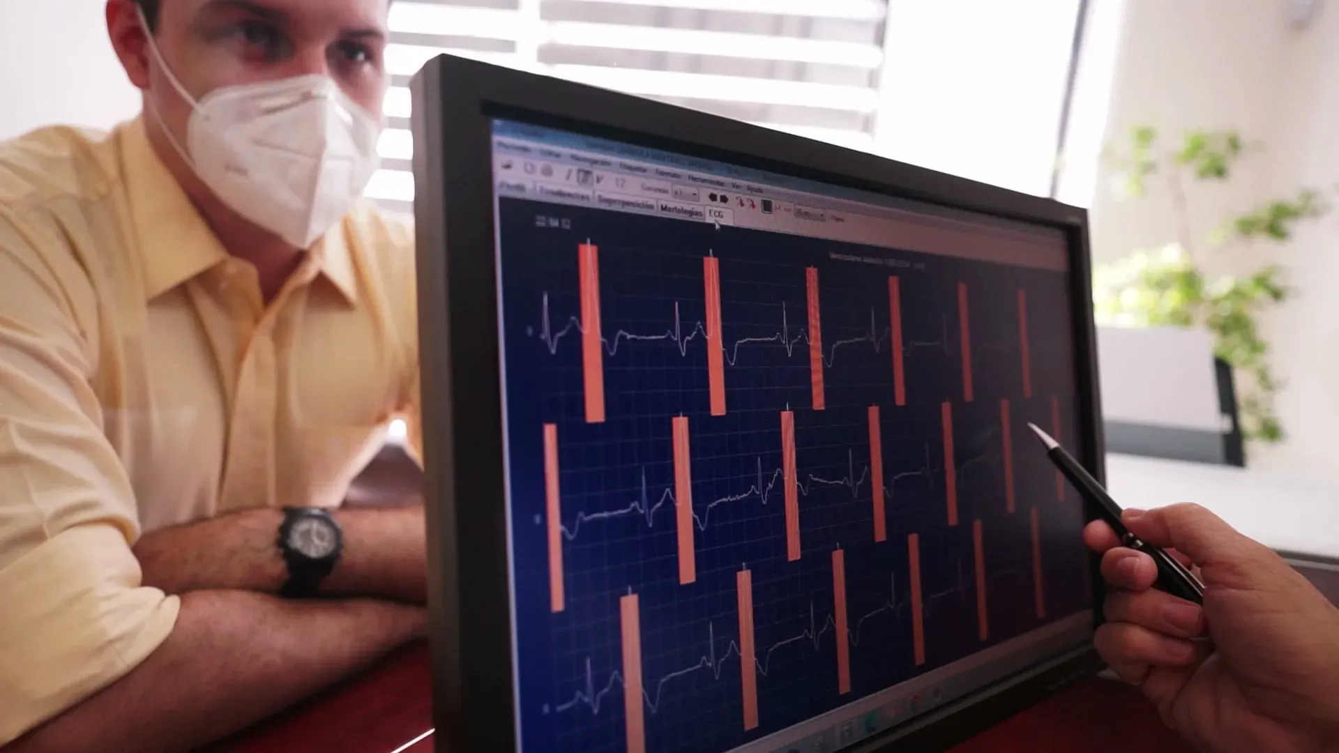
Echocardiogram in Dubai: Heart Ultrasound Imaging
See your heart beating in real-time with zero radiation
Нулевое излучение
Безопасное, неинвазивное сканирование с использованием магнитных полей (без рентгеновских лучей и ионизирующего излучения).
Fast Results
Expert cardiologist interpretation within 24 hours
Non-Invasive
Painless. No injections or contrast required
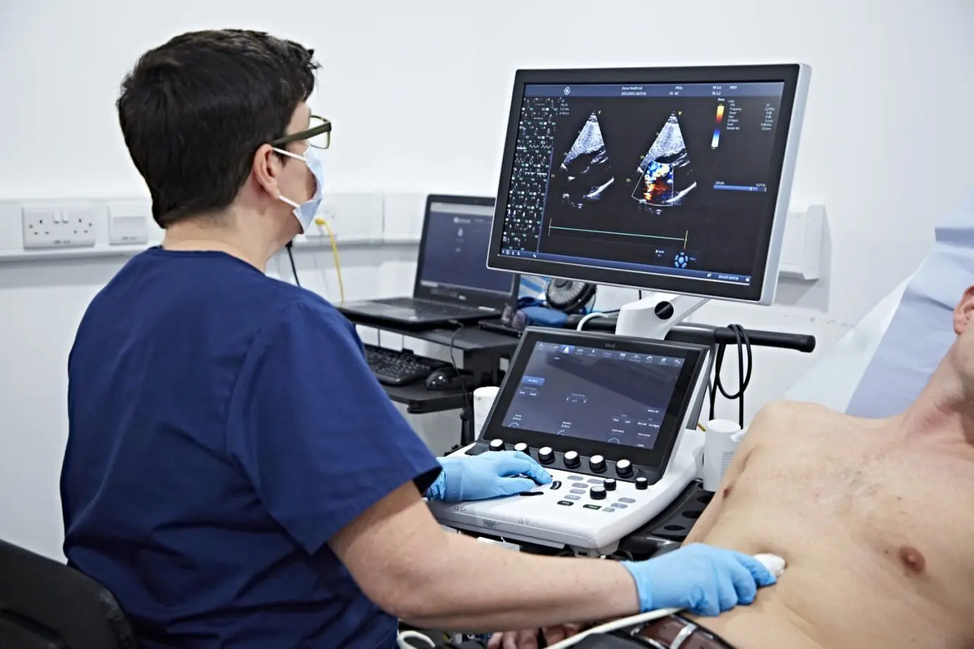
An echocardiogram uses sound waves to create live images of your heart. You can see your heart beating, valves opening and closing, and blood flowing through the chambers. It's completely painless with no radiation. making it ideal for repeated monitoring.
Your cardiologist may recommend an echo for heart murmurs, shortness of breath, chest pain, valve disease, or to assess heart function after a cardiac event. The test takes 20-40 minutes and provides detailed information about your heart's structure and pumping ability.
Every echo is reviewed by board-certified specialist cardiologists at our echocardiography suite near Oud Metha Metro with same-day appointments and results typically within 24 hours.
The echocardiogram is one of the most versatile tools in cardiology. It measures ejection fraction, the percentage of blood your heart pumps out with each beat, which is a critical indicator of heart function. A normal ejection fraction ranges from 50 to 70 percent. The test also evaluates heart valve function, identifies areas of the heart wall that may not be contracting properly, and detects fluid around the heart (pericardial effusion).
Unlike many cardiac imaging tests, echocardiography uses no radiation and requires no contrast injection for standard studies. This makes it safe for repeated monitoring over time, which is particularly valuable for patients with chronic heart conditions, those recovering from cardiac surgery, and individuals on medications that may affect heart function. It is also safe during pregnancy when cardiac assessment is needed.
An echocardiogram can detect a wide range of cardiac conditions that might otherwise go undiagnosed until they become serious. Heart valve disease, including mitral valve prolapse, aortic stenosis, and regurgitation, is identified by visualizing valve leaflet movement and measuring blood flow velocity across each valve. Cardiomyopathies, where the heart muscle becomes enlarged, thickened, or stiffened, are diagnosed through chamber measurements and wall thickness assessment. Pericardial effusion, the accumulation of fluid around the heart, is immediately visible on echo and can be monitored for progression toward the dangerous condition of cardiac tamponade. Congenital heart defects such as atrial and ventricular septal defects are reliably detected, even in adults who may have been unaware of these conditions for decades. The echocardiogram is often the first test that reveals the underlying cause of symptoms like unexplained breathlessness, fatigue, palpitations, or leg swelling.
Several types of echocardiogram exist, each suited to specific clinical questions. The standard transthoracic echocardiogram (TTE) is the most common type, performed by placing the transducer on the chest wall to obtain images through the rib spaces. A stress echocardiogram combines resting echo images with images captured immediately after exercise on a treadmill or pharmacological stress, revealing areas of the heart muscle that may not receive adequate blood flow during exertion, which can indicate coronary artery disease. Doppler echocardiography measures blood flow velocity and direction through the heart chambers and across the valves, essential for grading valve stenosis and regurgitation severity. Tissue Doppler imaging assesses the speed of heart muscle contraction and relaxation, providing sensitive markers of diastolic dysfunction that standard measurements may miss. Your cardiologist will select the type of echo most appropriate for your clinical situation.
A cardiologist typically recommends an echocardiogram when clinical findings or symptoms suggest a structural or functional heart problem. Common triggers include a newly detected heart murmur on physical examination, unexplained shortness of breath that worsens with activity, chest pain that may be cardiac in origin, swelling in the legs or abdomen suggesting fluid retention, abnormal ECG findings that need structural correlation, and a family history of cardiomyopathy or sudden cardiac death. An echo is also routinely ordered before and after cardiac surgery, to monitor the effects of chemotherapy drugs that can damage the heart, and to evaluate patients with poorly controlled hypertension for signs of left ventricular hypertrophy. If your doctor has recommended an echo test in Dubai, DCDC offers same-day appointments with board-certified cardiologist interpretation and results within 24 hours.
Нулевое излучение
Безопасное, неинвазивное сканирование с использованием магнитных полей (без рентгеновских лучей и ионизирующего излучения).
Fast Results
Expert cardiologist interpretation within 24 hours
Non-Invasive
Painless. No injections or contrast required
Echocardiogram in Dubai: Heart Ultrasound Imaging
See your heart beating in real-time with zero radiation
An echocardiogram uses sound waves to create live images of your heart. You can see your heart beating, valves opening and closing, and blood flowing through the chambers. It's completely painless with no radiation. making it ideal for repeated monitoring.
Your cardiologist may recommend an echo for heart murmurs, shortness of breath, chest pain, valve disease, or to assess heart function after a cardiac event. The test takes 20-40 minutes and provides detailed information about your heart's structure and pumping ability.
Every echo is reviewed by board-certified specialist cardiologists at our echocardiography suite near Oud Metha Metro with same-day appointments and results typically within 24 hours.
The echocardiogram is one of the most versatile tools in cardiology. It measures ejection fraction, the percentage of blood your heart pumps out with each beat, which is a critical indicator of heart function. A normal ejection fraction ranges from 50 to 70 percent. The test also evaluates heart valve function, identifies areas of the heart wall that may not be contracting properly, and detects fluid around the heart (pericardial effusion).
Unlike many cardiac imaging tests, echocardiography uses no radiation and requires no contrast injection for standard studies. This makes it safe for repeated monitoring over time, which is particularly valuable for patients with chronic heart conditions, those recovering from cardiac surgery, and individuals on medications that may affect heart function. It is also safe during pregnancy when cardiac assessment is needed.
An echocardiogram can detect a wide range of cardiac conditions that might otherwise go undiagnosed until they become serious. Heart valve disease, including mitral valve prolapse, aortic stenosis, and regurgitation, is identified by visualizing valve leaflet movement and measuring blood flow velocity across each valve. Cardiomyopathies, where the heart muscle becomes enlarged, thickened, or stiffened, are diagnosed through chamber measurements and wall thickness assessment. Pericardial effusion, the accumulation of fluid around the heart, is immediately visible on echo and can be monitored for progression toward the dangerous condition of cardiac tamponade. Congenital heart defects such as atrial and ventricular septal defects are reliably detected, even in adults who may have been unaware of these conditions for decades. The echocardiogram is often the first test that reveals the underlying cause of symptoms like unexplained breathlessness, fatigue, palpitations, or leg swelling.
Several types of echocardiogram exist, each suited to specific clinical questions. The standard transthoracic echocardiogram (TTE) is the most common type, performed by placing the transducer on the chest wall to obtain images through the rib spaces. A stress echocardiogram combines resting echo images with images captured immediately after exercise on a treadmill or pharmacological stress, revealing areas of the heart muscle that may not receive adequate blood flow during exertion, which can indicate coronary artery disease. Doppler echocardiography measures blood flow velocity and direction through the heart chambers and across the valves, essential for grading valve stenosis and regurgitation severity. Tissue Doppler imaging assesses the speed of heart muscle contraction and relaxation, providing sensitive markers of diastolic dysfunction that standard measurements may miss. Your cardiologist will select the type of echo most appropriate for your clinical situation.
A cardiologist typically recommends an echocardiogram when clinical findings or symptoms suggest a structural or functional heart problem. Common triggers include a newly detected heart murmur on physical examination, unexplained shortness of breath that worsens with activity, chest pain that may be cardiac in origin, swelling in the legs or abdomen suggesting fluid retention, abnormal ECG findings that need structural correlation, and a family history of cardiomyopathy or sudden cardiac death. An echo is also routinely ordered before and after cardiac surgery, to monitor the effects of chemotherapy drugs that can damage the heart, and to evaluate patients with poorly controlled hypertension for signs of left ventricular hypertrophy. If your doctor has recommended an echo test in Dubai, DCDC offers same-day appointments with board-certified cardiologist interpretation and results within 24 hours.

Dr. Shahoo Mazhari
Наши услуги
Комплексные услуги echocardiogram в DCDC Dubai Healthcare City.


Консультация кардиолога
Expert review of your echo results and cardiac care.
Все услуги выполняются специалистами с лицензией DHA
Кому рекомендуется Echocardiogram?
An echocardiogram is recommended when your cardiologist needs to visualize your heart's structure, pumping function, or valves. either for diagnosis or monitoring.
Heart murmurs or abnormal heart sounds
Unexplained chest pain or shortness of breath
Monitoring after heart attack or heart disease
Valve disease assessment (stenosis, regurgitation)
Heart function check after treatment or surgery
Hypertension effects on the heart
Why Choose DCDC for Your Echocardiogram?
MOHAP-licensed facility with advanced cardiac imaging, specialist cardiologists, and results within 24 hours.
MOHAP Licensed
Accredited medical center in Dubai Healthcare City. License NIMY7VY5-240925.
Fast Results
Reports ready within 18-24 hours. Same-day for urgent cases.
Insurance Support
Direct billing with 20+ insurers. We handle pre-authorization for you.
Convenient Location
Building 64, Block A in Dubai Healthcare City. Free parking on-site.
Expert Cardiologists
Board-certified specialists in cardiac imaging and echocardiography.
Patient Experience
4.8/5 Google rating. Comfortable, non-invasive cardiac imaging.
Ваш комфортный визит
Простой пошаговый процесс Echocardiogram сканирования, разработанный для комфорта, скорости и точности.
Echocardiogram Pricing
Стоимость Echocardiogram в Дубае зависит от типа теста и страхового покрытия для стандартная и стресс-эхокардиография.
- Цены варьируются в зависимости от типа сканирования и необходимости контраста
- Страховое покрытие принимается при наличии направления и подтверждения
- Прозрачные тарифы при самостоятельной оплате с мгновенным расчётом по запросу
Проверка страховки за минуты • Без скрытых платежей • Быстрый ответ в WhatsApp
Руководство для пациента
What to Expect During Your Echocardiogram
A painless, radiation-free test that takes 20-40 minutes.
Страхование и местоположение
Страховые партнёры
- •Более 20 страховых компаний в Дубае включая Daman, AXA, ADNIC и другие
- •Поддержка предварительного одобрения и прямой расчёт (при наличии)
- •Проверка покрытия перед вашим визитом в нашу клинику в Dubai Healthcare City
- •Прозрачные цены без скрытых платежей за услуги echocardiogram


Посетите нас в Медицинском городе Дубая
Докторс Клиник Диагностик Центр
Здание 64, Блок A, Медицинский комплекс Аль-Рази, Медицинский город Дубая, Дубай, ОАЭ
Рядом с улицей Уд-Мета · Удобный доступ из Бур-Дубая, центра Дубая, Бизнес-Бей · Бесплатная выделенная парковка
Часы работы
Пн-Чт, Сб: 8:00 - 22:30 | Вс: 8:30 - 22:30 | Пт: 9:00 - 22:00
Как работает страхование в DCDC
Проверьте покрытие
Убедитесь, что ваш план покрывает Echocardiogram
Получите направление
Некоторые страховщики требуют направление от терапевта — мы поможем
Предварительное одобрение
Мы оформляем предварительное одобрение напрямую с вашей страховой
Прямой расчёт
Без предоплаты — мы выставляем счёт напрямую вашей страховой
Только доплата
Вы оплачиваете только применимую доплату в клинике
Ваш специалист
Dr. Shahoo Mazhari
Consultant Cardiologist
MD, Consultant Cardiologist
Персидский · Английский · Курдский
Руководство для пациента
Understanding Echocardiography: How Heart Ultrasound Works
Echocardiography uses high-frequency sound waves (ultrasound) to create real-time moving images of your heart. A transducer placed on your chest emits sound waves that bounce off your heart structures and return to the transducer as echoes. A computer processes these echoes into detailed images that show your heart beating, valves opening and closing, and blood flowing through the chambers. This is the same safe ultrasound technology used during pregnancy.
Several modes of echocardiography provide different types of information. Two-dimensional (2D) echo shows real-time cross-sectional views of the heart. M-mode provides a single-beam view that is excellent for precise measurements of chamber sizes and wall thickness. Doppler echocardiography measures the speed and direction of blood flow through the heart, which is essential for assessing valve function and detecting abnormal blood flow patterns such as regurgitation or stenosis.
Color flow Doppler adds a visual map of blood flow direction and velocity overlaid on the 2D image, making it easy to see areas of turbulent or abnormal flow. Tissue Doppler imaging measures the velocity of the heart muscle itself, providing information about how well the heart muscle relaxes and contracts. Together, these techniques give cardiologists a comprehensive assessment of cardiac structure and function in a single non-invasive examination.
Ejection fraction (EF) is one of the most important measurements obtained from an echocardiogram. It represents the percentage of blood that leaves the left ventricle with each heartbeat. A normal EF is 50 to 70 percent. An EF below 40 percent typically indicates significant heart muscle weakness and may be classified as heart failure with reduced ejection fraction (HFrEF). Serial echocardiograms allow cardiologists to track changes in EF over time, monitor response to treatment, and adjust medications accordingly.
Heart Conditions Assessed by Echocardiogram
Heart Valve Disease
Echocardiography is the primary tool for diagnosing and grading valve stenosis (narrowing) and regurgitation (leaking) affecting the mitral, aortic, tricuspid, and pulmonary valves. It guides decisions about medical management versus surgical repair or replacement.
Heart Failure
Echo measures ejection fraction and other parameters that define the type and severity of heart failure. It distinguishes between heart failure with reduced ejection fraction (HFrEF) and heart failure with preserved ejection fraction (HFpEF), which require different treatment approaches.
Cardiomyopathy
Echocardiography detects dilated cardiomyopathy (enlarged, weakened heart), hypertrophic cardiomyopathy (thickened heart muscle), and restrictive cardiomyopathy (stiffened heart muscle). It helps differentiate between types and monitor disease progression.
Pericardial Effusion
Echo can identify fluid accumulation in the sac surrounding the heart (pericardium) and assess whether the effusion is large enough to compress the heart chambers, a potentially life-threatening condition called cardiac tamponade.
Congenital Heart Defects
Echocardiography is essential for diagnosing structural heart abnormalities present from birth, such as atrial septal defect (ASD), ventricular septal defect (VSD), and patent ductus arteriosus (PDA), both in children and adults.
Left Ventricular Hypertrophy
Long-standing high blood pressure can cause the heart muscle to thicken. Echocardiography measures wall thickness and left ventricular mass, providing an objective assessment of hypertensive heart disease that helps guide blood pressure treatment intensity.
Связанные статьи
Best Cardiologist Dubai: Heart Tests, ECG & Cardiac Screening
A comprehensive overview of cardiac diagnostic services available in Dubai, including echocardiography, stress testing, and when to see a cardiologist.
Cardiac Screening Dubai: Heart Checkup & Prevention Guide
How echocardiography fits into a preventive cardiac screening program, alongside ECG, blood tests, and lifestyle risk assessment.
Hypertension: The Silent Killer You Need to Know About
Understanding how long-standing high blood pressure affects the heart and why echocardiography is used to assess hypertensive heart disease.
Calcium Score Test: What Your Score Means
Learn how cardiac calcium scoring complements echocardiography in evaluating overall heart health and coronary artery disease risk.
Связанные услуги
Изучите другие диагностические и консультационные услуги, доступные в нашей клинике в Dubai Healthcare City.
Часто задаваемые вопросы
Часто задаваемые вопросы о Echocardiogram в Дубае.
No, an echocardiogram is completely painless. The test involves applying a warm water-based gel to your chest and moving a small handheld device called a transducer across different positions on your chest wall. The transducer emits sound waves that bounce off your heart structures to create images. You may feel light pressure from the transducer, but there are no needles, injections, or radiation involved. Some patients find lying on their left side for an extended period mildly uncomfortable, but the test itself causes no pain. The sonographer may ask you to change positions or hold your breath briefly to obtain specific views of your heart.
A standard transthoracic echocardiogram typically takes 20 to 40 minutes, with most patients done in about 30 minutes. The duration depends on the complexity of findings and the number of views the sonographer needs to obtain. If your cardiologist has requested specific measurements or your heart anatomy requires additional imaging angles, the test may take slightly longer. A stress echocardiogram, which combines echo imaging with exercise or medication-induced stress, takes approximately 45 to 60 minutes in total. There is no recovery time after a standard echocardiogram, and you can resume all normal activities, including driving and exercise, immediately afterward.
No fasting is required for a standard transthoracic echocardiogram. You can eat, drink, and take your regular medications as normal before the test. However, if you are scheduled for a stress echocardiogram, you may be asked to avoid heavy meals, caffeine, and certain medications for a specified period before the test, as these can affect your heart rate response during exercise. Your booking team will provide specific preparation instructions when you schedule the appointment. For a transesophageal echocardiogram (TEE), which involves passing a probe through the esophagus, fasting for several hours beforehand is required. This type is less common and is arranged separately.
An echocardiogram provides a comprehensive assessment of your heart. It measures the size of your heart chambers (left and right atria and ventricles), the thickness of your heart walls, and how strongly your heart contracts with each beat (ejection fraction). It evaluates all four heart valves for stenosis (narrowing) or regurgitation (leaking) and uses Doppler technology to measure the speed and direction of blood flow through your heart. The test can detect structural abnormalities, fluid around the heart (pericardial effusion), blood clots within the chambers, and signs of conditions such as cardiomyopathy, congenital heart defects, and the effects of high blood pressure on heart structure.
At DCDC, echocardiogram results are typically available within 18 to 24 hours. For urgent or clinically critical cases, same-day reporting is available. After your scan, the images and measurements are reviewed by a board-certified specialist cardiologist who prepares a detailed report including all key measurements, findings, and clinical interpretation. This report is sent directly to your referring physician and is also accessible through our patient portal. If your cardiologist identifies any immediately concerning findings during the review, they may contact your referring doctor sooner. We recommend scheduling a follow-up consultation with your cardiologist or referring doctor to discuss the results in detail.
Yes, a referral from a licensed physician is required for an echocardiogram, both for medical appropriateness and for insurance coverage. The referral provides the sonographer and interpreting cardiologist with important clinical context, such as your symptoms, medical history, and the specific clinical question being asked. This information is essential for accurate interpretation of the images. If you do not have a referral, our general practitioners or cardiologists at DCDC can assess your symptoms during a consultation and provide one on the same day if an echocardiogram is clinically indicated. For self-pay patients, we can also arrange a same-day consultation and echocardiogram.
Ejection fraction (EF) is the percentage of blood that your left ventricle pumps out with each heartbeat. It is one of the most important measurements obtained from an echocardiogram and serves as a key indicator of how well your heart is functioning as a pump. A normal ejection fraction ranges from 50 to 70 percent. An EF of 41 to 49 percent is considered mildly reduced, while an EF of 40 percent or below indicates significantly reduced heart function and may be classified as heart failure with reduced ejection fraction. Your cardiologist will interpret your EF in the context of your overall clinical picture, symptoms, and other echocardiographic findings.
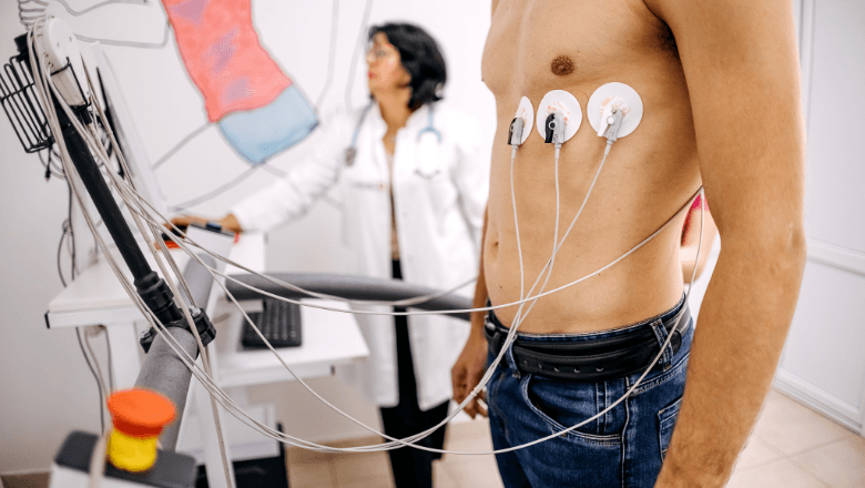
A standard echocardiogram images your heart at rest, assessing structure, valve function, chamber sizes, and ejection fraction under normal resting conditions. A stress echocardiogram adds a physical or pharmacological stress component. You either exercise on a treadmill or receive a medication that increases your heart rate, and echo images are taken before and immediately after the stress. This comparison reveals areas of the heart muscle that may not receive adequate blood supply during exertion, which can indicate coronary artery disease. A stress echo is particularly useful for evaluating chest pain during exercise, assessing known coronary artery disease, and pre-surgical cardiac risk evaluation.
Ключевые факты
Медицинский контроль
Все услуги предоставляются под наблюдением лицензированных медицинских специалистов в нашем аккредитованном MOHAP учреждении.