Ключевые выводы
- Допплер почечных артерий оценивает кровоток к почкам и внутри них с помощью звуковых волн
- Это основной неинвазивный тест для выявления стеноза почечной артерии — ведущей причины вторичной гипертензии
- Исследование играет ключевую роль в мониторинге пересаженных почек на предмет отторжения и сосудистых осложнений
- Пациенты с резистентной гипертензией, необъяснимым снижением функции почек или диабетом получают наибольшую пользу от допплера почек
- Для сканирования требуется голодание 6-8 часов, а длится оно приблизительно 20-30 минут
Допплер почечных артерий — это специализированное визуализационное исследование, которое оценивает кровоток через почечные артерии и вены, предоставляя детальную информацию о том, насколько хорошо кровь поступает и циркулирует внутри почек. Это неинвазивное сканирование необходимо для выявления состояний, влияющих на кровоснабжение почек, включая стеноз почечной артерии, и играет важную роль в мониторинге пациентов с пересаженными почками, резистентной гипертензией и хронической болезнью почек.


Что такое допплер почечных артерий?
Допплер почечных артерий сочетает стандартную визуализацию ультразвуком с допплеровским анализом кровотока для оценки кровеносных сосудов, снабжающих почки. В то время как обычное УЗИ почек показывает размер, форму и структуру почек, допплеровское исследование добавляет критически важное измерение кровотока. Оно может обнаружить сужение, блокировку или аномальные паттерны кровотока в почечных артериях и венах, которые стандартная визуализация не способна выявить.
Исследование измеряет несколько важных параметров, включая пиковую систолическую скорость (PSV) в почечных артериях, ренально-аортальное соотношение (RAR) и индекс резистентности (RI) внутри самой почки. Эти измерения помогают врачам определить, получают ли почки достаточный кровоток и присутствуют ли какие-либо сосудистые аномалии.
Что выявляет допплер почечных артерий?
Допплер почечных артерий может обнаружить и оценить несколько важных состояний, влияющих на сосудистое здоровье почек. Понимание того, что выявляет этот тест, помогает пациентам оценить его диагностическую ценность.
Стеноз почечной артерии
Стеноз почечной артерии (СПА) — это сужение одной или обеих артерий, снабжающих почки кровью. Это одна из наиболее частых причин вторичной гипертензии, то есть повышенного артериального давления, вызванного определяемым основным заболеванием, а не первичной гипертензией. СПА чаще всего вызывается атеросклерозом (накоплением бляшек в артериях) у пожилых людей или фибромускулярной дисплазией у молодых женщин.
Во время допплера почек стеноз выявляется по увеличению скорости кровотока в месте сужения. Пиковая систолическая скорость более 200 см/с в почечной артерии или ренально-аортальное соотношение более 3,5 настоятельно указывают на значительный стеноз. Тест также может обнаружить полную окклюзию при отсутствии кровотока, что помогает принимать неотложные решения о лечении.
Мониторинг пересаженной почки
Для пациентов, перенёсших пересадку почки, допплер почек является одним из важнейших инструментов мониторинга. Тест может обнаружить сосудистые осложнения трансплантации, включая стеноз почечной артерии в месте хирургического соединения, тромбоз почечной вены и артериовенозные фистулы. Изменения индекса резистентности в пересаженной почке также могут сигнализировать о раннем отторжении до появления клинических симптомов.
Реципиенты трансплантата обычно проходят допплеровскую оценку почек в первые несколько дней после операции, затем с регулярными интервалами в течение первого года и далее. Любое внезапное снижение функции почек требует немедленного допплеровского обследования для исключения сосудистых причин.
Оценка кровотока почек
Помимо стеноза и мониторинга трансплантата, допплер почек предоставляет ценную информацию об общей перфузии почек. Индекс резистентности, измеренный в интраренальных артериях, отражает состояние мелких кровеносных сосудов внутри почки. Повышенный индекс резистентности (выше 0,7) может указывать на повреждение почки от таких состояний, как диабетическая нефропатия, хроническая болезнь почек или гипертензивное повреждение почек.
Тромбоз почечной вены
Допплер почек может обнаружить тромбы в почечных венах — состояние, известное как тромбоз почечной вены. Это состояние может вызвать внезапную боль в боку, кровь в моче и быстрое снижение функции почек. Оно чаще встречается у пациентов с нефротическим синдромом, определёнными видами рака или нарушениями свёртываемости крови. Допплер выявляет тромб, демонстрируя отсутствие или обратный кровоток в поражённой почечной вене.
Кому необходим допплер почечных артерий?
Допплер почек не является рутинным скрининговым исследованием для всех. Он конкретно показан пациентам с определёнными клиническими данными или факторами риска. Ваш врач может рекомендовать допплер почек, если у вас есть:
- Резистентная гипертензия: повышенное артериальное давление, которое недостаточно реагирует на три или более лекарственных препарата
- Внезапное начало повышенного артериального давления, особенно у молодых людей (до 30 лет) или пожилых (старше 55 лет)
- Необъяснимое снижение функции почек, особенно если оно ухудшается после начала приёма ингибиторов АПФ или блокаторов рецепторов ангиотензина
- Абдоминальный шум (необычный звук, выслушиваемый стетоскопом над областью почки)
- Пересадка почки в анамнезе, особенно при изменении функции почек или артериального давления
- Повторные эпизоды молниеносного отёка лёгких (внезапного накопления жидкости в лёгких) без явной кардиологической причины
- Асимметричный размер почек, обнаруженный на других исследованиях визуализации (одна почка меньше другой)
- Подозрение на тромбоз почечной вены с симптомами боли в боку, крови в моче и белка в моче
- Диабет с ухудшением функции почек — для оценки как крупных, так и мелких сосудистых заболеваний почек
Нужна допплеровская оценка почек?
В Doctors Clinic Diagnostic Center в Дубай Хелскеа Сити мы проводим экспертный допплер почечных артерий для оценки кровотока почек, скрининга стеноза почечной артерии и мониторинга трансплантата.
Связь между стенозом почечной артерии и гипертензией
Стеноз почечной артерии является причиной приблизительно 1-5% всех случаев гипертензии и значительного процента случаев резистентной гипертензии. Когда одна или обе почечные артерии сужаются, поражённая почка воспринимает снижение кровотока и активирует ренин-ангиотензин-альдостероновую систему (РААС), которая повышает артериальное давление во всём организме.
Это создаёт порочный круг, при котором организм повышает давление, чтобы протолкнуть больше крови через суженную артерию, но высокое давление повреждает кровеносные сосуды по всему телу. Обнаружение СПА с помощью допплера почек позволяет врачам устранить первопричину гипертензии, а не просто лечить повышенные показатели дополнительными лекарствами. Варианты лечения могут включать ангиопластику со стентированием или медикаментозное лечение в зависимости от тяжести и причины стеноза.
Допплер почек в контексте Дубая
Ряд факторов, специфичных для населения ОАЭ, делают допплер почек особенно актуальным для жителей Дубая:
- Высокая распространённость диабета: ОАЭ имеют одну из самых высоких в мире показателей заболеваемости диабетом, а диабетическая нефропатия является ведущей причиной почечной недостаточности. Допплер почек помогает оценить сосудистые изменения в диабетических почках до развития значительного повреждения.
- Бремя гипертензии: гипертензия поражает значительную часть населения ОАЭ, и скрининг на вторичные причины, такие как СПА, необходим для пациентов с трудноконтролируемым артериальным давлением.
- Активные программы трансплантации: в ОАЭ развиваются программы пересадки почек, и допплер почек необходим для послеоперационного мониторинга сосудов.
- Разнообразное экспатриантское население: мультикультурное население Дубая включает людей из регионов с различной распространённостью факторов риска заболеваний почек, что делает индивидуальный скрининг важным.
- Обезвоживание из-за жары: хроническое обезвоживание в жарком климате может способствовать нагрузке на почки и ускорять существующие заболевания почек, что обосновывает более тщательный мониторинг у восприимчивых пациентов.
Допплер почек vs КТ-ангиография vs МР-ангиография
Несколько методов визуализации могут оценить почечные артерии. В таблице ниже сравниваются допплер почек, КТ-ангиография и МР-ангиография, чтобы помочь пациентам и врачам понять преимущества и компромиссы каждого подхода.
| Характеристика | Допплер почечных артерий | КТ-ангиография (КТА) | МР-ангиография (МРА) |
|---|---|---|---|
| Излучение | Нет | Да (ионизирующее излучение) | Нет |
| Контрастное вещество | Не требуется | Йодированный контраст (в/в) | Контраст с гадолинием (в/в) |
| Чувствительность для СПА | 85% – 95% | 95% – 100% | 90% – 100% |
| Лучше всего подходит для | Скрининг, наблюдение после трансплантации, серийный мониторинг | Окончательное предоперационное картирование | Пациенты, которые не могут получать йодированный контраст |
| Влияние на функцию почек | Безопасен независимо от функции почек | Контраст может ухудшить заболевание почек | Риск гадолиния при тяжёлой болезни почек |
| Длительность сканирования | 20 – 30 минут | 5 – 10 минут | 30 – 45 минут |
| Повторяемость | Неограниченная, без кумулятивного риска | Ограничена излучением и нагрузкой контрастом | Ограничена контрастом и доступностью |
| Стоимость (AED, приблизительно) | 600 – 1 200 | 1 500 – 3 000 | 2 500 – 5 000 |
Допплер почек является предпочтительным тестом первой линии. КТА и МРА обычно применяются в случаях, требующих более детального анатомического картирования, или когда результаты допплера неубедительны.
Как подготовиться к допплеру почечных артерий
Допплер почек — один из немногих типов допплеровского исследования, требующих голодания перед сканированием. Правильная подготовка к допплеровскому УЗИ обеспечивает наилучшее качество изображений и точные результаты.
- Не ешьте за 6-8 часов до приёма. Воду можно пить в небольших количествах; уточните у клиники.
- Избегайте газообразующих продуктов накануне (бобовые, чечевица, газированные напитки, сырые овощи).
- Продолжайте приём всех регулярных лекарств с небольшим глотком воды, если не указано иное.
- Наденьте удобную, свободную одежду, желательно комплект из двух предметов для лёгкого доступа к области живота.
- Возьмите направление, страховую карту и все предыдущие результаты визуализации почек или анализов крови.
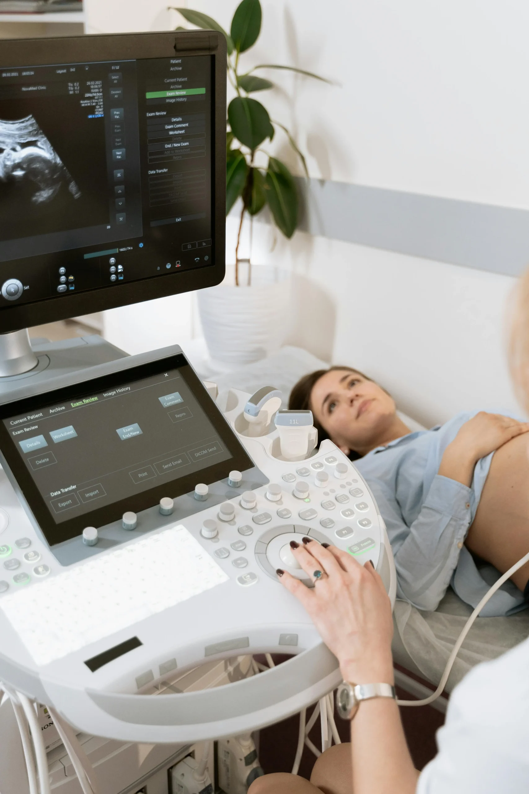
Что происходит во время сканирования
Во время допплера почечных артерий вы лежите на спине на кушетке. Сонографист наносит тёплый гель на живот и помещает датчик над областью почек. Датчик посылает и принимает звуковые волны, создающие изображения почек и их кровеносных сосудов на мониторе в реальном времени.
Вас могут попросить лечь на бок, глубоко вдохнуть или задержать дыхание в различные моменты сканирования. Эти изменения положения помогают сонографисту получить оптимальные изображения почечных артерий, которые сложно визуализировать из-за их глубины и наличия кишечных газов. Всё сканирование обычно занимает от 20 до 30 минут.
Радиолог просматривает изображения и измерения после сканирования и составляет подробный отчёт для вашего направившего врача. Результаты обычно доступны в течение 24 часов.
Допплер почек в DCDC Дубай Хелскеа Сити
В Doctors Clinic Diagnostic Center мы предоставляем комплексные услуги допплера почечных артерий, выполняемые опытными радиологами с использованием современного оборудования для допплеровского УЗИ. Наша команда имеет обширный опыт оценки нативных и пересаженных почек с детальной отчётностью, помогающей нефрологам и терапевтам принимать обоснованные решения о лечении.
Расположенный в Дубай Хелскеа Сити, мы обслуживаем пациентов, направленных нефрологами, терапевтами, кардиологами и трансплантационными командами по всему Дубаю. Мы принимаем большинство основных страховых планов и предлагаем прозрачные варианты оплаты для незастрахованных пациентов.
Нужен допплер почечных артерий?
В Doctors Clinic Diagnostic Center в Дубай Хелскеа Сити мы предоставляем экспертные услуги допплера почечных артерий для оценки кровотока почек, скрининга стеноза почечной артерии и мониторинга трансплантата.
Связанные услуги в DCDC
Квалифицированная помощь и современная диагностика в Dubai Healthcare City
Doppler Ultrasound
Specialized renal vascular imaging to assess kidney blood flow
Записаться на приёмChronic Kidney Disease
Expert management of kidney function decline and renal conditions
Записаться на приёмHypertension Treatment
Comprehensive blood pressure management including secondary causes
Записаться на приёмFrequently Asked Questions
Заключение
Допплер почечных артерий — это незаменимый диагностический инструмент, предоставляющий информацию о сосудистом здоровье почек, которую не может дать ни один другой неинвазивный тест. От обнаружения стеноза почечной артерии как причины резистентной гипертензии до мониторинга пересаженных почек на предмет сосудистых осложнений — этот тест играет критически важную роль в заботе о здоровье почек. Его безопасность, доступность и возможность повторения без риска делают его краеугольным камнем почечной диагностики.
Для жителей Дубая, где распространены диабет и гипертензия, допплер почек предлагает проактивный способ оценки сосудистого здоровья почек до развития необратимых повреждений. Для получения информации о ценах ознакомьтесь с нашим руководством по стоимости допплеровского УЗИ в Дубае. В Doctors Clinic Diagnostic Center в Дубай Хелскеа Сити наша опытная команда радиологов проводит тщательные допплеровские обследования почек с чёткими и применимыми отчётами, поддерживающими ваш путь к здоровью почек.
Источники и ссылки
Эта статья проверена нашей медицинской командой и ссылается на следующие источники:
- Американская коллегия радиологии — Руководство по визуализации стеноза почечной артерии
- Национальный фонд почек — Ренальные сосудистые заболевания
- Европейское общество радиологии — Стандарты допплера почек
- Трансплантационное общество — Руководство по мониторингу после трансплантации
Медицинский контент на этом сайте проверяется врачами, лицензированными DHA. См. нашу редакционную политику для получения дополнительной информации.

Автор
Dr. Osama Elzamzami
Консультант-радиолог
MD, Радиология
Д-р Осама Эльзамзами — консультант-радиолог, специализирующийся на диагностической визуализации, включая МРТ, КТ, ультразвук и допплеровские исследования в DCDC Дубай Хелскеа Сити.
Связанные статьи

Что такое допплеровское УЗИ? Полное руководство
Стоимость допплеровского УЗИ в Дубае: цены по видам
Как подготовиться к допплеровскому УЗИ

Что такое ультразвуковое исследование? Полное руководство
More in Diagnostic Imaging

X-Ray at Home Dubai: Cost & Guide (2026)
Читать далее
Ultrasound at Home Dubai: From AED 599 (2026)
Читать далее
Neck MRI Dubai: What It Shows & Cost (2026)
Читать далее
X-Ray Scan Dubai: Complete Guide (2026)
Читать далее
MRI vs Ultrasound Dubai: Which Scan? (2026)
Читать далее
MRI vs X-Ray Dubai: Which Scan Do You Need? (2026)
Читать далее© 2026 Doctors Clinic Diagnostic Center (DCDC), Dubai Healthcare City. Originally published at https://doctorsclinicdubai.ae/blog/renal-doppler-ultrasound-kidney. All rights reserved. Unauthorized reproduction is prohibited.