Ключевые выводы
- УЗИ малого таза — это безопасное, безболезненное исследование, которое использует звуковые волны для получения изображений матки, яичников, маточных труб, шейки матки и мочевого пузыря в реальном времени без какого-либо облучения
- Существуют два основных вида: трансабдоминальное УЗИ (выполняется по нижней части живота с наполненным мочевым пузырём) и трансвагинальное УЗИ (с использованием тонкого внутреннего датчика для получения изображений более высокого разрешения репродуктивных органов)
- Распространённые причины, по которым женщинам назначают УЗИ малого таза: аномальные кровотечения, боли в области таза, оценка кист яичников, оценка миом, обследование фертильности и подтверждение ранней беременности
- Подготовка зависит от типа исследования: трансабдоминальное УЗИ требует выпить 1–1,5 литра воды за час до приёма для наполнения мочевого пузыря, тогда как трансвагинальное УЗИ требует пустого мочевого пузыря
- DCDC в Дубай Хелскеа Сити предлагает трансабдоминальное и трансвагинальное УЗИ малого таза с результатами в тот же день от опытных врачей-рентгенологов
УЗИ малого таза — одно из наиболее часто выполняемых диагностических исследований для женщин. Оно использует высокочастотные звуковые волны для создания детальных изображений органов малого таза в реальном времени, включая матку, яичники, маточные трубы, шейку матки и мочевой пузырь. В отличие от рентгена или КТ, УЗИ малого таза не предполагает ионизирующего излучения, что делает его одним из самых безопасных методов визуализации. Независимо от того, назначил ли ваш врач обследование малого таза для исследования аномальных кровотечений, оценки болей в области таза, мониторинга кист яичников, оценки миом или проверки ранней беременности — понимание того, что включает в себя обследование, поможет вам быть информированной и подготовленной.
Это руководство охватывает всё, что нужно знать женщинам об УЗИ малого таза: два основных вида (трансабдоминальное и трансвагинальное), когда рекомендуется УЗИ малого таза, как подготовиться к обследованию, что может выявить сканирование, как понять результаты и стоимость УЗИ малого таза в Дубае в Doctors Clinic Diagnostic Center (DCDC) в Дубай Хелскеа Сити.
Health Screening Packages
Save with our bundled screening packages — specialist consultation included


Что такое УЗИ малого таза?
УЗИ малого таза — это неинвазивное визуализирующее исследование, использующее устройство, называемое датчиком (трансдюсером), для отправки высокочастотных звуковых волн в тело. Эти звуковые волны отражаются от внутренних органов и тканей, а возвращающиеся эхо-сигналы преобразуются компьютером в изображения в реальном времени, отображаемые на мониторе. Техника позволяет сонографу и рентгенологу визуализировать размер, форму, положение и внутреннюю структуру органов малого таза без какого-либо разреза, инъекции или облучения.
УЗИ малого таза является исследованием первой линии для большинства гинекологических заболеваний. Американский колледж акушеров и гинекологов (ACOG) рекомендует УЗИ малого таза как начальный метод визуализации для оценки аномальных маточных кровотечений, образований придатков, болей в области таза и бесплодия. Обследование широко доступно, относительно недорого по сравнению с МРТ или КТ, совершенно безболезненно для большинства женщин и предоставляет немедленную диагностическую информацию для клинического принятия решений.
Существуют два основных подхода к проведению УЗИ малого таза у женщин: трансабдоминальное УЗИ, при котором датчик располагается на поверхности нижней части живота, и трансвагинальное УЗИ, при котором тонкий специально разработанный зонд аккуратно вводится во влагалищный канал. Во многих случаях оба подхода используются во время одного приёма для получения дополняющей информации. Трансабдоминальное сканирование даёт общий обзор анатомии малого таза, тогда как трансвагинальное сканирование обеспечивает превосходное разрешение крупного плана матки, эндометрия, яичников и окружающих структур.
«УЗИ малого таза остаётся краеугольным камнем гинекологической визуализации, потому что оно безопасно, высокоинформативно и может быть выполнено быстро на месте оказания помощи», говорит доктор Усама Аль-Замзами, заведующий отделением рентгенологии DCDC. «Для большинства женщин, обращающихся с симптомами в области таза, ультразвук предоставляет всю диагностическую информацию, необходимую для определения следующего шага в их лечении».
Трансабдоминальное vs трансвагинальное УЗИ
Понимание разницы между трансабдоминальным и трансвагинальным УЗИ помогает женщинам знать, чего ожидать, и почему врач может рекомендовать один подход вместо другого или оба. Каждая методика имеет свои сильные стороны, и выбор зависит от клинического вопроса, анатомии пациентки и требуемого уровня детализации.
Трансабдоминальное УЗИ малого таза
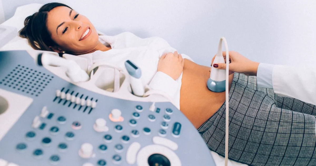
При трансабдоминальном УЗИ малого таза сонограф наносит водную основу геля на нижнюю часть живота и перемещает изогнутый датчик по поверхности кожи. Гель устраняет воздух между датчиком и кожей, позволяя звуковым волнам эффективно проникать в тело. Исследование требует наполненного мочевого пузыря, который служит акустическим окном: заполненный жидкостью мочевой пузырь отодвигает петли кишечника от таза и более эффективно передаёт ультразвуковые волны, обеспечивая более чёткое изображение матки и яичников за ним.
Трансабдоминальное УЗИ обеспечивает широкое поле зрения, которое особенно полезно для оценки крупных образований малого таза, больших миом или органов, выходящих за пределы малого таза. Это также предпочтительный подход для женщин, не имевших половой активности, подростков и любой пациентки, предпочитающей избежать внутреннего обследования. Компромисс заключается в том, что разрешение изображения ниже, чем при трансвагинальном сканировании, поскольку звуковые волны должны пройти большее расстояние через брюшную стенку.
Трансвагинальное УЗИ малого таза
Трансвагинальное УЗИ использует тонкий цилиндрический датчик (диаметром примерно 2–3 сантиметра), покрытый одноразовым безлатексным чехлом и небольшим количеством смазывающего геля. Зонд аккуратно вводится во влагалищный канал, располагаясь в непосредственной близости от матки, яичников и маточных труб. Поскольку датчик находится ближе к исследуемым органам, трансвагинальное УЗИ даёт значительно более высокое разрешение изображений, чем трансабдоминальное сканирование.
Трансвагинальное УЗИ является золотым стандартом для оценки эндометриальной выстилки (толщина, паттерн и очаговые аномалии), обнаружения мелких кист яичников, выявления ранних внутриматочных беременностей (уже с 5-й недели гестации), диагностики внематочной беременности и характеристики образований придатков. Оно не требует наполненного мочевого пузыря; фактически, мочевой пузырь должен быть пустым для оптимального качества изображения. Большинство женщин описывают процедуру как слегка дискомфортную, но не болезненную, и обычно она длится 10–20 минут.
«Мы часто выполняем и трансабдоминальное, и трансвагинальное УЗИ в одном сеансе», говорит доктор Усама Аль-Замзами, заведующий отделением рентгенологии DCDC. «Трансабдоминальное сканирование даёт нам общую картину, а трансвагинальное сканирование — мелкие детали. Вместе они обеспечивают всестороннюю оценку, максимизирующую диагностическую точность».
| Характеристика | Трансабдоминальное УЗИ | Трансвагинальное УЗИ |
|---|---|---|
| Расположение датчика | На поверхности нижней части живота | Аккуратно вводится во влагалищный канал |
| Требования к мочевому пузырю | Наполненный мочевой пузырь | Пустой мочевой пузырь |
| Разрешение изображения | Хороший обзор, но более низкое разрешение | Превосходное разрешение крупного плана |
| Лучше всего для | Крупные образования, миомы, общий обзор малого таза | Оценка эндометрия, мелкие кисты, ранняя беременность, внематочная беременность |
| Комфорт пациентки | Без внутреннего обследования; давление от наполненного мочевого пузыря | Лёгкое внутреннее давление; без дискомфорта от мочевого пузыря |
| Продолжительность | 10–15 минут | 10–20 минут |
| Подходит для девственниц | Да | Нет (вместо этого используется трансабдоминальное) |
| Облучение | Нет | Нет |
Сравнение подходов трансабдоминального и трансвагинального УЗИ малого таза. Ваш рентгенолог порекомендует наиболее подходящую методику на основе ваших клинических потребностей.
Когда женщинам нужно УЗИ малого таза?
УЗИ малого таза рекомендуется всегда, когда медицинскому работнику необходимо визуализировать женские репродуктивные органы или мочевой пузырь для исследования симптомов, мониторинга известных состояний или принятия решений о лечении. Наиболее распространённые клинические показания для УЗИ малого таза у женщин:
Аномальные маточные кровотечения
Аномальные маточные кровотечения — самая распространённая причина направления женщин на УЗИ малого таза. Это включает обильные менструации (меноррагия), межменструальные кровотечения (метроррагия), нерегулярные циклы и постменопаузальные кровотечения. Ультразвук может выявить структурные причины, такие как миомы матки, полипы эндометрия, утолщение эндометрия (гиперплазия) и аденомиоз.
Боли в области таза
Острая или хроническая боль в области таза — ещё одно частое показание. УЗИ помогает выявить кисты яичников (простые, сложные или геморрагические), перекрут яичника, разрыв кист, воспалительные заболевания органов малого таза, тубоовариальный абсцесс, эндометриомы (шоколадные кисты, связанные с эндометриозом) и внематочную беременность.
Мониторинг кист яичников
Многие кисты яичников обнаруживаются случайно или при плановых обследованиях. Большинство простых кист у пременопаузальных женщин являются функциональными и разрешаются самостоятельно в течение одного-трёх менструальных циклов. УЗИ малого таза используется для мониторинга этих кист через контрольные интервалы, обычно через 6–12 недель после первоначального сканирования.
Миомы матки
Миомы матки (лейомиомы) — доброкачественные мышечные опухоли матки, поражающие до 70 процентов женщин к 50 годам. УЗИ малого таза является основным методом визуализации для диагностики миом, определения их количества, размера и расположения (подслизистые, интрамуральные или субсерозные) и мониторинга роста во времени.
Оценка фертильности
Для женщин, проходящих оценку фертильности, УЗИ малого таза играет центральную роль. Оно используется для подсчёта антральных фолликулов (AFC) — ключевого маркера яичникового резерва. УЗИ также мониторирует фолликулярное развитие во время циклов индукции овуляции, подтверждает овуляцию, оценивает толщину и паттерн эндометрия перед переносом эмбриона при ЭКО и выявляет структурные аномалии.
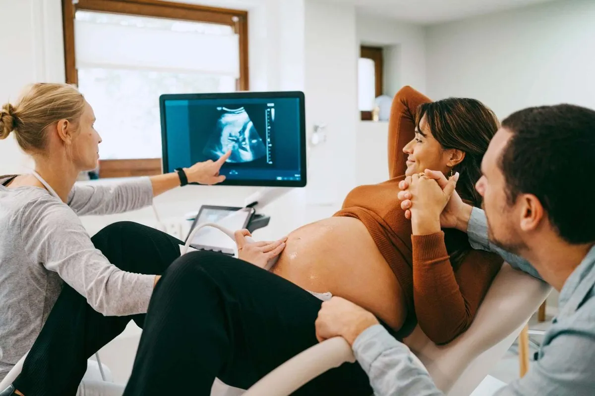
Ранняя беременность
УЗИ малого таза, особенно трансвагинальное, используется на ранних сроках беременности для подтверждения внутриматочной беременности, определения гестационного возраста, обнаружения сердцебиения плода, выявления многоплодной беременности и исключения внематочной и пузырной беременности. Самый ранний срок, когда беременность можно увидеть на трансвагинальном УЗИ — обычно около 5 недель гестации.
Проверка внутриматочной спирали (ВМС)
УЗИ регулярно используется для подтверждения правильного положения внутриматочной спирали после установки или для исследования предполагаемого смещения или миграции. Сканирование подтверждает, что ВМС правильно расположена в полости матки и не перфорировала стенку матки.
Запишитесь на УЗИ малого таза в DCDC
В Doctors Clinic Diagnostic Center в Дубай Хелскеа Сити наша команда рентгенологов предоставляет экспертные услуги УЗИ малого таза с трансабдоминальной и трансвагинальной методиками. Получите точные результаты от опытных врачей-рентгенологов с доступностью в тот же день.
Как подготовиться к УЗИ малого таза
Правильная подготовка к УЗИ малого таза полностью зависит от назначенного типа исследования. Правильная подготовка обеспечивает наилучшее качество изображения и позволяет избежать необходимости повторного обследования. Вот что нужно сделать для каждого типа:
Подготовка к трансабдоминальному УЗИ малого таза
- Пейте воду: Выпейте примерно 1–1,5 литра (4–6 стаканов) воды, начиная за час до приёма. Не опорожняйте мочевой пузырь после питья. Наполненный мочевой пузырь необходим, так как он служит акустическим окном.
- Время имеет значение: Начните пить воду примерно за 60 минут до назначенного времени сканирования. Если мочевой пузырь наполнится слишком рано, вы можете испытать значительный дискомфорт.
- Носите удобную одежду: Наденьте одежду из двух частей, чтобы легко обнажить нижнюю часть живота без полного раздевания.
- Голодание не требуется: Перед УЗИ малого таза можно нормально питаться. Необходимость в голодании отсутствует, если только на тот же день не назначено УЗИ органов брюшной полости.
Подготовка к трансвагинальному УЗИ малого таза
- Опорожните мочевой пузырь: В отличие от трансабдоминального подхода, трансвагинальное УЗИ требует пустого мочевого пузыря.
- Специальная подготовка не требуется: Нет необходимости делать спринцевание или использовать специальные очищающие средства. Просто приходите с пустым мочевым пузырём.
- Менструация — не препятствие: Трансвагинальное УЗИ может быть выполнено во время менструации, если это клинически необходимо.
- Сообщите команде: Сообщите персоналу на рецепции или сонографу, если у вас аллергия на латекс, так как чехол датчика может быть заменён на безлатексный.
Когда оба исследования выполняются вместе
Когда трансабдоминальное и трансвагинальное УЗИ выполняются в одном сеансе, сначала проводится трансабдоминальное сканирование с наполненным мочевым пузырём, затем трансвагинальное после опорожнения мочевого пузыря. Эта последовательность — наиболее эффективный способ получить оба набора изображений за одно посещение.
«Одна из самых распространённых причин, по которым нам нужно повторить УЗИ малого таза, — недостаточная подготовка мочевого пузыря», говорит доктор Усама Аль-Замзами. «Потратить время на то, чтобы выпить достаточно воды и прийти с комфортно наполненным мочевым пузырём, имеет существенное значение для качества изображений».
Что показывает УЗИ малого таза?
УЗИ малого таза предоставляет детальную информацию о структуре, размере и состоянии женских органов малого таза. Рентгенолог оценивает множество параметров во время обследования, и результаты документируются в подробном заключении. Вот что может показать УЗИ малого таза:
- Матка: Размер, форма, положение (антеверсия или ретроверсия), текстура миометрия и наличие миом
- Эндометрий: Толщина эндометрия, паттерн и очаговые аномалии
- Яичники: Размер, объём, подсчёт фолликулов, наличие кист, солидных образований и кровотока по допплеру
- Маточные трубы: Нормальные маточные трубы обычно не видны на УЗИ. Однако расширенные трубы и трубная внематочная беременность могут быть обнаружены
- Шейка матки: Длина шейки, цервикальный канал и наличие наботовых кист или образований
- Мочевой пузырь: Толщина стенки, остаточный объём мочи и наличие образований или камней
- Свободная жидкость: Наличие и количество свободной жидкости в дугласовом пространстве
- Образования малого таза: Любое аномальное образование в малом тазу с оценкой его происхождения, размера, внутренних характеристик и кровотока
История пациентки: УЗИ малого таза выявило скрытую миому
38-летняя женщина в Дубае более года страдала от постепенно усиливающихся менструальных кровотечений. Её гинеколог провёл клинический осмотр и отметил, что матка казалась слегка увеличенной, но причина была неясна. Она была направлена в DCDC на УЗИ малого таза.
Трансабдоминальное сканирование подтвердило увеличение матки, но именно трансвагинальное УЗИ выявило критическую находку: субмукозную миому размером 4 сантиметра, выступающую в полость матки. Вооружённая точными ультразвуковыми измерениями и подробным заключением DCDC, её гинеколог смогла рекомендовать целенаправленный план лечения: гистероскопическую миомэктомию.
Пациентка успешно прошла процедуру и сообщила о возвращении к нормальному менструальному потоку в течение двух циклов.
Понимание результатов УЗИ малого таза
После УЗИ малого таза рентгенолог просматривает все полученные изображения, измеряет соответствующие структуры и готовит письменное заключение, которое направляется вашему лечащему врачу. Понимание значения ключевых результатов поможет вам более продуктивно обсудить их с врачом.
Нормальные результаты
Нормальное заключение УЗИ малого таза обычно описывает матку нормального размера (примерно 7–8 см в длину у женщин репродуктивного возраста) с гладкой текстурой миометрия, толщину эндометрия, соответствующую фазе менструального цикла, два яичника нормального размера с нормальным количеством мелких фолликулов, отсутствие свободной жидкости и нормальный мочевой пузырь.
Распространённые патологические находки
- Миомы матки: Описываются по количеству, размеру и расположению. Мелкие бессимптомные миомы могут требовать только наблюдения.
- Кисты яичников: Простые кисты с тонкими стенками и прозрачной жидкостью почти всегда доброкачественные.
- Утолщение эндометрия: Толщина эндометрия более 4–5 мм у постменопаузальной женщины может потребовать дополнительного обследования.
- Полипы эндометрия: Проявляются как очаговые утолщения или мелкие образования в полости эндометрия.
- Аденомиоз: Характеризуется диффузно увеличенной, шаровидной маткой с неоднородной текстурой миометрия.
- Свободная жидкость: Небольшое количество свободной жидкости в малом тазу после овуляции — норма. Большие объёмы могут указывать на разрыв кисты, внематочную беременность или инфекцию.
Когда рекомендуется дополнительная визуализация
В некоторых случаях заключение УЗИ малого таза может рекомендовать дополнительную визуализацию. МРТ может быть предложена для сложных миом, неопределённых образований яичников или при подозрении на глубокий эндометриоз. Ваш лечащий врач обсудит эти рекомендации и объяснит следующие шаги.
«Заключение УЗИ — это важнейший инструмент коммуникации между рентгенологом и лечащим врачом», говорит доктор Усама Аль-Замзами. «В DCDC мы структурируем наши заключения так, чтобы предоставить вашему врачу чёткую, практически применимую информацию».
Стоимость УЗИ малого таза в Дубае
Стоимость УЗИ малого таза в Дубае варьируется в зависимости от типа исследования, учреждения и страхового покрытия. Вот общее руководство по ценам:
- Трансабдоминальное УЗИ малого таза: от 300 до 800 AED в большинстве диагностических центров Дубая
- Трансвагинальное УЗИ: от 400 до 1 000 AED
- Комбинированное трансабдоминальное и трансвагинальное УЗИ: от 500 до 1 200 AED
В DCDC в Дубай Хелскеа Сити стоимость УЗИ малого таза включает сканирование, интерпретацию рентгенолога и подробное письменное заключение. Большинство крупных страховых планов покрывают УЗИ малого таза при наличии медицинских показаний и направления врача.
Несколько факторов влияют на общую стоимость: тип сканирования, включение допплеровской оценки, квалификация рентгенолога и расположение учреждения. Стоит отметить, что самая низкая цена не всегда означает лучшее качество.
Для более широкого обзора цен на ультразвук см. наше руководство по стоимости УЗИ в Дубае.
Экспертное УЗИ малого таза в DCDC
DCDC в Дубай Хелскеа Сити предлагает комплексные услуги УЗИ малого таза для женщин, включая трансабдоминальную, трансвагинальную и допплеровскую оценки. Наши опытные рентгенологи предоставляют результаты в тот же день с подробными заключениями.
Или позвоните нам напрямую, чтобы записаться на УЗИ малого таза.
Связанные услуги в DCDC
Квалифицированная помощь и современная диагностика в Dubai Healthcare City
Frequently Asked Questions
Заключительные мысли
УЗИ малого таза — это безопасное, без облучения и высокоинформативное исследование, которое играет центральную роль в диагностике и лечении широкого спектра гинекологических заболеваний. Понимание разницы между трансабдоминальным и трансвагинальным подходами, правильная подготовка к обследованию и знание того, что означают результаты, ставит вас в более сильную позицию как активного участника собственного здравоохранения.
В Doctors Clinic Diagnostic Center в Дубай Хелскеа Сити женщины получают экспертные услуги УЗИ малого таза от опытных сонографов и врачей-рентгенологов, специализирующихся на женской визуализации. С результатами в тот же день, комфортной обстановкой и приверженностью чётким заключениям, DCDC обеспечивает каждой пациентке необходимую диагностическую ясность. Подробнее об ультразвуке — в нашем руководстве что такое УЗИ и как подготовиться к УЗИ.
Источники и ссылки
Эта статья проверена нашей медицинской командой и ссылается на следующие источники:
- American College of Obstetricians and Gynecologists (ACOG) - Practice Bulletin: Evaluation of the Uterus in Patients With Abnormal Uterine Bleeding
- Royal College of Obstetricians and Gynaecologists - Management of Postmenopausal Bleeding
- RadiologyInfo.org - Ultrasound: Pelvis
- American Institute of Ultrasound in Medicine (AIUM) - Practice Parameter for the Performance of Ultrasound of the Female Pelvis
Медицинский контент на этом сайте проверяется врачами, лицензированными DHA. См. нашу редакционную политику для получения дополнительной информации.

Автор
Dr. Osama Elzamzami
Диагностическая рентгенология
MD, FRCR
Доктор Усама Аль-Замзами — заведующий отделением рентгенологии DCDC в Дубай Хелскеа Сити, специализируется на диагностической визуализации, включая УЗИ, КТ, МРТ и интервенционную рентгенологию.
Связанные статьи
Что такое УЗИ? Полное руководство
Как подготовиться к УЗИ
Допплер УЗИ при беременности
More in Women's Health

AMH Test Cost Dubai: Pricing Guide (2026)
Читать далее
Perimenopause vs Menopause Dubai: Signs (2026)
Читать далее
Mother & Baby Home Care Dubai: Guide (2026)
Читать далее
Gestational Diabetes Dubai: Screening (2026)
Читать далее
Mammogram Preparation Dubai: What to Expect (2026)
Читать далее
CA-125 Test Dubai: Ovarian Cancer Marker (2026)
Читать далее© 2026 Doctors Clinic Diagnostic Center (DCDC), Dubai Healthcare City. Originally published at https://doctorsclinicdubai.ae/blog/pelvic-ultrasound-women-guide. All rights reserved. Unauthorized reproduction is prohibited.