Ключевые выводы
- Normal HSG results show a smooth triangular uterine cavity with free spill of contrast dye from both fallopian tubes
- Abnormal findings can include tubal blockage, hydrosalpinx, uterine septum, polyps, fibroids, or adhesions
- Proximal tubal blockage (near the uterus) may be caused by temporary spasm and might require repeat testing
- Distal tubal blockage (near the ovary) is more commonly associated with previous infection or endometriosis
- HSG results should always be interpreted by your doctor in the context of your complete clinical picture
After your HSG test (hysterosalpingography), you will receive a radiology report describing the findings. Understanding what these results mean can be confusing, especially when the report uses medical terminology. This guide explains how to read your HSG results, what normal and abnormal findings look like, and what the next steps might be based on different outcomes.
While your gynecologist will review your HSG results with you in detail during a follow-up consultation, having a general understanding of what the findings mean can help you feel more informed and prepared for that conversation. This article covers the most common findings seen on HSG reports.
How HSG Results Are Reported
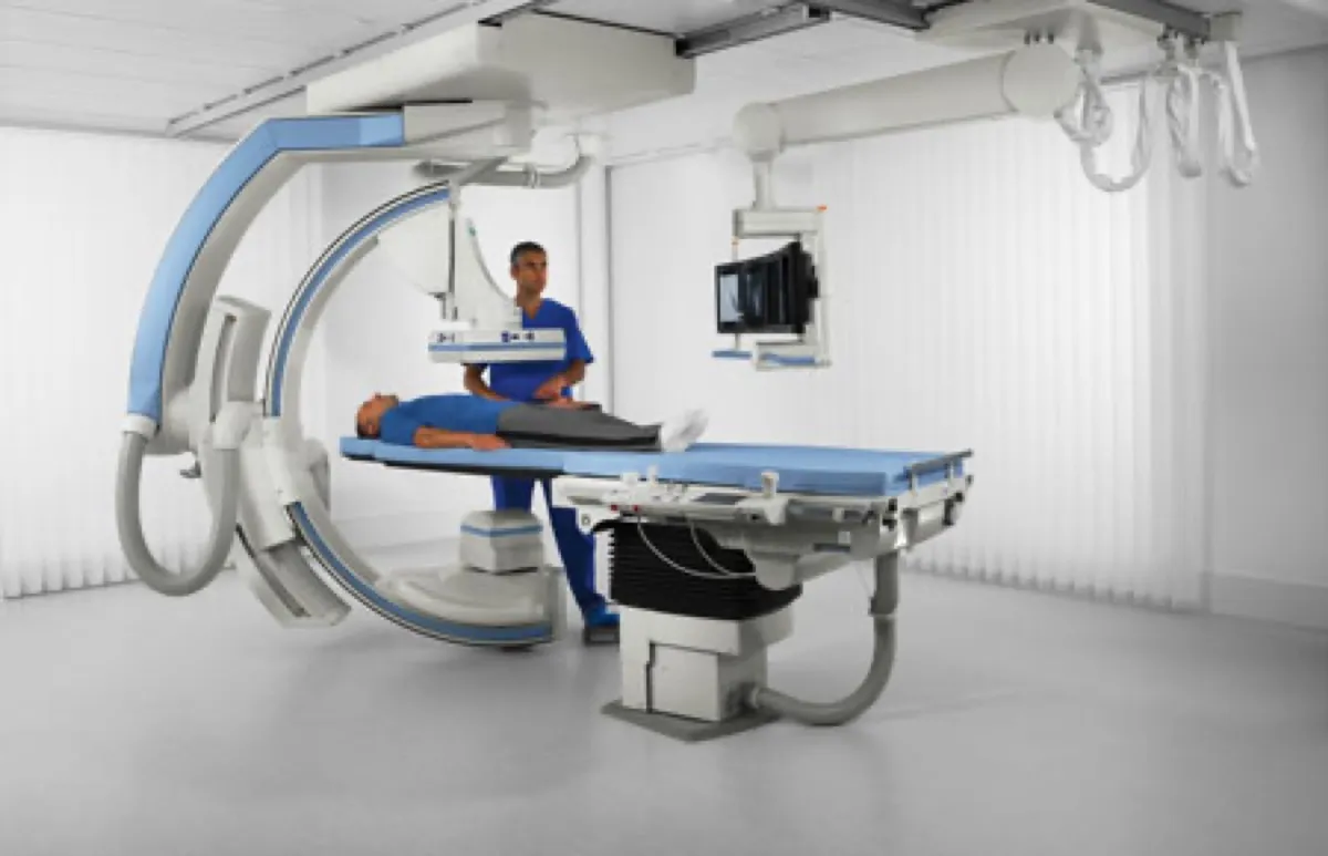
After the HSG procedure, the radiologist analyzes the fluoroscopic images captured during the test and writes a detailed report. This report typically describes two main areas: the uterine cavity (its shape, size, and any abnormalities) and the fallopian tubes (whether each tube fills with contrast and whether the dye spills freely into the pelvic cavity). The report is then sent to your referring gynecologist, who interprets the findings in relation to your specific clinical situation.
Most HSG reports are available within 24 to 48 hours after the procedure. In some cases, the radiologist may share preliminary findings with you immediately after the test, though the official written report requires more careful analysis of all the images captured.
"I never rush the reporting process, because every detail matters," says Dr. Osama Elzamzami, Consultant Radiologist at DCDC. "I review each image methodically, looking not just at whether the tubes are open, but at the shape of the uterine cavity, the caliber of each tube, the pattern of dye flow, and whether there are subtle findings that could be clinically relevant. A thorough report gives the gynecologist the information she needs to make the right decisions for her patient."
Normal HSG Results: What They Look Like
A normal HSG result is reassuring and indicates that the structural pathway for conception appears intact. Here is what a normal HSG report typically describes.
Normal Uterine Cavity
On a normal HSG, the uterine cavity appears as a smooth, inverted triangle. The contrast dye fills the cavity evenly, with smooth, regular borders and no filling defects. The cavity should be symmetrical, with no evidence of polyps, fibroids, adhesions, or congenital abnormalities. The report may describe this as "normal uterine cavity" or "uterine cavity of normal shape and contour."
Normal Fallopian Tubes (Free Spill)
The hallmark of normal fallopian tube function on HSG is "free spill" or "free peritoneal spillage." This means the contrast dye flowed through the entire length of each fallopian tube and spilled out freely into the pelvic (peritoneal) cavity. Free spill from both tubes confirms bilateral tubal patency, meaning both tubes are open and unobstructed. The report may describe the tubes as "patent bilaterally with free peritoneal spillage."
Abnormal HSG Findings: What They Mean
When the HSG reveals findings that deviate from normal, the report will describe the specific abnormalities observed. Understanding these findings helps you have a more meaningful discussion with your doctor about next steps.
Proximal Tubal Blockage
A proximal blockage occurs at the point where the fallopian tube connects to the uterus (the cornual region). On the HSG image, the contrast dye enters the uterine cavity but does not pass into the affected tube. This finding can be caused by actual scarring or adhesions, but it can also result from tubal spasm, a temporary muscular contraction of the tube. Because spasm can mimic a true blockage, doctors may recommend repeating the test or using antispasmodic medication before the procedure to rule out this possibility.
Distal Tubal Blockage
A distal blockage occurs at the far end of the fallopian tube, near the ovary (the fimbrial end). On the HSG image, the contrast dye fills the tube but does not spill into the pelvic cavity. The tube may appear dilated or swollen at the end, which is a sign of hydrosalpinx. Distal blockages are more commonly associated with previous pelvic infections, endometriosis, or prior pelvic surgery.
Hydrosalpinx
Hydrosalpinx is a specific condition where the blocked fallopian tube fills with fluid and becomes dilated. On the HSG image, the tube appears significantly enlarged and club-shaped at the distal end, with no contrast spillage. Hydrosalpinx is clinically significant because research has shown it can reduce IVF success rates. Many fertility specialists recommend treatment of hydrosalpinx (often by removing or clipping the affected tube) before proceeding with IVF.
Uterine Septum
A uterine septum is a band of tissue that partially or completely divides the uterine cavity. On HSG, it appears as an indentation or a dividing wall within the cavity, making the normally triangular shape look more like a heart shape or two separate chambers. A septum can interfere with embryo implantation and increase the risk of miscarriage. Depending on its size and location, surgical correction may be recommended.
Filling Defects (Polyps and Fibroids)
Filling defects appear as areas within the uterine cavity where the contrast dye does not fill evenly. These may indicate endometrial polyps or submucosal fibroids that protrude into the cavity. On the HSG image, they appear as dark spots or irregular shapes within the dye-filled cavity. These findings may require further evaluation with hysteroscopy or ultrasound to confirm the diagnosis and determine whether removal is necessary.
Intrauterine Adhesions (Asherman Syndrome)
Intrauterine adhesions are bands of scar tissue within the uterine cavity, often resulting from previous uterine surgery, particularly dilation and curettage (D&C). On HSG, adhesions appear as irregular filling defects or angular outlines within the cavity. Significant adhesions can reduce the available surface area for embryo implantation and may need to be surgically divided through hysteroscopy.
Congenital Uterine Anomalies
HSG can identify congenital uterine shape abnormalities such as a bicornuate uterus (heart-shaped), unicornuate uterus (one-sided), didelphys uterus (double uterus), or T-shaped uterus. These are structural variations that a woman is born with. While many women with congenital anomalies conceive naturally, some variants are associated with higher rates of pregnancy complications or recurrent miscarriage.
Normal vs. Abnormal HSG Findings: Comparison Table
| Finding | Normal | Abnormal |
|---|---|---|
| Uterine cavity shape | Smooth, inverted triangle | Irregular, heart-shaped, divided, or distorted |
| Cavity borders | Smooth and regular | Irregular, with filling defects or indentations |
| Right fallopian tube | Fills completely with free spill | Blocked (no spill), dilated, or does not fill |
| Left fallopian tube | Fills completely with free spill | Blocked (no spill), dilated, or does not fill |
| Tubal caliber | Normal thin caliber throughout | Dilated (especially distally), suggesting hydrosalpinx |
| Peritoneal spillage | Present bilaterally | Absent on one or both sides |
| Filling defects | None | Present (suggesting polyps, fibroids, or adhesions) |
This table provides a simplified comparison. Actual reports contain more detailed descriptions specific to each case.
Understanding Tubal Spasm vs. True Blockage
One of the most important concepts when reading HSG results is the distinction between tubal spasm and true tubal blockage. A tubal spasm is a temporary muscular contraction of the fallopian tube, usually at the proximal (cornual) end, that can prevent contrast from entering the tube. On the HSG image, this looks identical to a genuine blockage.
Spasm is relatively common and can be triggered by anxiety, pain during the procedure, or the catheter touching the uterine wall near the tubal opening. It is estimated that 10 to 20 percent of apparent proximal blockages on initial HSG are actually due to spasm rather than true obstruction. For this reason, if your HSG shows a proximal blockage, your doctor may recommend one of the following approaches before concluding that the tube is truly blocked.
- Repeating the HSG on a different day, as spasm may not occur during a second attempt
- Administering an antispasmodic medication before the repeat procedure
- Performing selective salpingography, a more targeted tubal catheterization under fluoroscopy
- Recommending a diagnostic laparoscopy with chromopertubation for definitive confirmation
Next Steps After Abnormal HSG Results
Receiving abnormal HSG results does not automatically mean you cannot conceive. The next steps depend on the specific findings and their severity. Your gynecologist or fertility specialist will create a tailored plan based on the complete picture, which includes HSG findings, blood test results, your partner's semen analysis, and your overall medical history.
A 38-year-old patient from Dubai came to DCDC after her initial HSG at another facility showed bilateral tubal blockage. She was told IVF was her only option. Her gynecologist recommended a repeat HSG at our center before proceeding. The second test, performed with antispasmodic medication and a slow injection technique, showed that her right tube was actually patent with free spill. The initial blockage had been caused by bilateral tubal spasm. With one confirmed open tube, her fertility specialist recommended IUI rather than IVF, and she conceived on her third cycle. Her case illustrates why accurate interpretation and, when indicated, repeat testing can change the course of treatment entirely.
| HSG Finding | Possible Next Steps |
|---|---|
| Unilateral tubal blockage (one tube blocked) | Natural conception possible through the open tube; timed intercourse or IUI may be recommended |
| Bilateral tubal blockage (both tubes blocked) | Further testing (laparoscopy) to confirm; IVF may be recommended if confirmed |
| Hydrosalpinx | Surgical evaluation; tube removal or clipping often recommended before IVF |
| Uterine septum | Hysteroscopic septum resection may be recommended before conception attempts |
| Polyps or fibroids | Hysteroscopy for further evaluation and possible removal |
| Intrauterine adhesions | Hysteroscopic adhesiolysis (surgical removal of adhesions) |
| Tubal spasm (suspected) | Repeat HSG with antispasmodic or selective salpingography |
Treatment plans are individualized. Your doctor will explain the best approach for your specific situation.
Can HSG Results Be Wrong?
While HSG is a reliable diagnostic test, it is not perfect. False-positive results (indicating a blockage that is not actually present) can occur due to tubal spasm, as discussed above. False-negative results (missing a real problem) are less common but can happen if a very small polyp or early adhesion is not visible on the X-ray images.
The accuracy of HSG depends significantly on the experience of the radiologist performing the test and the quality of the fluoroscopic equipment. At Doctors Clinic Diagnostic Center, our experienced radiology team uses modern fluoroscopy equipment and careful technique to maximize diagnostic accuracy. When findings are equivocal, we clearly communicate this in the report so that the referring physician can decide whether additional testing is warranted.
Understanding Your HSG Report at DCDC
At Doctors Clinic Diagnostic Center in Dubai Healthcare City, we believe patients should understand their test results. With over 13 years of operation and more than 1,000 diagnostic scans performed every month, our radiologists have the depth of experience to identify even subtle findings that less experienced teams might overlook. After your HSG, the referring gynecologist will schedule a follow-up consultation to review the radiology report and images with you. During this consultation, you will receive a clear explanation of what the findings mean for your specific fertility journey and what the recommended next steps are.
Our HSG test services include detailed reporting with image documentation, making it easy for your treating physician to explain the findings visually. If further evaluation is needed, our clinic offers a range of complementary diagnostic services under one roof, minimizing delays in your care.
Need Help Understanding Your HSG Results?
At Doctors Clinic Diagnostic Center, our radiology and gynecology teams work together to provide clear, accurate HSG testing with detailed reports. Book a follow-up consultation to discuss your results and plan your next steps.
Book a ConsultationЧасто задаваемые вопросы
Final Thoughts
Understanding your HSG results is an important step in your fertility journey. Whether your results are normal or show abnormalities, having a clear picture of what the findings mean empowers you to make informed decisions about your next steps. Normal results provide reassurance that the structural pathway for conception is intact, while abnormal findings point toward specific issues that can often be addressed with targeted treatment.
Remember that HSG is one piece of a larger diagnostic puzzle. Your doctor considers HSG findings alongside other test results and your complete medical history to develop a comprehensive treatment plan. At Doctors Clinic Diagnostic Center in Dubai Healthcare City, our team is committed to providing accurate diagnostics and clear communication so that you can navigate your fertility journey with confidence and clarity. To learn about pricing, visit our HSG test cost in Dubai page.
Источники и ссылки
Эта статья проверена нашей медицинской командой и ссылается на следующие источники:
- American Society for Reproductive Medicine - HSG Interpretation
- Radiological Society of North America - Hysterosalpingography Findings
- Fertility and Sterility - Tubal Factor Assessment
- American College of Radiology - HSG Reporting Standards
- Dubai Health Authority - Diagnostic Imaging Practices
Медицинский контент на этом сайте проверяется врачами, лицензированными DHA. См. нашу редакционную политику для получения дополнительной информации.

Автор
Dr. Osama Elzamzami
Consultant Radiologist
MD, Radiology
Dr. Osama Elzamzami is a Consultant Radiologist specializing in diagnostic imaging including MRI, CT, ultrasound, and fluoroscopy procedures such as HSG at DCDC Dubai Healthcare City.