मुख्य बातें
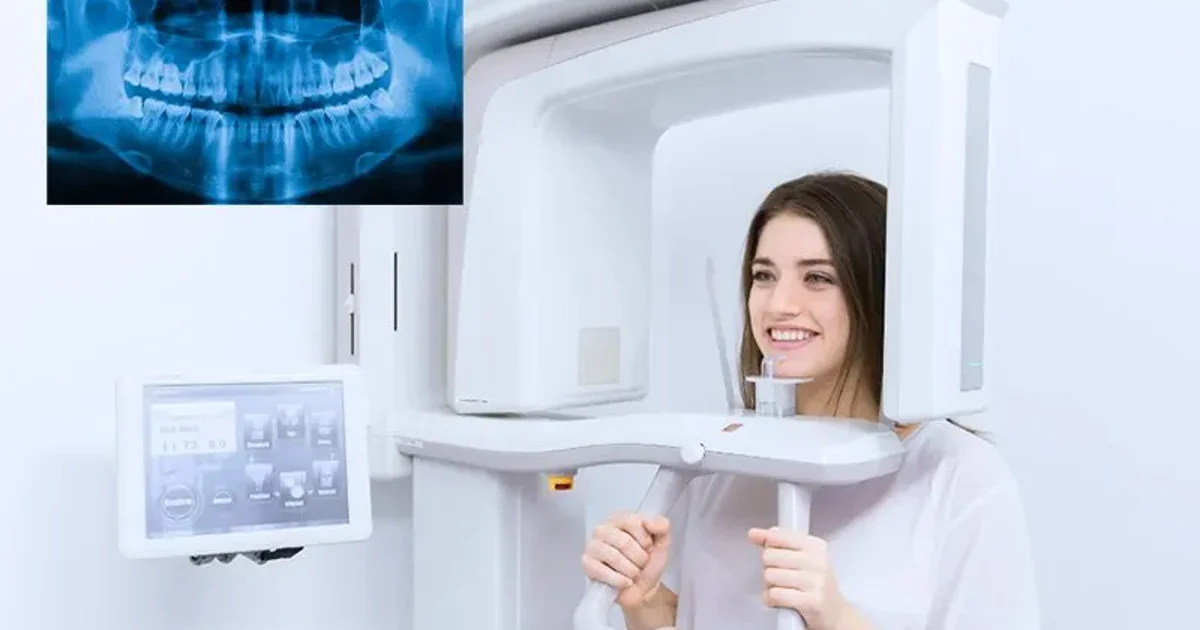
- OPG (ऑर्थोपैंटोमोग्राम) एक पैनोरामिक एक्स-रे है जो दोनों जबड़े, सभी दांत, TMJ जोड़, साइनस और तंत्रिका नलिकाओं को एक 15-सेकंड के स्कैन में बिना दर्द और मुंह में सेंसर के कैप्चर करता है
- स्कैन के लिए लगभग कोई तैयारी नहीं चाहिए - बस सिर और गर्दन क्षेत्र से धातु के आभूषण हटाएं; उपवास, इंजेक्शन या कंट्रास्ट डाई की जरूरत नहीं
- OPG केवल 10-20 माइक्रोसीवर्ट विकिरण देता है, जो 1-2 दिन की प्राकृतिक पृष्ठभूमि विकिरण के बराबर और एक लंबी दूरी की उड़ान से कम है
- दंत चिकित्सक OPG परिणामों को व्यवस्थित रूप से पढ़ते हैं, दांतों, हड्डी के स्तर, जबड़े के जोड़ों, साइनस और तंत्रिका नलिकाओं का कैविटी, हड्डी हानि, इम्पैक्टेड दांत, सिस्ट, फ्रैक्चर और TMJ विकारों के लिए मूल्यांकन करते हैं
- OPG डेंटल इम्प्लांट प्लानिंग, ऑर्थोडॉन्टिक मूल्यांकन, विजडम टूथ मूल्यांकन, जबड़े के दर्द का निदान और नियमित डेंटल स्क्रीनिंग के लिए आवश्यक प्रथम-पंक्ति इमेजिंग अध्ययन है
- जब OPG ऐसा कोई निष्कर्ष पहचानता है जिसके लिए त्रि-आयामी विवरण चाहिए, तो CBCT स्कैन सर्जिकल प्लानिंग के लिए आवश्यक पूरक सटीकता प्रदान करता है
OPG एक्स-रे (ऑर्थोपैंटोमोग्राम) दुनिया में सबसे व्यापक रूप से उपयोग की जाने वाली एक्स्ट्राओरल डेंटल इमेजिंग तकनीक है। 15 सेकंड से कम के एक दर्दरहित स्कैन में, यह दोनों जबड़ों, सभी दांतों, टेम्पोरोमैंडिबुलर जोड़ों, मैक्सिलरी साइनस और आसपास की हड्डी का एक कान से कान तक एक सपाट, द्वि-आयामी पैनोरामिक चित्र कैप्चर करता है। यह व्यापक गाइड वह सब कुछ कवर करती है जो आपको जानना चाहिए।
यह 2026 मेगा-गाइड डॉ. ओसामा एलज़मज़मी, सलाहकार रेडियोलॉजिस्ट (MD, FRCR) द्वारा डॉक्टर्स क्लिनिक डायग्नोस्टिक सेंटर (DCDC) दुबई हेल्थकेयर सिटी में समीक्षित है। प्रत्येक अनुभाग चिकित्सकीय रूप से सत्यापित है और इसमें तुलना तालिकाएं, रिपोर्ट शब्दावली स्पष्टीकरण और विशेषज्ञ मार्गदर्शन शामिल हैं।


OPG एक्स-रे क्या है?
OPG का अर्थ है ऑर्थोपैंटोमोग्राम, ग्रीक शब्दों "ऑर्थो" (सीधा), "पैन" (सभी) और "टोमोग्राम" (अनुभागीय चित्र) से मिलकर बना एक यौगिक शब्द - जिसका सामूहिक अर्थ है जबड़ों और दांतों का एक सीधा, सर्वव्यापी अनुभागीय चित्र। दैनिक नैदानिक अभ्यास में, स्कैन को पैनोरामिक रेडियोग्राफ या बस "पैनो" भी कहा जाता है।
इस अवधारणा को सबसे पहले फिनिश रेडियोलॉजिस्ट यर्जो वेली पाटेरो ने 1940 के दशक में विकसित किया था। आज, डिजिटल OPG सिस्टम ने अधिकांश आधुनिक क्लीनिकों में फिल्म-आधारित इकाइयों को बदल दिया है।
OPG एक्स-रे कैसे काम करता है?
OPG एक्स-रे रोटेशनल टोमोग्राफी नामक तकनीक का उपयोग करता है, जिसमें एक एक्स-रे ट्यूब और एक डिजिटल डिटेक्टर एक साथ रोगी के सिर के चारों ओर एक समन्वित अर्धवृत्ताकार चाप में घूमते हैं। सॉफ्टवेयर फिर इन स्लाइस को एक पैनोरामिक छवि में पुनर्निर्मित करता है।
मुख्य सिद्धांत को नैरो-बीम टोमोग्राफी कहा जाता है। पूरे सिर को एक साथ विकिरण के संपर्क में लाने के बजाय, OPG मशीन एक पतली, केंद्रित बीम का उपयोग करती है।
चूंकि एक्स-रे स्रोत पूरी प्रक्रिया के दौरान मुंह के बाहर रहता है, OPG को एक एक्स्ट्राओरल इमेजिंग तकनीक के रूप में वर्गीकृत किया जाता है। जिन रोगियों को तीव्र गैग रिफ्लेक्स, सीमित मुंह खुलना या दंत चिंता है, वे OPG को इंट्राओरल विकल्पों की तुलना में काफी अधिक आरामदायक पाते हैं।
"OPG दंत चिकित्सा की दुनिया का सबसे बहुमुखी स्क्रीनिंग उपकरण है," डॉ. ओसामा एलज़मज़मी बताते हैं। "एक 15-सेकंड के स्कैन में, हम दोनों जबड़े, सभी दांत, टेम्पोरोमैंडिबुलर जोड़ और आसपास की हड्डी को कैप्चर करते हैं।"
OPG बनाम इंट्राओरल एक्स-रे बनाम CBCT
अंतर को समझने का सबसे सरल तरीका इन्हें वाइड-एंगल लेंस बनाम मैक्रो लेंस के रूप में सोचना है। OPG आपके सिर के बाहर घूमता है और एक सपाट छवि बनाता है। इंट्राओरल एक्स-रे आपके मुंह में एक छोटा सेंसर रखता है और केवल 2-4 दांतों की तस्वीर लेता है। CBCT त्रि-आयामी वॉल्यूमेट्रिक छवियां बनाता है।
इंट्राओरल एक्स-रे तीन उपप्रकारों में आते हैं। पेरिएपिकल एक्स-रे पूरे दांत को क्राउन से रूट टिप तक कैप्चर करता है। बाइटविंग एक्स-रे ऊपरी और निचले दांतों के क्राउन एक साथ कैप्चर करता है। ऑक्लूसल एक्स-रे काटने की सतह पर एक बड़े सेंसर का उपयोग करता है।
| विशेषता | OPG (पैनोरामिक) | पेरिएपिकल | बाइटविंग | CBCT |
|---|---|---|---|---|
| कवरेज | दोनों जबड़े कान से कान, TMJ, साइनस | 2-3 दांत + रूट एपिसेस | ऊपरी/निचले प्रीमोलर और मोलर के क्राउन | चयनित क्षेत्र या दोनों जबड़ों का 3D वॉल्यूम |
| सेंसर की स्थिति | मुंह के बाहर (एक्स्ट्राओरल) | मुंह के अंदर | मुंह के अंदर | मुंह के बाहर (एक्स्ट्राओरल) |
| विवरण स्तर | मध्यम (व्यापक अवलोकन) | बहुत उच्च (दांत-विशिष्ट) | बहुत उच्च (क्राउन और ऊपरी रूट) | बहुत उच्च (3D क्रॉस-सेक्शन) |
| विकिरण खुराक | 10-20 μSv | 1-8 μSv प्रति छवि | 1-5 μSv प्रति छवि | 30-200 μSv |
| पूरे मुंह के लिए छवियां | 1 छवि | 14-20 छवियां (पूर्ण-मुंह श्रृंखला) | 4 छवियां (मानक श्रृंखला) | 1 स्कैन |
| स्कैन समय | 15-20 सेकंड | < 1 सेकंड प्रति एक्सपोजर | < 1 सेकंड प्रति एक्सपोजर | 15-30 सेकंड |
| रोगी आराम | बहुत आरामदायक; मुंह में कोई सेंसर नहीं | असहज हो सकता है; गैग रिफ्लेक्स का जोखिम | मध्यम; बाइट टैब मदद करता है | आरामदायक; मुंह में कोई सेंसर नहीं |
| सबसे उपयुक्त | स्क्रीनिंग, इम्पैक्टेड दांत, फ्रैक्चर, ऑर्थोडॉन्टिक्स, इम्प्लांट अवलोकन, TMJ | रूट कैनाल, पेरिएपिकल संक्रमण, रूट फ्रैक्चर | प्रारंभिक कैविटी पहचान, रेस्टोरेशन जांच | इम्प्लांट प्लानिंग, जटिल सर्जरी, 3D एनाटॉमी |
| अनुमानित दुबई लागत (AED) | 150-300 | 50-150 प्रति छवि | 50-100 प्रति छवि | 400-1,200 |
OPG पैनोरामिक एक्स-रे, इंट्राओरल एक्स-रे उपप्रकारों और CBCT की तुलना। विस्तृत CBCT तुलना के लिए, <a href="/blog/cbct-vs-opg-xray" class="text-primary-600 hover:underline">CBCT बनाम OPG</a> पर हमारी गाइड देखें।
लागत-प्रति-कवरेज के दृष्टिकोण से, OPG काफी अधिक किफायती है। विस्तृत मूल्य के लिए, दुबई में OPG एक्स-रे लागत पर हमारी गाइड देखें।
OPG क्या दिखाता है?
OPG एक्स-रे एक छवि में पूरी दंत और मैक्सिलोफेशियल एनाटॉमी दिखाता है।
- सभी निकले हुए दांत दोनों जबड़ों में उनकी जड़ों, क्राउन और आसपास की हड्डी सहित
- न निकले और इम्पैक्टेड दांत, विशेष रूप से विजडम टीथ
- विकसित हो रहे दांत बच्चों और किशोरों में
- दांतों की सड़न (कैरीज), विशेष रूप से बड़ी इंटरप्रॉक्सिमल कैविटी
- पेरिएपिकल पैथोलॉजी जैसे डेंटल एब्सेस और ग्रैनुलोमा
- हड्डी हानि पीरियडॉन्टल (मसूड़ों की) बीमारी से
- जबड़े की हड्डी की असामान्यताएं जिसमें सिस्ट, ट्यूमर और सौम्य वृद्धि शामिल हैं
- जबड़े के फ्रैक्चर आघात से
- टेम्पोरोमैंडिबुलर जोड़ (TMJ) गठिया, क्षरण या विस्थापन के संकेत दिखाते हुए
- मैक्सिलरी साइनस, साइनसाइटिस, पॉलिप्स या रिटेंशन सिस्ट प्रकट करते हुए
- नाक गुहा और नाक सेप्टम विचलन या बाधा के लिए
- दंत पुनर्स्थापन जैसे फिलिंग, क्राउन, ब्रिज और इम्प्लांट
- अवर एल्वियोलर नर्व कैनाल, इम्प्लांट प्लानिंग और विजडम टूथ निष्कर्षण के लिए महत्वपूर्ण
जबकि OPG एक उत्कृष्ट अवलोकन प्रदान करता है, इसकी द्वि-आयामी प्रकृति का अर्थ है कि कुछ बारीक विवरणों को पुष्टि के लिए पूरक इंट्राओरल एक्स-रे या CBCT इमेजिंग की आवश्यकता हो सकती है।
OPG की तैयारी कैसे करें
OPG एक्स-रे की तैयारी सीधी है क्योंकि स्कैन के लिए लगभग कोई विशेष तैयारी नहीं चाहिए। कोई उपवास, रक्त कार्य, इंजेक्शन, कंट्रास्ट डाई या रिकवरी समय नहीं। सबसे महत्वपूर्ण कदम सिर और गर्दन क्षेत्र से सभी धातु की वस्तुओं को हटाना है।
"सबसे आम सवाल जो मरीज पूछते हैं वह है कि क्या उन्हें कुछ तैयार करना है," डॉ. एलज़मज़मी कहते हैं। "जवाब ताज़गी से सरल है: अपना धातु हटाएं, 15 सेकंड खड़े रहें, और स्कैन हो गया।"
| हटाने की वस्तु | इसे क्यों हटाना चाहिए |
|---|---|
| कान की बालियां (स्टड, हुप्स, ईयर कफ) | धातु की बालियां स्कैन पथ में होती हैं और चमकीले सफेद धब्बे बनाती हैं |
| हार और चेन | हार रैखिक आर्टिफैक्ट बनाते हैं जो फ्रैक्चर की नकल कर सकते हैं |
| चेहरे और मुंह की पियर्सिंग | पियर्सिंग फोकल सफेद आर्टिफैक्ट बनाती हैं जो दांतों और हड्डी को ढकती हैं |
| चश्मा और धूप का चश्मा | धातु के फ्रेम ऊपरी जबड़े और नाक गुहा के दृश्य को अवरुद्ध करते हैं |
| श्रवण यंत्र | TMJ क्षेत्र में सघन आर्टिफैक्ट बनाते हैं |
| हटाने योग्य डेंचर और रिटेनर | धातु के क्लैस्प प्राकृतिक दांतों पर ओवरलैप होते हैं |
| हेयरपिन और धातु की क्लिप | पूरी छवि में बिखरे आर्टिफैक्ट बनाते हैं |
| धातु की पिन वाले स्कार्फ | धातु की पिन बिंदु-जैसे आर्टिफैक्ट बनाती हैं; कपड़ा रह सकता है अगर सभी धातु हटा दी जाए |
सिर, गर्दन और ऊपरी छाती से सभी धातु की वस्तुएं हटाएं। फिक्स्ड डेंटल वर्क (क्राउन, ब्रिज, ब्रेसेज, इम्प्लांट) हटाने की जरूरत नहीं।
खाना, पीना और दवाइयां
OPG एक्स-रे से पहले आप सामान्य रूप से खा-पी सकते हैं। कोई उपवास जरूरी नहीं। सभी नियमित दवाइयां निर्धारित अनुसार लें।
गर्भावस्था और OPG तैयारी
यदि आप गर्भवती हैं या गर्भवती होने की संभावना है, तो स्कैन से पहले रेडियोग्राफर को सूचित करें। ऐच्छिक दंत इमेजिंग को गर्भावस्था के दौरान नियमित रूप से स्थगित किया जाता है, विशेष रूप से पहली तिमाही में। हालांकि, नैदानिक आपातकाल में, लेड एप्रन के साथ OPG सुरक्षित रूप से किया जा सकता है।
रेडियोग्राफर को क्या बताएं
- गर्भावस्था या संभावित गर्भावस्था
- स्कैन का कारण - रेडियोलॉजिस्ट को रिपोर्ट केंद्रित करने में मदद करता है
- पिछली जबड़े की सर्जरी या आघात
- स्थिर खड़े रहने में कठिनाई
- पिछली दंत इमेजिंग - तुलना के लिए
व्यावहारिक सुझाव: आभूषण घर पर छोड़ें और बिना ऊंचे धातु के ज़िपर वाला साधारण शीर्ष पहनें।
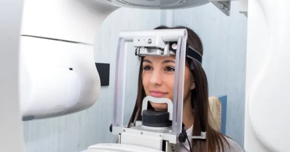
OPG के दौरान क्या होता है
पूरी OPG एक्स-रे प्रक्रिया 5 मिनट से कम लेती है, जिसमें वास्तविक स्कैन केवल 15 से 20 सेकंड लेता है। कोई तैयारी, रिकवरी या दुष्प्रभाव नहीं।
- चरण 1 - आगमन और पंजीकरण: एमिरेट्स आईडी या पासपोर्ट के साथ रिसेप्शन पर चेक-इन करें। 2-3 मिनट।
- चरण 2 - धातु की वस्तुएं हटाएं: रेडियोग्राफर आपसे सभी धातु की वस्तुएं हटाने को कहेगा। लगभग 30 सेकंड।
- चरण 3 - सुरक्षात्मक ढाल पहनें: लेड थायरॉइड कॉलर और/या लेड एप्रन लगाई जाती है।
- चरण 4 - मशीन में स्थिति: आप अपनी ठोड़ी को चिन रेस्ट पर रखते हैं। सही स्थिति उच्च गुणवत्ता वाली छवि के लिए सबसे महत्वपूर्ण कारक है।
- चरण 5 - पोजिशनिंग गाइड को काटें: आप धीरे से एक निष्फल प्लास्टिक बाइट ब्लॉक पर काटते हैं। अपने होंठ बंद करें और जीभ को तालू के खिलाफ सपाट दबाएं।
- चरण 6 - स्कैन: C-आकार का आर्म आपके सिर के चारों ओर धीरे-धीरे घूमता है। आपको पूरी तरह स्थिर रहना होगा। 15 से 20 सेकंड।
- चरण 7 - स्कैन पूरा: डिजिटल छवि सेकंडों में स्क्रीन पर दिखाई देती है। कमरे में कुल समय: 3 से 5 मिनट।
कोई रिकवरी समय नहीं। स्कैन पूरी तरह दर्दरहित है - कोई सुई, इंजेक्शन, कंट्रास्ट डाई या मुंह में सेंसर नहीं।
बच्चों के लिए OPG
5 साल की उम्र के बच्चे OPG करवा सकते हैं। आधुनिक डिजिटल OPG मशीनों में स्वचालित बाल खुराक-कमी प्रोटोकॉल शामिल हैं। बाल OPG लगभग 5 से 14 μSv देता है।
7 वर्षीय यूसुफ अपनी माँ के साथ अपने पहले OPG स्कैन के लिए DCDC आया। रेडियोग्राफर ने मशीन दिखाने में समय लिया। यूसुफ 16 सेकंड पूरी तरह स्थिर खड़ा रहा और स्कैन हो गया। OPG ने दो स्थायी दांत दिखाए जो असामान्य कोणों पर विकसित हो रहे थे।
DCDC दुबई हेल्थकेयर सिटी में डिजिटल OPG एक्स-रे
डॉक्टर्स क्लिनिक डायग्नोस्टिक सेंटर में उसी दिन रेडियोलॉजिस्ट रिपोर्ट के साथ उच्च-रिज़ॉल्यूशन डिजिटल OPG स्कैन प्राप्त करें। वॉक-इन स्वागत है।
स्व-भुगतान रोगियों के लिए कोई रेफरल आवश्यक नहीं
अपने OPG परिणाम पढ़ना
दंत चिकित्सक और रेडियोलॉजिस्ट एक संरचित, व्यवस्थित दृष्टिकोण का पालन करते हैं जो सुनिश्चित करता है कि प्रत्येक शारीरिक क्षेत्र का मूल्यांकन हो।
व्यवस्थित पठन दृष्टिकोण
- समग्र छवि गुणवत्ता जांच: रेडियोलॉजिस्ट पहले सही एक्सपोजर और पोजिशनिंग की पुष्टि करता है।
- समरूपता मूल्यांकन: बाएं और दाएं पक्ष लगभग सममित दिखने चाहिए।
- दांतों का मूल्यांकन (चतुर्थांश दर चतुर्थांश): प्रत्येक दांत की व्यक्तिगत रूप से जांच की जाती है।
- हड्डी स्तर मूल्यांकन: प्रत्येक दांत के आसपास एल्वियोलर हड्डी की ऊंचाई का मूल्यांकन।
- TMJ मूल्यांकन: दोनों जबड़े के जोड़ों की आकार, आकृति और समरूपता की जांच।
- मैक्सिलरी साइनस समीक्षा: साइनस की अपारदर्शिता और पॉलिप्स के लिए जांच।
- तंत्रिका नलिका ट्रेसिंग: अवर एल्वियोलर नर्व कैनाल को ट्रेस किया जाता है।
- पैथोलॉजी पहचान: सभी असामान्य अंधेरे या उज्ज्वल क्षेत्रों को दस्तावेज किया जाता है।
DCDC में, प्रत्येक OPG को एक सलाहकार रेडियोलॉजिस्ट द्वारा पढ़ा जाता है।
OPG एक्स-रे पर सामान्य निष्कर्ष
| निष्कर्ष | OPG पर दिखावट | इसका क्या अर्थ है |
|---|---|---|
| दंत कैरीज | दांत के क्राउन में अंधेरे क्षेत्र | दांत सड़न; फिलिंग से रूट कैनाल तक उपचार |
| हड्डी हानि (पीरियडॉन्टल रोग) | जड़ों के आसपास कम हड्डी ऊंचाई | मसूड़ों की बीमारी से हड्डी का पीछे हटना |
| इम्पैक्टेड दांत | मसूड़े या हड्डी के नीचे आंशिक या पूर्ण रूप से फंसा दांत | विजडम टीथ में सामान्य; दर्द या सिस्ट कर सकते हैं |
| पेरिएपिकल पैथोलॉजी | जड़ की नोक पर अंधेरा क्षेत्र | संक्रमण जिसके लिए रूट कैनाल या निष्कर्षण चाहिए |
| सिस्ट | जबड़े की हड्डी में अच्छी तरह परिभाषित गोल अंधेरा क्षेत्र | तरल से भरी थैली; सर्जिकल हटाने की जरूरत |
| TMJ असामान्यताएं | कॉन्डाइल्स का चपटापन, क्षरण या विषमता | जबड़े के जोड़ में अपक्षयी परिवर्तन |
| जड़ पुनर्अवशोषण | छोटी या अनियमित आकार की जड़ | विभिन्न कारणों से जड़ का घुलना |
| साइनस असामान्यताएं | मैक्सिलरी साइनस में अपारदर्शिता | साइनसाइटिस या रिटेंशन सिस्ट |
सामान्य OPG निष्कर्ष और उनका नैदानिक महत्व।
सामान्य बनाम असामान्य OPG परिणाम
एक सामान्य OPG दिखाता है: बिना अंधेरे धब्बों वाले अक्षुण्ण दांत, स्वस्थ जड़ें, समान हड्डी स्तर, सममित कॉन्डाइल्स, साफ वायु-भरे साइनस और दृश्यमान तंत्रिका नलिकाएं।
असामान्य निष्कर्ष में शामिल हैं: जबड़े की हड्डी में अंधेरे क्षेत्र, चमकीले सफेद द्रव्यमान, महत्वपूर्ण हड्डी हानि, जड़ का छोटा होना और अपारदर्शी साइनस।
OPG रिपोर्ट शब्दावली समझना
| रिपोर्ट शब्द | सरल भाषा में अर्थ |
|---|---|
| रेडियोल्यूसेंट / रेडियोल्यूसेंसी | अंधेरा क्षेत्र - हड्डी विनाश, सिस्ट, एब्सेस या ट्यूमर इंगित करता है |
| रेडियोपेक / रेडियोपेसिटी | चमकीला सफेद क्षेत्र - धातु फिलिंग, इम्प्लांट या सघन हड्डी |
| पेरिएपिकल पैथोलॉजी | दांत की जड़ की नोक पर रोग |
| पुनर्अवशोषण | दांत की जड़ घुलना - छोटापन या अनियमित रूपरेखा के रूप में दिखता है |
| क्षैतिज हड्डी हानि | पुरानी मसूड़ों की बीमारी से हड्डी की ऊंचाई में समान कमी |
| ऊर्ध्वाधर (कोणीय) हड्डी हानि | दांत की जड़ के एक तरफ स्थानीय V-आकार का हड्डी दोष |
| इम्पैक्शन | दांत जो अपनी सामान्य स्थिति में नहीं निकला |
| अच्छी तरह परिभाषित बनाम खराब परिभाषित किनारे | एक घाव की सीमा: स्पष्ट = आमतौर पर सौम्य; धुंधला = आक्रामक प्रक्रिया की चिंता |
| ऑस्टियोस्क्लेरोसिस | बढ़ी हुई हड्डी घनत्व का क्षेत्र; आमतौर पर सौम्य |
| पेरिकोरोनाइटिस | आंशिक रूप से निकले दांत के आसपास नरम ऊतक संक्रमण |
| डेंटिजेरस सिस्ट | न निकले दांत के क्राउन के आसपास तरल-भरी थैली |
सामान्य OPG रिपोर्ट शब्दावली का अनुवाद।
डेंटल इम्प्लांट के लिए OPG
OPG डेंटल इम्प्लांट उम्मीदवारों के लिए मानक प्रथम-पंक्ति स्क्रीनिंग एक्स-रे है।
OPG इम्प्लांट मूल्यांकन के लिए क्या प्रकट करता है
- ऊर्ध्वाधर हड्डी ऊंचाई: OPG एल्वियोलर रिज की पूरी ऊंचाई दिखाता है।
- तंत्रिका नलिका स्थिति: OPG तंत्रिका नलिका को रेडियोल्यूसेंट बैंड के रूप में दिखाता है।
- साइनस फ्लोर स्थिति: दिखाता है कि साइनस लिफ्ट प्रक्रिया की जरूरत होगी या नहीं।
- पड़ोसी दांतों की स्थिति: कैरीज, पेरिएपिकल पैथोलॉजी और हड्डी हानि।
- मौजूदा पैथोलॉजी: सिस्ट, ट्यूमर, बचे हुए जड़ के टुकड़े।
- समग्र जबड़ा वास्तुकला: एकाधिक इम्प्लांट की आवश्यकता वाले रोगियों के लिए।
OPG सीमाएं और CBCT में अपग्रेड कब करें
OPG हड्डी की चौड़ाई, हड्डी घनत्व या सटीक 3D तंत्रिका स्थिति नहीं दिखा सकता। जिन परिदृश्यों में लगभग हमेशा फॉलो-अप CBCT स्कैन की जरूरत होती है उनमें शामिल हैं: तंत्रिका नलिका के पास पिछले जबड़े के इम्प्लांट, साइनस के पास ऊपरी पिछले इम्प्लांट और एकाधिक इम्प्लांट।
ऑर्थोडॉन्टिक्स के लिए OPG
OPG पहली इमेजिंग स्टडी है जो हर ऑर्थोडॉन्टिस्ट ब्रेसेज या एलाइनर्स की सिफारिश करने से पहले मांगता है।
| संरचना / स्थिति | OPG क्या प्रकट करता है | ऑर्थोडॉन्टिक्स के लिए क्यों महत्वपूर्ण |
|---|---|---|
| निकले हुए दांत | स्थिति, संरेखण और अंतर | नैदानिक निष्कर्षों की पुष्टि; ब्रैकेट प्लेसमेंट में मदद |
| इम्पैक्टेड दांत | स्थान, कोण और गहराई | इम्पैक्टेड कैनाइन को सर्जिकल एक्सपोजर की जरूरत हो सकती है |
| जन्मजात अनुपस्थित दांत | दांत कलियों का अभाव | उपचार लक्ष्य बदलता है |
| अतिरिक्त दांत | सामान्य निकलने को रोकने वाले अतिरिक्त दांत | संरेखण से पहले निकालने की जरूरत |
| जड़ लंबाई और आकृति विज्ञान | प्रत्येक जड़ की लंबाई, आकार और अखंडता | छोटी जड़ों में पुनर्अवशोषण का अधिक जोखिम |
| हड्डी स्तर | हड्डी समर्थन की ऊंचाई | कम हड्डी सुरक्षित दांत गति को सीमित करती है |
| जबड़ा समरूपता | बाएं और दाएं जबड़े की शाखाओं का सापेक्ष आकार | विषमता कंकाल समस्या इंगित कर सकती है |
| TMJ जोड़ | कॉन्डाइल्स का आकार और स्थिति | मौजूदा अपक्षय ऑर्थोडॉन्टिक्स के दौरान बिगड़ सकता है |
| पिछला दंत कार्य | क्राउन, ब्रिज, रूट कैनाल, इम्प्लांट | इम्प्लांट पर ब्रैकेट नहीं चिपकाए जा सकते |
OPG ऑर्थोडॉन्टिस्ट को एक व्यापक नैदानिक अवलोकन प्रदान करता है।
विजडम टीथ के लिए OPG
OPG विजडम टीथ (तीसरे मोलर) के मूल्यांकन के लिए मानक इमेजिंग स्टडी है।
| इम्पैक्शन प्रकार | OPG दिखावट | आवृत्ति | निष्कर्षण कठिनाई | मुख्य निष्कर्ष |
|---|---|---|---|---|
| मीसियोएंगुलर | दूसरे मोलर की ओर आगे झुका | 40-45% | मध्यम | क्राउन दूसरे मोलर पर दबा रहा है |
| ऊर्ध्वाधर | सीधा लेकिन नहीं निकला | 25-30% | मध्यम से कठिन | सही अक्ष लेकिन अवरुद्ध |
| क्षैतिज | करवट लेटा हुआ | 10-15% | कठिन | क्राउन दूसरे मोलर की जड़ों की ओर |
| डिस्टोएंगुलर | पीछे की ओर झुका | 5-10% | सबसे कठिन (निचला जबड़ा) | निष्कर्षण पथ के लिए सीमित पहुंच |
| बक्कल/लिंगुअल (अनुप्रस्थ) | गाल या जीभ की ओर विस्थापित | दुर्लभ | परिवर्तनशील | OPG विस्थापन को कम आंक सकता है; CBCT अक्सर जरूरी |
OPG आधारित विजडम टूथ इम्पैक्शन वर्गीकरण।
डेंटल एक्स-रे सुरक्षा
हां, डेंटल एक्स-रे सुरक्षित हैं। मानक OPG की विकिरण खुराक लगभग 10 से 20 माइक्रोसीवर्ट (μSv) है - लगभग 1 से 2 दिन की प्राकृतिक पृष्ठभूमि विकिरण के बराबर।
| विकिरण स्रोत | अनुमानित खुराक (μSv) | OPG स्कैन में समतुल्य |
|---|---|---|
| 1 केला खाना | 0.1 | एक OPG का 1/140वां |
| एकल पेरिएपिकल एक्स-रे | 1-8 | < 1 OPG |
| एक दिन की प्राकृतिक पृष्ठभूमि विकिरण | 6.5 | लगभग आधा OPG |
| OPG (पैनोरामिक डेंटल एक्स-रे) | 10-20 | 1 OPG |
| छाती का एक्स-रे | 20 | ~1 OPG |
| दुबई-लंदन उड़ान | 40-80 | 3-6 OPGs |
| CBCT | 30-200 | 2-14 OPGs |
| सिर का CT स्कैन | 2,000 | ~140 OPGs |
| कुल वार्षिक पृष्ठभूमि विकिरण | 2,400 | ~170 OPGs |
डेंटल एक्स-रे चिकित्सा विकिरण स्पेक्ट्रम के सबसे निचले स्तर पर हैं।
गर्भावस्था के दौरान OPG सुरक्षा
ACOG और ADA दोनों कहते हैं कि नैदानिक रूप से संकेत होने पर गर्भावस्था के दौरान डेंटल एक्स-रे सुरक्षित रूप से किए जा सकते हैं। तत्काल नैदानिक इमेजिंग में देरी नहीं करनी चाहिए।
बच्चों के लिए OPG सुरक्षा
उचित तकनीक के साथ डेंटल एक्स-रे बच्चों के लिए सुरक्षित हैं। आधुनिक मशीनों में स्वचालित बाल खुराक-कमी प्रोटोकॉल हैं।
DCDC में सुरक्षित, कम-खुराक डेंटल एक्स-रे
DCDC नवीनतम डिजिटल OPG और डिजिटल एक्स-रे सिस्टम का उपयोग करता है। DCDC दुबई हेल्थकेयर सिटी में वॉक-इन स्वागत है।
स्व-भुगतान रोगियों के लिए कोई रेफरल आवश्यक नहीं
DCDC दुबई हेल्थकेयर सिटी में OPG एक्स-रे
डॉक्टर्स क्लिनिक डायग्नोस्टिक सेंटर (DCDC) दुबई हेल्थकेयर सिटी में व्यापक डिजिटल OPG एक्स-रे सेवाएं प्रदान करता है।
- एक छत के नीचे पूर्ण इमेजिंग: OPG, CBCT, पेरिएपिकल एक्स-रे, सेफैलोमेट्रिक इमेजिंग, CT, MRI और अल्ट्रासाउंड साइट पर उपलब्ध।
- नवीनतम डिजिटल OPG तकनीक: कम विकिरण खुराक पर स्पष्ट छवियां।
- बाल खुराक प्रोटोकॉल: बच्चों के लिए स्वचालित खुराक कमी।
- उसी दिन सलाहकार रेडियोलॉजिस्ट रिपोर्ट: प्रत्येक OPG की विशेषज्ञ रेडियोलॉजिस्ट द्वारा समीक्षा।
- वॉक-इन उपलब्धता: स्व-भुगतान रोगियों के लिए कोई रेफरल आवश्यक नहीं।
- बीमा स्वीकृत: DCDC दुबई के प्रमुख बीमा प्रदाताओं के साथ काम करता है।
- सुविधाजनक DHCC स्थान: ऊद मेठा, उम्म हुरैर 2, कराम से आसानी से पहुंचा जा सकता है।
मूल्य विवरण के लिए, दुबई में OPG एक्स-रे लागत पर हमारी गाइड देखें।
आज ही अपना OPG स्कैन बुक करें
DCDC दुबई हेल्थकेयर सिटी में डिजिटल OPG एक्स-रे के लिए वॉक इन करें या पहले से बुक करें। सलाहकार रेडियोलॉजिस्ट से उसी दिन परिणाम।
DCDC में संबंधित सेवाएं
दुबई हेल्थकेयर सिटी में विशेषज्ञ देखभाल और उन्नत निदान
Frequently Asked Questions
अंतिम विचार
OPG एक्स-रे डेंटल और मैक्सिलोफेशियल इमेजिंग में सबसे मूल्यवान और बहुमुखी उपकरणों में से एक बना हुआ है। 15 सेकंड से कम में, यह एक व्यापक पैनोरामिक दृश्य प्रदान करता है। बहुत कम विकिरण (10-20 μSv), शून्य दर्द और कोई विशेष तैयारी नहीं के साथ, यह रोगी-अनुकूल इमेजिंग अध्ययनों में से एक है।
चाहे आपके दंत चिकित्सक ने OPG की सिफारिश की हो या आपको व्यापक दंत स्क्रीनिंग चाहिए, दुबई हेल्थकेयर सिटी में डॉक्टर्स क्लिनिक डायग्नोस्टिक सेंटर उसी दिन सलाहकार रेडियोलॉजिस्ट रिपोर्टिंग और वॉक-इन उपलब्धता के साथ डिजिटल OPG स्कैन प्रदान करता है। CBCT, CT और MRI सहित सभी फॉलो-अप इमेजिंग साइट पर उपलब्ध है। विस्तृत मूल्य के लिए, दुबई में OPG एक्स-रे लागत पर हमारी गाइड देखें।
स्रोत एवं संदर्भ
यह लेख हमारी चिकित्सा टीम द्वारा समीक्षित है और निम्नलिखित स्रोतों का संदर्भ देता है:
- American Dental Association - Dental Radiographic Examinations: Recommendations for Patient Selection and Limiting Radiation Exposure
- European Commission - Radiation Protection 136: European Guidelines on Radiation Protection in Dental Radiology
- NCRP Report No. 177: Radiation Protection in Dentistry and Oral & Maxillofacial Imaging
- AAOMS - White Paper on Third Molar Data
- AAO - Clinical Practice Guidelines for Orthodontic Radiography
- EAO - Consensus Statements on Radiographic Examination for Implant Patients
- RadiologyInfo.org - Panoramic Dental X-ray
- ACOG - Guidelines for Diagnostic Imaging During Pregnancy and Lactation
इस साइट पर चिकित्सा सामग्री DHA-लाइसेंस प्राप्त चिकित्सकों द्वारा समीक्षित है। हमारी देखें संपादकीय नीति अधिक जानकारी के लिए।

लेखक
Dr. Osama Elzamzami
नैदानिक रेडियोलॉजी
MD, FRCR
डॉ. ओसामा एलज़मज़मी DCDC दुबई हेल्थकेयर सिटी में रेडियोलॉजी विभाग के प्रमुख हैं, जो एक्स-रे, CT, MRI, OPG, CBCT और अल्ट्रासाउंड सहित नैदानिक इमेजिंग में विशेषज्ञ हैं। उनके पास MD और FRCR (रॉयल कॉलेज ऑफ रेडियोलॉजिस्ट्स की फेलोशिप) है और वे दंत, मैक्सिलोफेशियल और सामान्य नैदानिक इमेजिंग अध्ययनों के लिए विशेषज्ञ रिपोर्टिंग प्रदान करते हैं।
संबंधित लेख

दुबई में OPG एक्स-रे लागत: अपडेटेड कीमतें और बीमा गाइड
CBCT बनाम OPG एक्स-रे: आपको कौन सी डेंटल इमेजिंग चाहिए?
© 2026 Doctors Clinic Diagnostic Center (DCDC), Dubai Healthcare City. Originally published at https://doctorsclinicdubai.ae/blog/opg-xray-results-explained. All rights reserved. Unauthorized reproduction is prohibited.