Prenez votre rendez-vous
Avis verifies des patients du centre de diagnostic DCDC a Dubai
Avis verifies et temoignages reels de patients de Dubai Healthcare City
Je suis très satisfaite de cette clinique médicale. J'ai fait mon premier dépistage de grossesse ici. C'était abordable et très professionnel. Le radiologue était accueillant, gentil et a rendu l'expérience formidable.
Aizhan Tokmanbetova
Je me suis blessée au genou vendredi soir et j'ai obtenu un rendez-vous IRM dimanche soir (réservé dimanche après-midi). J'ai attendu environ 2 minutes et c'était terminé en un peu plus de 30 minutes. L'équipe était incroyable, sympathique et efficace.
Kirsten Evans
L'équipe du Dr Osama était douce et incroyable. Après une mauvaise expérience ailleurs, mon examen pelvien ici était beaucoup plus professionnel. Excellent rapport et très précis.
Mary


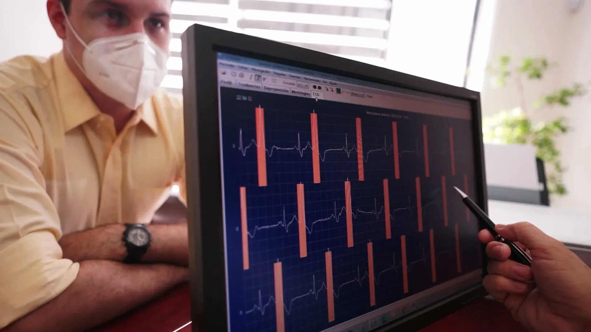
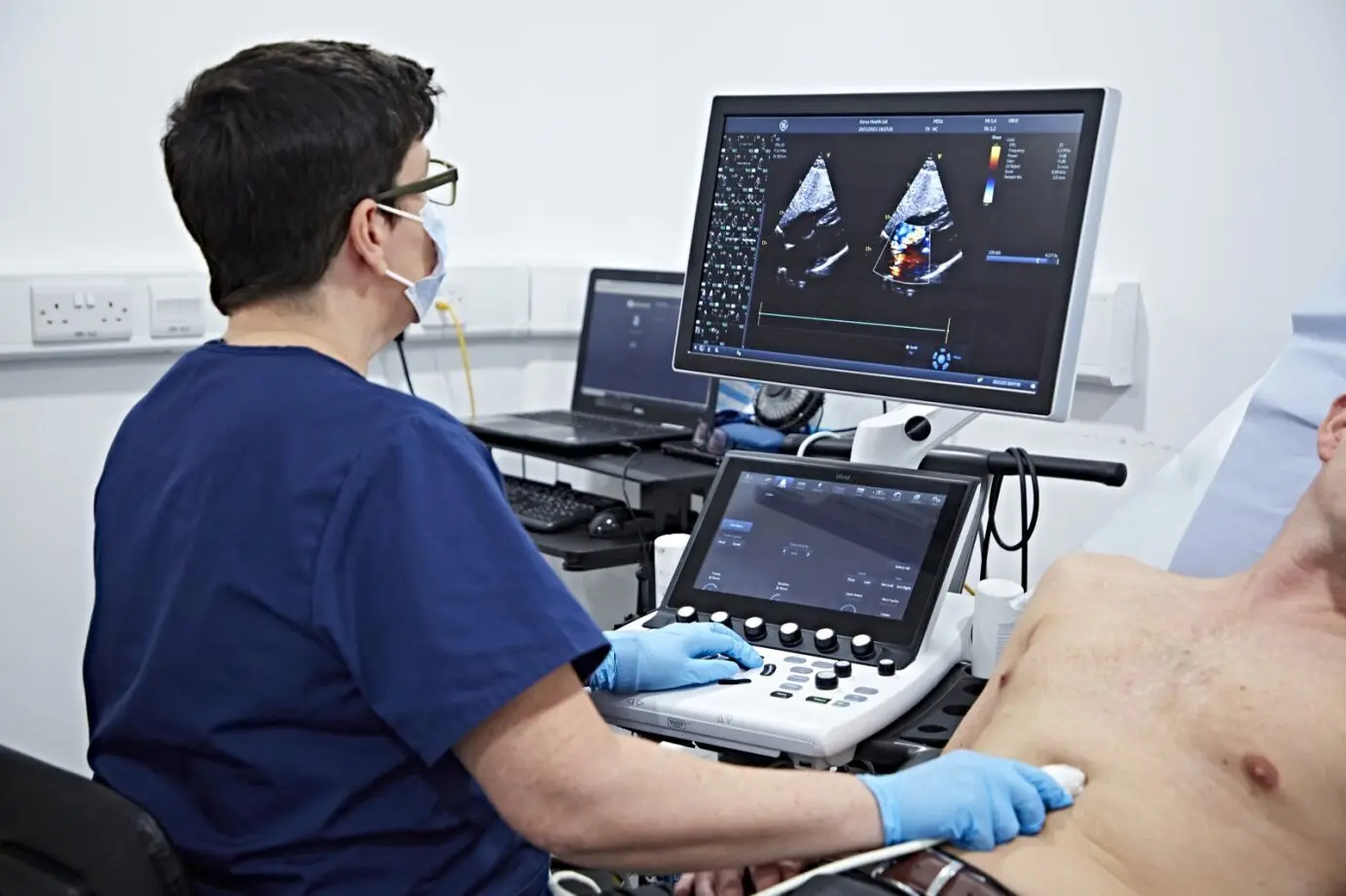
Echocardiogram in Dubai: Heart Ultrasound Imaging
See your heart beating in real-time with zero radiation
Zero radiation
Examen sur et non invasif utilisant des champs magnetiques (pas de rayons X ni de radiation ionisante).
Fast Results
Expert cardiologist interpretation within 24 hours
Non-Invasive
Painless. No injections or contrast required
An echocardiogram uses sound waves to create live images of your heart. You can see your heart beating, valves opening and closing, and blood flowing through the chambers. It's completely painless with no radiation. making it ideal for repeated monitoring.
Your cardiologist may recommend an echo for heart murmurs, shortness of breath, chest pain, valve disease, or to assess heart function after a cardiac event. The test takes 20-40 minutes and provides detailed information about your heart's structure and pumping ability.
Every echo is reviewed by board-certified specialist cardiologists at our echocardiography suite near Oud Metha Metro with same-day appointments and results typically within 24 hours.
The echocardiogram is one of the most versatile tools in cardiology. It measures ejection fraction, the percentage of blood your heart pumps out with each beat, which is a critical indicator of heart function. A normal ejection fraction ranges from 50 to 70 percent. The test also evaluates heart valve function, identifies areas of the heart wall that may not be contracting properly, and detects fluid around the heart (pericardial effusion).
Unlike many cardiac imaging tests, echocardiography uses no radiation and requires no contrast injection for standard studies. This makes it safe for repeated monitoring over time, which is particularly valuable for patients with chronic heart conditions, those recovering from cardiac surgery, and individuals on medications that may affect heart function. It is also safe during pregnancy when cardiac assessment is needed.
An echocardiogram can detect a wide range of cardiac conditions that might otherwise go undiagnosed until they become serious. Heart valve disease, including mitral valve prolapse, aortic stenosis, and regurgitation, is identified by visualizing valve leaflet movement and measuring blood flow velocity across each valve. Cardiomyopathies, where the heart muscle becomes enlarged, thickened, or stiffened, are diagnosed through chamber measurements and wall thickness assessment. Pericardial effusion, the accumulation of fluid around the heart, is immediately visible on echo and can be monitored for progression toward the dangerous condition of cardiac tamponade. Congenital heart defects such as atrial and ventricular septal defects are reliably detected, even in adults who may have been unaware of these conditions for decades. The echocardiogram is often the first test that reveals the underlying cause of symptoms like unexplained breathlessness, fatigue, palpitations, or leg swelling.
Several types of echocardiogram exist, each suited to specific clinical questions. The standard transthoracic echocardiogram (TTE) is the most common type, performed by placing the transducer on the chest wall to obtain images through the rib spaces. A stress echocardiogram combines resting echo images with images captured immediately after exercise on a treadmill or pharmacological stress, revealing areas of the heart muscle that may not receive adequate blood flow during exertion, which can indicate coronary artery disease. Doppler echocardiography measures blood flow velocity and direction through the heart chambers and across the valves, essential for grading valve stenosis and regurgitation severity. Tissue Doppler imaging assesses the speed of heart muscle contraction and relaxation, providing sensitive markers of diastolic dysfunction that standard measurements may miss. Your cardiologist will select the type of echo most appropriate for your clinical situation.
A cardiologist typically recommends an echocardiogram when clinical findings or symptoms suggest a structural or functional heart problem. Common triggers include a newly detected heart murmur on physical examination, unexplained shortness of breath that worsens with activity, chest pain that may be cardiac in origin, swelling in the legs or abdomen suggesting fluid retention, abnormal ECG findings that need structural correlation, and a family history of cardiomyopathy or sudden cardiac death. An echo is also routinely ordered before and after cardiac surgery, to monitor the effects of chemotherapy drugs that can damage the heart, and to evaluate patients with poorly controlled hypertension for signs of left ventricular hypertrophy. If your doctor has recommended an echo test in Dubai, DCDC offers same-day appointments with board-certified cardiologist interpretation and results within 24 hours.
Zero radiation
Examen sur et non invasif utilisant des champs magnetiques (pas de rayons X ni de radiation ionisante).
Fast Results
Expert cardiologist interpretation within 24 hours
Non-Invasive
Painless. No injections or contrast required
Echocardiogram in Dubai: Heart Ultrasound Imaging
See your heart beating in real-time with zero radiation
An echocardiogram uses sound waves to create live images of your heart. You can see your heart beating, valves opening and closing, and blood flowing through the chambers. It's completely painless with no radiation. making it ideal for repeated monitoring.
Your cardiologist may recommend an echo for heart murmurs, shortness of breath, chest pain, valve disease, or to assess heart function after a cardiac event. The test takes 20-40 minutes and provides detailed information about your heart's structure and pumping ability.
Every echo is reviewed by board-certified specialist cardiologists at our echocardiography suite near Oud Metha Metro with same-day appointments and results typically within 24 hours.
The echocardiogram is one of the most versatile tools in cardiology. It measures ejection fraction, the percentage of blood your heart pumps out with each beat, which is a critical indicator of heart function. A normal ejection fraction ranges from 50 to 70 percent. The test also evaluates heart valve function, identifies areas of the heart wall that may not be contracting properly, and detects fluid around the heart (pericardial effusion).
Unlike many cardiac imaging tests, echocardiography uses no radiation and requires no contrast injection for standard studies. This makes it safe for repeated monitoring over time, which is particularly valuable for patients with chronic heart conditions, those recovering from cardiac surgery, and individuals on medications that may affect heart function. It is also safe during pregnancy when cardiac assessment is needed.
An echocardiogram can detect a wide range of cardiac conditions that might otherwise go undiagnosed until they become serious. Heart valve disease, including mitral valve prolapse, aortic stenosis, and regurgitation, is identified by visualizing valve leaflet movement and measuring blood flow velocity across each valve. Cardiomyopathies, where the heart muscle becomes enlarged, thickened, or stiffened, are diagnosed through chamber measurements and wall thickness assessment. Pericardial effusion, the accumulation of fluid around the heart, is immediately visible on echo and can be monitored for progression toward the dangerous condition of cardiac tamponade. Congenital heart defects such as atrial and ventricular septal defects are reliably detected, even in adults who may have been unaware of these conditions for decades. The echocardiogram is often the first test that reveals the underlying cause of symptoms like unexplained breathlessness, fatigue, palpitations, or leg swelling.
Several types of echocardiogram exist, each suited to specific clinical questions. The standard transthoracic echocardiogram (TTE) is the most common type, performed by placing the transducer on the chest wall to obtain images through the rib spaces. A stress echocardiogram combines resting echo images with images captured immediately after exercise on a treadmill or pharmacological stress, revealing areas of the heart muscle that may not receive adequate blood flow during exertion, which can indicate coronary artery disease. Doppler echocardiography measures blood flow velocity and direction through the heart chambers and across the valves, essential for grading valve stenosis and regurgitation severity. Tissue Doppler imaging assesses the speed of heart muscle contraction and relaxation, providing sensitive markers of diastolic dysfunction that standard measurements may miss. Your cardiologist will select the type of echo most appropriate for your clinical situation.
A cardiologist typically recommends an echocardiogram when clinical findings or symptoms suggest a structural or functional heart problem. Common triggers include a newly detected heart murmur on physical examination, unexplained shortness of breath that worsens with activity, chest pain that may be cardiac in origin, swelling in the legs or abdomen suggesting fluid retention, abnormal ECG findings that need structural correlation, and a family history of cardiomyopathy or sudden cardiac death. An echo is also routinely ordered before and after cardiac surgery, to monitor the effects of chemotherapy drugs that can damage the heart, and to evaluate patients with poorly controlled hypertension for signs of left ventricular hypertrophy. If your doctor has recommended an echo test in Dubai, DCDC offers same-day appointments with board-certified cardiologist interpretation and results within 24 hours.

Dr. Shahoo Mazhari
Découvrez nos services
Services echocardiogram complets au DCDC Dubai Healthcare City.


Tous les services realises par des specialistes agrees DHA
Qui devrait beneficier de Echocardiogram ?
An echocardiogram is recommended when your cardiologist needs to visualize your heart's structure, pumping function, or valves. either for diagnosis or monitoring.
Heart murmurs or abnormal heart sounds
Unexplained chest pain or shortness of breath
Monitoring after heart attack or heart disease
Valve disease assessment (stenosis, regurgitation)
Heart function check after treatment or surgery
Hypertension effects on the heart
Why Choose DCDC for Your Echocardiogram?
MOHAP-licensed facility with advanced cardiac imaging, specialist cardiologists, and results within 24 hours.
MOHAP Licensed
Accredited medical center in Dubai Healthcare City. License NIMY7VY5-240925.
Fast Results
Reports ready within 18-24 hours. Same-day for urgent cases.
Insurance Support
Direct billing with 20+ insurers. We handle pre-authorization for you.
Convenient Location
Building 64, Block A in Dubai Healthcare City. Free parking on-site.
Expert Cardiologists
Board-certified specialists in cardiac imaging and echocardiography.
Patient Experience
4.8/5 Google rating. Comfortable, non-invasive cardiac imaging.
Votre visite en tout confort
Un processus d'examen Echocardiogram simple, etape par etape, concu pour le confort, la rapidite et la precision.
Echocardiogram Pricing
Les tarifs Echocardiogram à Dubaï varient selon le type de test et la couverture d'assurance pour échocardiogrammes standard et de stress.
- Les tarifs varient selon le type d'examen et la necessite de produit de contraste
- Couverture d'assurance acceptee avec ordonnance et verification
- Tarifs transparents pour paiement direct avec devis instantane sur demande
Verification d'assurance en quelques minutes - Pas de frais caches - Reponse rapide sur WhatsApp
Guide du patient
What to Expect During Your Echocardiogram
A painless, radiation-free test that takes 20-40 minutes.
Assurance et localisation
Partenaires d'assurance
- •Plus de 20 assureurs à Dubaï dont Daman, AXA, ADNIC et d'autres
- •Assistance pré-autorisation et facturation directe (le cas échéant)
- •Vérification de la couverture avant votre rendez-vous dans notre clinique de Dubai Healthcare City
- •Tarification transparente sans frais cachés pour les services de echocardiogram


Rendez-nous visite a Dubai Healthcare City
Doctors Clinic Diagnostic Center
Batiment 64, Bloc A, Al Razi Medical Complex, Dubai Healthcare City, Dubai, EAU
Pres de Oud Metha Road - Acces facile depuis Bur Dubai, Downtown Dubai, Business Bay - Parking gratuit dedie disponible
Horaires d'ouverture
Lun-Jeu, Sam : 8h - 22h30 | Dim : 8h30 - 22h30 | Ven : 9h - 22h
Comment fonctionne l'assurance chez DCDC
Vérifier la couverture
Vérifiez que votre plan couvre Echocardiogram
Obtenir une référence
Certains assureurs exigent une référence du médecin — nous pouvons vous guider
Pré-autorisation
Nous gérons la pré-autorisation directement avec votre assureur
Facturation directe
Pas de paiement initial — nous facturons directement votre assureur
Co-paiement uniquement
Vous ne payez que le co-paiement applicable à la clinique
Votre spécialiste
Dr. Shahoo Mazhari
Consultant Cardiologist
MD, Consultant Cardiologist
Persan · Anglais · Kurde
Guide du patient
Understanding Echocardiography: How Heart Ultrasound Works
Echocardiography uses high-frequency sound waves (ultrasound) to create real-time moving images of your heart. A transducer placed on your chest emits sound waves that bounce off your heart structures and return to the transducer as echoes. A computer processes these echoes into detailed images that show your heart beating, valves opening and closing, and blood flowing through the chambers. This is the same safe ultrasound technology used during pregnancy.
Several modes of echocardiography provide different types of information. Two-dimensional (2D) echo shows real-time cross-sectional views of the heart. M-mode provides a single-beam view that is excellent for precise measurements of chamber sizes and wall thickness. Doppler echocardiography measures the speed and direction of blood flow through the heart, which is essential for assessing valve function and detecting abnormal blood flow patterns such as regurgitation or stenosis.
Color flow Doppler adds a visual map of blood flow direction and velocity overlaid on the 2D image, making it easy to see areas of turbulent or abnormal flow. Tissue Doppler imaging measures the velocity of the heart muscle itself, providing information about how well the heart muscle relaxes and contracts. Together, these techniques give cardiologists a comprehensive assessment of cardiac structure and function in a single non-invasive examination.
Ejection fraction (EF) is one of the most important measurements obtained from an echocardiogram. It represents the percentage of blood that leaves the left ventricle with each heartbeat. A normal EF is 50 to 70 percent. An EF below 40 percent typically indicates significant heart muscle weakness and may be classified as heart failure with reduced ejection fraction (HFrEF). Serial echocardiograms allow cardiologists to track changes in EF over time, monitor response to treatment, and adjust medications accordingly.
Heart Conditions Assessed by Echocardiogram
Heart Valve Disease
Echocardiography is the primary tool for diagnosing and grading valve stenosis (narrowing) and regurgitation (leaking) affecting the mitral, aortic, tricuspid, and pulmonary valves. It guides decisions about medical management versus surgical repair or replacement.
Heart Failure
Echo measures ejection fraction and other parameters that define the type and severity of heart failure. It distinguishes between heart failure with reduced ejection fraction (HFrEF) and heart failure with preserved ejection fraction (HFpEF), which require different treatment approaches.
Cardiomyopathy
Echocardiography detects dilated cardiomyopathy (enlarged, weakened heart), hypertrophic cardiomyopathy (thickened heart muscle), and restrictive cardiomyopathy (stiffened heart muscle). It helps differentiate between types and monitor disease progression.
Pericardial Effusion
Echo can identify fluid accumulation in the sac surrounding the heart (pericardium) and assess whether the effusion is large enough to compress the heart chambers, a potentially life-threatening condition called cardiac tamponade.
Congenital Heart Defects
Echocardiography is essential for diagnosing structural heart abnormalities present from birth, such as atrial septal defect (ASD), ventricular septal defect (VSD), and patent ductus arteriosus (PDA), both in children and adults.
Left Ventricular Hypertrophy
Long-standing high blood pressure can cause the heart muscle to thicken. Echocardiography measures wall thickness and left ventricular mass, providing an objective assessment of hypertensive heart disease that helps guide blood pressure treatment intensity.
Articles associés
Best Cardiologist Dubai: Heart Tests, ECG & Cardiac Screening
A comprehensive overview of cardiac diagnostic services available in Dubai, including echocardiography, stress testing, and when to see a cardiologist.
Cardiac Screening Dubai: Heart Checkup & Prevention Guide
How echocardiography fits into a preventive cardiac screening program, alongside ECG, blood tests, and lifestyle risk assessment.
Hypertension: The Silent Killer You Need to Know About
Understanding how long-standing high blood pressure affects the heart and why echocardiography is used to assess hypertensive heart disease.
Calcium Score Test: What Your Score Means
Learn how cardiac calcium scoring complements echocardiography in evaluating overall heart health and coronary artery disease risk.
Services connexes
Decouvrez d'autres services de diagnostic et de consultation disponibles dans notre clinique de Dubai Healthcare City.
Questions frequentes
Questions courantes sur Echocardiogram a Dubai.
No, an echocardiogram is completely painless. The test involves applying a warm water-based gel to your chest and moving a small handheld device called a transducer across different positions on your chest wall. The transducer emits sound waves that bounce off your heart structures to create images. You may feel light pressure from the transducer, but there are no needles, injections, or radiation involved. Some patients find lying on their left side for an extended period mildly uncomfortable, but the test itself causes no pain. The sonographer may ask you to change positions or hold your breath briefly to obtain specific views of your heart.
A standard transthoracic echocardiogram typically takes 20 to 40 minutes, with most patients done in about 30 minutes. The duration depends on the complexity of findings and the number of views the sonographer needs to obtain. If your cardiologist has requested specific measurements or your heart anatomy requires additional imaging angles, the test may take slightly longer. A stress echocardiogram, which combines echo imaging with exercise or medication-induced stress, takes approximately 45 to 60 minutes in total. There is no recovery time after a standard echocardiogram, and you can resume all normal activities, including driving and exercise, immediately afterward.
No fasting is required for a standard transthoracic echocardiogram. You can eat, drink, and take your regular medications as normal before the test. However, if you are scheduled for a stress echocardiogram, you may be asked to avoid heavy meals, caffeine, and certain medications for a specified period before the test, as these can affect your heart rate response during exercise. Your booking team will provide specific preparation instructions when you schedule the appointment. For a transesophageal echocardiogram (TEE), which involves passing a probe through the esophagus, fasting for several hours beforehand is required. This type is less common and is arranged separately.
An echocardiogram provides a comprehensive assessment of your heart. It measures the size of your heart chambers (left and right atria and ventricles), the thickness of your heart walls, and how strongly your heart contracts with each beat (ejection fraction). It evaluates all four heart valves for stenosis (narrowing) or regurgitation (leaking) and uses Doppler technology to measure the speed and direction of blood flow through your heart. The test can detect structural abnormalities, fluid around the heart (pericardial effusion), blood clots within the chambers, and signs of conditions such as cardiomyopathy, congenital heart defects, and the effects of high blood pressure on heart structure.
At DCDC, echocardiogram results are typically available within 18 to 24 hours. For urgent or clinically critical cases, same-day reporting is available. After your scan, the images and measurements are reviewed by a board-certified specialist cardiologist who prepares a detailed report including all key measurements, findings, and clinical interpretation. This report is sent directly to your referring physician and is also accessible through our patient portal. If your cardiologist identifies any immediately concerning findings during the review, they may contact your referring doctor sooner. We recommend scheduling a follow-up consultation with your cardiologist or referring doctor to discuss the results in detail.
Yes, a referral from a licensed physician is required for an echocardiogram, both for medical appropriateness and for insurance coverage. The referral provides the sonographer and interpreting cardiologist with important clinical context, such as your symptoms, medical history, and the specific clinical question being asked. This information is essential for accurate interpretation of the images. If you do not have a referral, our general practitioners or cardiologists at DCDC can assess your symptoms during a consultation and provide one on the same day if an echocardiogram is clinically indicated. For self-pay patients, we can also arrange a same-day consultation and echocardiogram.
Ejection fraction (EF) is the percentage of blood that your left ventricle pumps out with each heartbeat. It is one of the most important measurements obtained from an echocardiogram and serves as a key indicator of how well your heart is functioning as a pump. A normal ejection fraction ranges from 50 to 70 percent. An EF of 41 to 49 percent is considered mildly reduced, while an EF of 40 percent or below indicates significantly reduced heart function and may be classified as heart failure with reduced ejection fraction. Your cardiologist will interpret your EF in the context of your overall clinical picture, symptoms, and other echocardiographic findings.
A standard echocardiogram images your heart at rest, assessing structure, valve function, chamber sizes, and ejection fraction under normal resting conditions. A stress echocardiogram adds a physical or pharmacological stress component. You either exercise on a treadmill or receive a medication that increases your heart rate, and echo images are taken before and immediately after the stress. This comparison reveals areas of the heart muscle that may not receive adequate blood supply during exertion, which can indicate coronary artery disease. A stress echo is particularly useful for evaluating chest pain during exercise, assessing known coronary artery disease, and pre-surgical cardiac risk evaluation.
Faits cles
Supervision medicale
Tous les services sont fournis sous la supervision de professionnels medicaux agrees dans notre etablissement accredite MOHAP.