Points cles
- A hip MRI in Dubai typically costs between AED 900 and AED 1,400 without contrast, with contrast-enhanced scans ranging from AED 1,200 to AED 1,800
- Hip MRI is the gold standard for detecting avascular necrosis (AVN) in its earliest stages, often months before X-ray shows any abnormality, enabling treatment that may prevent joint collapse
- MRI provides detailed visualization of the hip labrum, articular cartilage, tendons, and bone marrow, making it essential for diagnosing labral tears, femoroacetabular impingement, and stress fractures
- Your doctor will typically order a hip MRI after persistent groin or hip pain, mechanical symptoms such as clicking or catching, or when X-rays appear normal despite ongoing symptoms
- The scan takes approximately 30 to 45 minutes, is completely painless, involves no radiation, and usually requires no special preparation
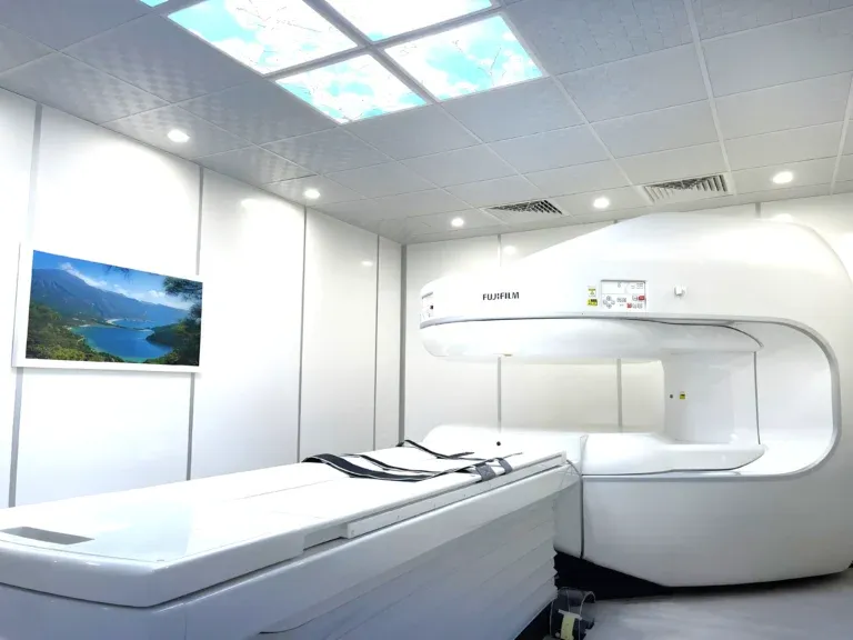
A hip MRI scan is the most detailed imaging tool for evaluating the hip joint and its surrounding structures. The hip is a deep ball-and-socket joint that bears the full weight of the body, making it susceptible to a range of conditions from sports injuries and labral tears to degenerative changes and avascular necrosis. Whether you are experiencing groin pain after exercise, deep hip pain that worsens with activity, or stiffness that limits your range of motion, a hip MRI can reveal the precise cause by visualizing the labrum, cartilage, bone marrow, tendons, and muscles in exquisite detail. In Dubai, hip MRI is available at advanced imaging centers and is the preferred diagnostic step when X-rays fail to explain the source of hip symptoms.


What Does a Hip MRI Show?
A hip MRI produces detailed cross-sectional images of the hip joint, including the femoral head, acetabulum (socket), labrum, articular cartilage, tendons, ligaments, bursae, and surrounding muscles. Unlike X-rays, which only show bone, MRI uses magnetic fields and radio waves to generate high-resolution images of both bone and soft tissue, making it the most comprehensive imaging modality for the hip.
The following conditions are commonly identified on a hip MRI:
- Avascular necrosis (AVN / osteonecrosis): AVN occurs when blood supply to the femoral head is disrupted, causing bone tissue to die and eventually collapse. MRI is the earliest and most sensitive imaging tool for detecting AVN, identifying characteristic bone marrow signal changes months before X-ray shows any abnormality. Early detection is critical because treatment options such as core decompression are most effective before the femoral head collapses. Risk factors include long-term steroid use, excessive alcohol consumption, sickle cell disease, and prior hip fracture or dislocation.
- Labral tears: The acetabular labrum is a ring of fibrocartilage that lines the rim of the hip socket, providing stability and a seal for the joint. Labral tears cause groin pain, clicking or catching with movement, and reduced range of motion. MRI, particularly with intra-articular contrast (MR arthrogram), provides excellent visualization of labral pathology, including tear location, size, and associated cartilage damage.
- Femoroacetabular impingement (FAI): FAI occurs when abnormal bone morphology of the femoral head (cam-type), acetabulum (pincer-type), or both (mixed) causes abnormal contact between the bones during movement, damaging the labrum and cartilage. MRI reveals the bony abnormalities and, more importantly, the resulting soft tissue damage including labral tears and cartilage delamination.
- Hip bursitis (trochanteric bursitis): Inflammation of the trochanteric bursa on the outer aspect of the hip causes lateral hip pain. MRI can confirm the diagnosis by showing fluid in the bursa and distinguishing bursitis from other causes of lateral hip pain such as gluteal tendinopathy or referred lumbar spine pain.
- Stress fractures: Stress fractures of the femoral neck are particularly important because they carry a risk of complete fracture and displacement if not detected early. MRI is far more sensitive than X-ray for detecting stress fractures, often identifying them weeks before they become visible on radiographs. This is especially relevant for runners, military personnel, and individuals with osteoporosis.
- Hip arthritis: MRI can detect early cartilage loss, bone marrow edema, synovial inflammation, and labral degeneration associated with osteoarthritis, providing a more complete picture than X-ray alone. This information helps determine whether conservative management, injections, or hip replacement is most appropriate.
- Snapping hip syndrome: Also known as coxa saltans, this condition involves a snapping sensation during hip movement caused by the iliotibial band, iliopsoas tendon, or labral pathology. MRI and dynamic assessment can identify the specific structure responsible for the snapping.
- Tendinopathy and muscle tears: The hip is surrounded by powerful muscles including the gluteals, hamstrings, hip flexors, and adductors. MRI detects tendinitis, partial tears, and complete ruptures of these tendons and muscles, including gluteal tendinopathy (a common cause of lateral hip pain, especially in women over 50) and proximal hamstring tears.
"One of the most important roles of hip MRI is early detection of avascular necrosis," explains Dr. Osama Elzamzami, Consultant Radiologist at DCDC. "AVN can be completely invisible on X-ray in its early stages, yet MRI detects it with near-perfect sensitivity. Catching AVN early — before the femoral head collapses — can mean the difference between a joint-preserving procedure and a total hip replacement."
How Much Does a Hip MRI Cost in Dubai?
The cost of a hip MRI in Dubai generally ranges from AED 900 to AED 1,400 for a standard non-contrast examination. The price varies depending on the imaging center, the MRI machine specifications, whether contrast dye is required, and whether the radiologist report is included. At Doctors Clinic Diagnostic Center in Dubai Healthcare City, hip MRI pricing is all-inclusive — covering the scan, a comprehensive radiologist report, and digital image copies.
| Hip MRI Type | Approximate Cost (AED) |
|---|---|
| Hip MRI without contrast | 900 – 1,400 |
| Hip MRI with contrast (gadolinium) | 1,200 – 1,800 |
| MR arthrogram (intra-articular contrast) | 1,500 – 2,200 |
| Both hips (bilateral) | 1,600 – 2,400 |
| Hip MRI + pelvis | 1,800 – 2,600 |
Prices are approximate and include the radiologist report at DCDC. Contact us for exact pricing.
Contrast-enhanced hip MRI is used when the referring physician suspects AVN, tumors, infection, or inflammatory conditions. An MR arthrogram, where contrast is injected directly into the hip joint, is sometimes requested for detailed evaluation of suspected labral tears. For most evaluations of hip pain, a standard non-contrast MRI provides excellent diagnostic information.
Most health insurance plans in the UAE cover hip MRI when ordered by a licensed physician with a documented clinical indication. DCDC assists patients with insurance pre-authorization to confirm coverage before the appointment.
When Does a Doctor Order a Hip MRI?
A hip MRI is typically ordered when clinical examination and X-rays do not fully explain a patient's symptoms, or when a specific soft tissue or bone marrow pathology is suspected. The following situations commonly lead to a hip MRI referral: For related information, see our guide on Shoulder MRI Dubai: Rotator Cuff & Sports Injuries.
- Persistent groin pain: Groin pain is the hallmark symptom of intra-articular hip problems including labral tears, early arthritis, and AVN. If groin pain persists for more than 4 to 6 weeks despite rest and conservative treatment, MRI is indicated.
- Suspected avascular necrosis: Patients with risk factors for AVN (steroid use, alcohol use, sickle cell disease, prior hip trauma) who develop hip or groin pain should have an MRI even if X-rays appear normal, as MRI detects AVN in its earliest, most treatable stage.
- Mechanical symptoms: Clicking, catching, locking, or a sensation of something moving inside the hip joint suggests a labral tear or loose body that requires MRI for confirmation.
- Hip pain in young active adults: Femoroacetabular impingement and labral tears are common in young adults, particularly those involved in sports requiring repetitive hip flexion (football, martial arts, dancing). MRI identifies the bony and soft tissue abnormalities.
- Normal X-ray but ongoing pain: When hip X-rays are normal but significant symptoms persist, MRI is the next step to identify soft tissue pathology, stress fractures, early AVN, or bone marrow edema invisible on radiographs.
- Pre-surgical planning: Before hip arthroscopy, hip replacement, or osteotomy, surgeons require detailed MRI to assess the extent of damage and plan the surgical approach.
- Lateral hip pain: Persistent pain on the outer aspect of the hip that does not respond to treatment may indicate gluteal tendinopathy, trochanteric bursitis, or iliotibial band pathology, all of which are clearly visible on MRI.
Book Your Hip MRI at DCDC
At Doctors Clinic Diagnostic Center in Dubai Healthcare City, we offer same-week appointments for hip MRI scans with comprehensive radiologist reports delivered within 24 to 48 hours.
Hip MRI vs X-Ray vs Ultrasound: Which Imaging Do You Need?
X-ray, ultrasound, and MRI each serve different roles in evaluating hip problems. Understanding their strengths and limitations helps patients appreciate why their doctor may recommend one test over another.
| Factor | X-Ray | Ultrasound | MRI |
|---|---|---|---|
| Cost | AED 150 – 300 | AED 400 – 700 | AED 900 – 1,400 |
| Radiation | Low dose | None | None |
| Scan time | 5 minutes | 15 – 20 minutes | 30 – 45 minutes |
| Bone visualization | Excellent | Limited | Excellent |
| AVN detection (early) | Not sensitive | Not reliable | Gold standard (near 100% sensitivity) |
| Labral tears | Not visible | Very limited | Excellent (especially MR arthrogram) |
| Cartilage damage | Indirect signs only | Not visible | Direct visualization |
| Stress fractures | Often missed early | Not visible | Highly sensitive |
| Bone marrow edema | Not visible | Not visible | Excellent |
| Best for | Fractures, advanced arthritis, joint space | Guided injections, effusion, bursitis | Complete hip evaluation, AVN, labral tears, FAI |
X-ray is typically the first-line test for hip pain. MRI is indicated when X-ray findings do not explain symptoms or specific pathology is suspected.
X-ray remains the appropriate first test for most hip complaints, as it effectively shows fractures, joint space narrowing, and advanced arthritis. However, X-ray is fundamentally limited — it cannot visualize the labrum, cartilage surfaces, bone marrow, or tendons. For conditions like AVN, labral tears, stress fractures, and femoroacetabular impingement, MRI is the only imaging modality that provides a definitive diagnosis. Ultrasound is useful for guided injections and assessing superficial structures like the trochanteric bursa, but cannot evaluate the deep intra-articular structures of the hip.
What to Expect During a Hip MRI
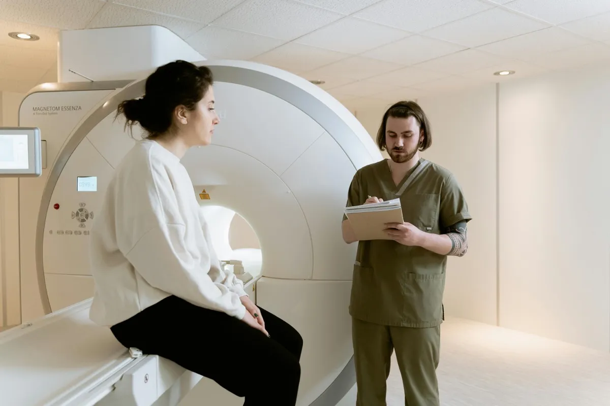
A hip MRI is a straightforward, painless procedure that requires minimal preparation. Here is what the process involves:
- Before the scan: Complete a safety screening questionnaire to confirm the absence of MRI-incompatible implants. Remove all metal objects. Wear comfortable clothing without metal components.
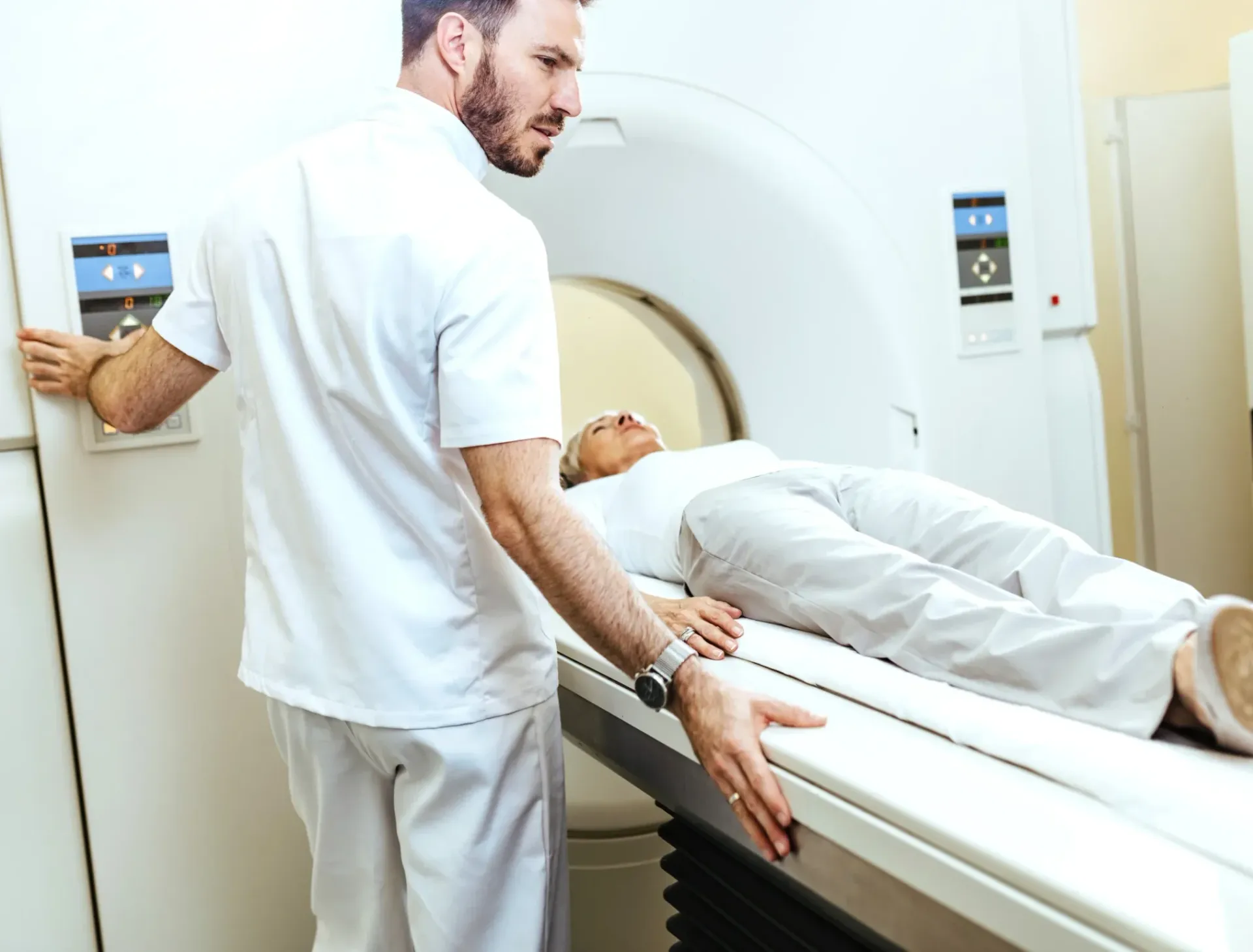
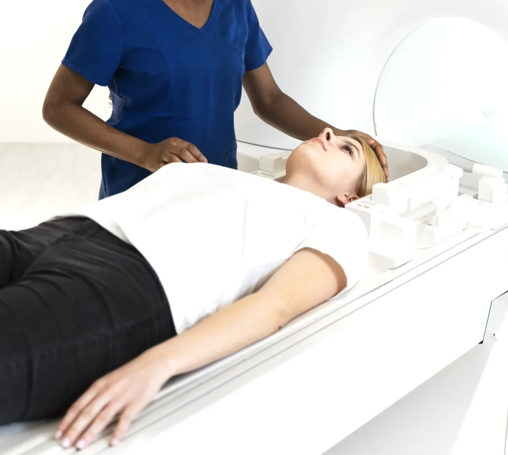
- Positioning: You will lie on your back on the MRI table. The affected hip (or both hips) will be positioned within the imaging field. A dedicated hip or body coil is placed over the hip area to optimize image quality. Your legs may be gently secured to minimize movement.
- During the scan: The table slides into the MRI machine. The machine produces characteristic knocking and humming sounds — earplugs or headphones are provided. Remaining still is essential for clear images. For hip imaging, your lower body enters the machine while your head remains near the opening.
- Duration: A standard hip MRI takes approximately 30 to 45 minutes. If an MR arthrogram is performed, additional time is needed for the injection procedure.
- After the scan: No recovery period is needed. You can return to normal activities immediately. Your radiologist report will typically be available within 24 to 48 hours at DCDC.
Get Expert Hip MRI Interpretation
At DCDC, every hip MRI is interpreted by a consultant radiologist with subspecialty musculoskeletal imaging experience. Our structured reports include detailed assessment of the labrum, cartilage, bone marrow, and surrounding soft tissues. Learn more about our MRI services.
Services associés au DCDC
Soins spécialisés et diagnostics avancés à Dubai Healthcare City
MRI Scan
High-resolution MRI imaging with expert radiologist interpretation
Prendre rendez-vousFull Body MRI
Comprehensive whole-body MRI screening for early detection of abnormalities
Prendre rendez-vousHealth Checkup
Combine MRI with full-panel blood work for complete health screening
Prendre rendez-vousFrequently Asked Questions
Final Thoughts
A hip MRI is the most comprehensive and sensitive imaging tool for evaluating the hip joint, particularly for detecting avascular necrosis, labral tears, femoroacetabular impingement, stress fractures, and early cartilage damage. If you are experiencing persistent hip or groin pain that has not responded to conservative treatment, or if your doctor suspects a condition that X-ray cannot adequately assess, a hip MRI provides the detailed information needed for accurate diagnosis and effective treatment planning.
At Doctors Clinic Diagnostic Center in Dubai Healthcare City, we combine advanced MRI technology with experienced consultant radiologist interpretation to deliver thorough, reliable hip MRI reports. Contact us to book your scan or to discuss whether a hip MRI is appropriate for your symptoms.
Sources et references
Cet article a ete revise par notre equipe medicale et fait reference aux sources suivantes :
- American College of Radiology - Appropriateness Criteria for Hip Pain
- Radiological Society of North America - Hip MRI
- American Academy of Orthopaedic Surgeons - Avascular Necrosis of the Hip
- European Radiology - MRI Sensitivity for AVN and Labral Tears
- Dubai Health Authority - Diagnostic Imaging Standards
Le contenu medical de ce site est revise par des medecins agrees DHA. Voir notre politique editoriale pour plus d'informations.

Redige par
Dr. Osama Elzamzami
Diagnostic Radiology
MD, FRCR
Dr. Osama Elzamzami is a Consultant Radiologist specializing in diagnostic imaging including MRI, CT, and ultrasound at DCDC Dubai Healthcare City.
Related Articles
MRI Scan Dubai: Complete Guide to Types, Cost & Full Body MRI
Knee MRI in Dubai: What It Shows, Cost & When You Need One
Shoulder MRI Dubai: Rotator Cuff, Labrum & Sports Injuries
Ankle MRI Dubai: What It Shows, Cost & When You Need One
Full Body MRI Cost Dubai: AED 5,000-15,000 (2026)
More in Diagnostic Imaging
Neck MRI Dubai: What It Shows & Cost (2026)
Lire la suite
X-Ray Scan Dubai: Complete Guide (2026)
Lire la suite
MRI vs Ultrasound Dubai: Which Scan? (2026)
Lire la suite
MRI vs X-Ray Dubai: Which Scan Do You Need? (2026)
Lire la suite
Kidney Ultrasound vs CT Dubai: Which Test? (2026)
Lire la suiteMRI Preparation Dubai: Complete Guide (2026)
Lire la suite© 2026 Doctors Clinic Diagnostic Center (DCDC), Dubai Healthcare City. Originally published at https://doctorsclinicdubai.ae/blog/hip-mri-dubai. All rights reserved. Unauthorized reproduction is prohibited.