Points cles
- Le Doppler est l'examen de reference de premiere intention pour detecter la thrombose veineuse profonde, offrant une visualisation en temps reel des caillots sanguins dans les veines des jambes sans radiation
- Les symptomes courants de TVP comprennent un gonflement soudain de la jambe, une douleur ou sensibilite au mollet, une chaleur et une rougeur, bien que certaines TVP ne produisent aucun symptome
- La technique d'echographie par compression, ou la sonde appuie sur la veine, est fiable a plus de 95% pour detecter la TVP au niveau de la cuisse et derriere le genou
- Les residents de Dubai font face a des facteurs de risque specifiques de TVP, notamment les vols long-courriers, le travail sedentaire au bureau et la deshydratation liee au climat chaud
- Si une TVP est detectee, un traitement anticoagulant rapide est essentiel pour empecher le caillot de grossir ou de se detacher et de migrer vers les poumons sous forme d'embolie pulmonaire
Un Doppler pour TVP est un examen d'imagerie non invasif qui utilise des ondes sonores pour detecter les caillots sanguins dans les veines profondes de vos jambes. La thrombose veineuse profonde, ou TVP, survient lorsqu'un caillot sanguin se forme dans une veine profonde, le plus souvent dans la partie inferieure de la jambe ou la cuisse. Non traitee, une TVP peut se detacher et migrer vers les poumons, provoquant une embolie pulmonaire potentiellement fatale. Le Doppler est l'outil diagnostique de premiere intention pour cette pathologie, fournissant des resultats rapides, precis et sans radiation.
La TVP touche environ 1 a 2 personnes pour 1 000 par an dans le monde, et son incidence augmente en raison de modes de vie de plus en plus sedentaires et du vieillissement des populations. Pour les residents de Dubai, des facteurs supplementaires tels que les voyages frequents en avion sur de longues distances, les heures de bureau prolongees et la deshydratation liee a la chaleur elevent davantage le risque. Ce guide complet explique comment le Doppler detecte la TVP, quels symptomes doivent inciter a un examen, quelle est la precision du test et ce a quoi s'attendre si un caillot est trouve.


Comment le Doppler Detecte les Caillots Sanguins dans les Veines Profondes
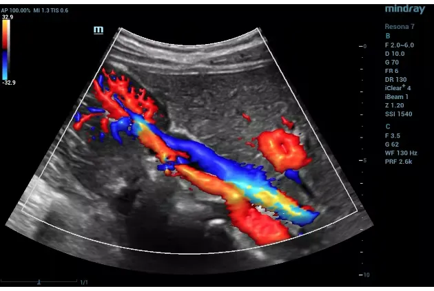
Le Doppler fonctionne en emettant des ondes sonores a haute frequence qui rebondissent sur les globules rouges en mouvement dans vos veines. Les echos de retour sont convertis en images et en cartes de flux colorees qui permettent au radiologue de voir exactement ou le sang circule normalement et ou il peut etre obstrue. Lorsqu'un caillot sanguin est present dans une veine profonde, le signal Doppler montre un flux absent ou reduit a cet endroit.
L'examen combine deux techniques complementaires. L'echographie en mode B produit une image en niveaux de gris de la structure veineuse, montrant les parois de la veine et tout materiel de caillot visible a l'interieur. Le Doppler couleur superpose une carte coloree sur cette image, avec differentes couleurs representant le sang circulant vers et loin de la sonde. Le Doppler spectral fournit une forme d'onde qui montre la vitesse et la direction du flux sanguin en un point specifique, revelant des perturbations subtiles du flux pouvant indiquer un caillot partiel.
Ensemble, ces techniques offrent au radiologue une vue complete de votre systeme veineux, permettant la detection des obstructions completes et partielles avec une grande precision. L'examen est realise en temps reel, ce qui signifie que le radiologue peut evaluer les veines de maniere dynamique pendant que vous respirez et que la sonde est positionnee sous differents angles.
« Quand un patient se presente avec un gonflement soudain de la jambe, chaque minute compte. Le Doppler de TVP est le chemin le plus rapide vers une reponse definitive, » declare le Dr Osama Elzamzami, Radiologue Consultant au DCDC. « Le test de compression est d'une elegante simplicite : une veine normale s'affaisse sous une pression douce tandis qu'une veine remplie de caillot ne le fait pas, et entre des mains experimentees, il offre une precision superieure a 95% pour la TVP proximale. J'insiste toujours aupres des patients sur le fait que cet examen peut etre la decision unique qui previent une embolie pulmonaire. »
Symptomes Declenchant un Examen de TVP
La TVP ne cause pas toujours des symptomes evidents, ce qui la rend particulierement dangereuse. Cependant, lorsque les symptomes apparaissent, ils affectent generalement une seule jambe et se developpent relativement rapidement. Reconnaitre ces signes d'alerte et consulter rapidement peut sauver des vies.
- Gonflement soudain d'une jambe, surtout s'il se developpe en quelques heures a quelques jours plutot que progressivement sur plusieurs semaines
- Douleur ou sensibilite au mollet ou a la cuisse pouvant ressembler a une crampe ou une douleur profonde, s'aggravant souvent en position debout ou a la marche
- Chaleur au niveau de la zone atteinte, perceptible au toucher en comparant une jambe a l'autre
- Rougeur ou decoloration de la peau, allant d'une teinte rosee subtile a un rouge-bleu plus evident
- Gonflement visible des veines superficielles pres de la surface de la peau
- Sensation de lourdeur ou de tension dans la jambe atteinte
Il est important de comprendre que la TVP peut aussi etre completement silencieuse. Certains patients decouvrent leur TVP uniquement lorsqu'ils developpent des symptomes d'embolie pulmonaire, tels qu'un essoufflement soudain, une douleur thoracique, une acceleration du rythme cardiaque ou des crachats de sang. Il s'agit d'une urgence medicale necessitant une prise en charge immediate.
Les medecins utilisent des outils d'evaluation clinique tels que le Score de Wells pour estimer la probabilite de TVP avant de prescrire un Doppler. Ce systeme de notation prend en compte des facteurs comme une chirurgie recente, des antecedents de cancer, un gonflement de la jambe, une sensibilite, une immobilisation et si un diagnostic alternatif est egalement probable. Un score de Wells eleve soutient fortement la necessite d'une evaluation echographique urgente.
La Technique d'Echographie par Compression
Le composant le plus critique d'un examen Doppler de TVP est le test de compression. Cette technique simple mais tres efficace est ce qui rend l'echographie si fiable pour detecter les caillots sanguins dans les veines.
Lors du test de compression, le manipulateur place la sonde echographique directement sur une veine et applique une pression douce vers le bas. Une veine normale, sans caillot, s'affaissera completement sous cette pression car les veines ont des parois fines et sont flexibles. Cependant, si un caillot sanguin est present a l'interieur de la veine, celle-ci ne se comprimera pas completement. Le caillot agit comme une masse solide qui empeche les parois de la veine de se rapprocher. Cette incompressibilite est le signe le plus important de TVP a l'echographie.
Le manipulateur effectue systematiquement la compression en plusieurs points le long des veines profondes de la jambe, commencant generalement par la veine femorale commune au niveau de l'aine et descendant a travers la veine femorale superficielle, la veine poplitee derriere le genou, et dans de nombreux cas les veines du mollet egalement. A chaque point, la veine est evaluee avec et sans compression, et le signal Doppler est evalue pour des patterns de flux normaux.
Un examen Doppler complet de TVP pour une jambe dure generalement 15 a 30 minutes, selon la complexite du cas. Dans certaines situations, les deux jambes sont examinees pour comparaison ou pour verifier une TVP bilaterale, ce qui peut prolonger la duree de l'examen.
Precision du Doppler par Rapport aux Autres Tests de TVP
Le Doppler est le test de premiere intention etabli pour la TVP en raison de son excellente combinaison de precision, de disponibilite, de securite et de rapport cout-efficacite. Il est cependant utile de comprendre comment il se compare aux autres methodes diagnostiques.
| Test | Sensibilite | Specificite | Principaux Avantages | Principales Limites |
|---|---|---|---|---|
| Echographie par compression (TVP proximale) | 95% – 99% | 95% – 99% | Sans radiation, portable, temps reel, repetable | Dependant de l'operateur ; veines du mollet plus difficiles a evaluer |
| Test sanguin D-dimeres | 95% – 97% | 40% – 60% | Valeur predictive negative tres elevee ; exclut la TVP | Faible specificite ; eleve dans de nombreuses situations (infection, chirurgie, grossesse) |
| Phlebographie par scanner | 95% – 100% | 95% – 100% | Excellent pour les veines pelviennes et abdominales | Exposition aux radiations, produit de contraste IV necessaire, cout plus eleve |
| Phlebographie par IRM | 92% – 100% | 95% – 100% | Sans radiation, bon pour les veines pelviennes | Couteux, temps d'examen plus long, disponibilite limitee |
Les valeurs de sensibilite et de specificite sont approximatives et basees sur des etudes cliniques publiees. La performance reelle depend de la localisation anatomique et de l'experience de l'operateur.
En pratique clinique, le parcours diagnostique commence generalement par une evaluation clinique (Score de Wells) combinee a un test sanguin de D-dimeres. Si les D-dimeres sont negatifs et la probabilite clinique faible, la TVP peut souvent etre ecartee sans imagerie. Cependant, si les D-dimeres sont eleves ou la suspicion clinique moderee a elevee, un Doppler est realise. La phlebographie par scanner est generalement reservee aux cas ou les resultats de l'echographie sont non concluants, ou lorsqu'une TVP des veines pelviennes ou abdominales est suspectee.
Un point important est que l'echographie est un peu moins sensible pour la TVP isolee des veines du mollet par rapport a la TVP proximale de la cuisse. Si la suspicion clinique reste elevee apres un Doppler negatif, un nouvel examen peut etre recommande dans 5 a 7 jours pour verifier l'extension du caillot du mollet vers les veines proximales.
Suspicion de TVP ? Faites-vous Tester Aujourd'hui
Au Doctors Clinic Diagnostic Center a Dubai Healthcare City, nos radiologues experimentes realisent des Doppler de TVP avec resultats le jour meme. Sans radiation, aucune preparation speciale requise.
Facteurs de Risque de TVP et Risques Specifiques a Dubai
Comprendre qui est a risque de TVP est essentiel pour la prevention et la detection precoce. Bien que n'importe qui puisse developper un caillot sanguin, certains facteurs augmentent considerablement la probabilite. Les residents de Dubai et la population des EAU au sens large font face a plusieurs facteurs de risque qui meritent une attention particuliere.
Voyages Aeriens Long-Courriers
Dubai est un hub de voyage international majeur, et de nombreux residents effectuent regulierement des vols long-courriers depassant 4 heures. L'immobilite prolongee dans un siege d'avion etroit restreint le flux sanguin dans les jambes, et la pression reduite en cabine ainsi que l'humidite plus faible contribuent a la deshydratation et a l'epaississement du sang. Le risque de TVP est environ deux a trois fois plus eleve apres des vols de plus de 4 heures. Les voyageurs frequents qui parcourent de longues distances plusieurs fois par an font face a un risque cumulatif souvent sous-estime.
Travail de Bureau Sedentaire
Le mode de vie professionnel moderne a Dubai implique souvent de rester assis a un bureau pendant 8 a 10 heures ou plus par jour. La position assise prolongee, que ce soit au travail ou lors de longs trajets en voiture dans les embouteillages, reduit la velocite du flux sanguin dans les veines des jambes et cree des conditions favorables a la formation de caillots. La combinaison d'un environnement interieur climatise et du manque de mouvement est un facteur de risque important mais sous-apprecie.
Chaleur et Deshydratation
Les temperatures estivales extremes de Dubai, qui depassent regulierement 45 degres Celsius, provoquent une perte importante de liquides par la transpiration. La deshydratation concentre le sang et le rend plus susceptible de coaguler. De nombreux residents ne boivent pas suffisamment d'eau, en particulier pendant les heures de jeune du Ramadan ou lorsqu'ils passent du temps a l'exterieur pendant les mois les plus chauds. Cette deshydratation chronique legere est un facteur de risque propre au climat regional.
Autres Facteurs de Risque Etablis
- Chirurgie ou hospitalisation recente, en particulier les interventions orthopediques concernant la hanche ou le genou
- Cancer actif ou traitement anticancereux, qui augmente la tendance a la coagulation sanguine
- Grossesse et periode post-partum, en raison des changements hormonaux et de la compression des veines pelviennes
- Pilules contraceptives orales ou traitement hormonal substitutif contenant de l'oestrogene
- Obesite, qui augmente la pression sur les veines pelviennes et des jambes et favorise l'inflammation
- Antecedents familiaux ou personnels de TVP ou d'embolie pulmonaire
- Troubles hereditaires de la coagulation tels que le facteur V Leiden ou la mutation du gene de la prothrombine
- Age superieur a 60 ans, car le risque de coagulation augmente avec l'age
- Tabagisme, qui endommage les parois des vaisseaux sanguins et favorise la formation de caillots
Que Se Passe-t-il Si une TVP Est Detectee ?
Lorsqu'un Doppler confirme la presence d'une TVP, le traitement commence generalement immediatement. L'objectif principal est d'empecher le caillot de grossir, d'empecher qu'il ne se detache et ne provoque une embolie pulmonaire, et de reduire le risque de complications a long terme telles que le syndrome post-thrombotique.
Traitement Anticoagulant
La pierre angulaire du traitement de la TVP est l'anticoagulation, communement appelee traitement par anticoagulants. Ces medicaments ne dissolvent pas les caillots existants mais empechent la formation de nouveaux caillots et arretent la croissance du caillot existant. Le systeme fibrinolytique propre du corps dissout ensuite progressivement le caillot au fil des semaines a des mois. Le traitement moderne implique generalement des anticoagulants oraux directs tels que le rivaroxaban ou l'apixaban, qui ne necessitent pas de surveillance sanguine reguliere. La duree du traitement va de 3 mois pour un premier episode avec une cause temporaire claire a un traitement indefini pour une TVP recurrente ou non provoquee.
Bas de Compression et Suivi
Les bas de compression gradues appliquent une pression douce sur les jambes, aidant le sang a remonter vers le coeur et reduisant le gonflement. Ils sont souvent recommandes pendant au moins deux ans apres un diagnostic de TVP pour reduire le risque de syndrome post-thrombotique. Des examens Doppler de controle sont generalement effectues pendant et apres le traitement pour surveiller la resolution du caillot, avec un examen de reference au moment du diagnostic suivi d'examens a 3 a 6 mois pour suivre la reponse au traitement.
Interventions Avancees
Dans les cas graves, comme une TVP ilio-femorale massive menacant la viabilite du membre, des traitements plus agressifs peuvent etre envisages. Ceux-ci incluent la thrombolyse dirigee par catheter (administration de medicaments thrombolytiques directement au niveau du caillot), la thrombectomie mecanique (retrait physique du caillot), ou la mise en place d'un filtre de veine cave inferieure chez les patients ne pouvant pas prendre d'anticoagulants. Ces interventions sont reservees a des situations cliniques specifiques et sont realisees dans des centres vasculaires specialises.
Strategies de Prevention pour les Residents de Dubai
La prevention est toujours preferable au traitement en matiere de TVP. Les residents de Dubai peuvent prendre plusieurs mesures pratiques pour reduire leur risque, en particulier compte tenu des facteurs lies au mode de vie et a l'environnement de la region.
- Restez bien hydrate en buvant au moins 2 a 3 litres d'eau par jour, en augmentant la quantite pendant les mois d'ete et les periodes de jeune
- Faites des pauses de mouvement regulieres toutes les 60 a 90 minutes lors de longues periodes assises au travail ; de simples levees de mollets et rotations de chevilles favorisent le retour veineux
- Pendant les vols long-courriers, levez-vous et marchez dans la cabine toutes les 1 a 2 heures, faites des exercices de jambes en position assise et portez des chaussettes de compression si vous avez des facteurs de risque
- Maintenez un poids sante grace a une activite physique reguliere, en visant au moins 150 minutes d'exercice modere par semaine
- Si vous prenez des contraceptifs oraux ou un traitement hormonal, discutez du risque de TVP avec votre medecin, notamment avant un long voyage
- Apres une chirurgie ou une maladie necessitant un alitement, suivez les instructions de votre medecin concernant la mobilisation precoce et les anticoagulants preventifs
- Evitez de croiser les jambes pendant de longues periodes, car cela comprime les veines et restreint le flux sanguin
- Si vous avez des antecedents personnels ou familiaux de caillots sanguins, demandez a votre medecin un depistage des troubles hereditaires de la coagulation
Doppler de TVP au DCDC Dubai
Au Doctors Clinic Diagnostic Center a Dubai Healthcare City, le Doppler de TVP est realise par des radiologues experimentes utilisant un equipement Doppler avance offrant une imagerie en mode B haute resolution ainsi que des capacites Doppler couleur et spectral sensibles. La clinique suit des protocoles standardises d'echographie par compression pour assurer une evaluation complete et precise du systeme veineux profond.
Les resultats sont generalement disponibles le jour meme, avec des rapports detailles incluant la localisation et l'etendue de tout caillot trouve, le degre d'obstruction veineuse et les recommandations de suivi. Une coordination etroite avec les medecins referents, y compris les internistes, les chirurgiens vasculaires et les hematologues, garantit que les patients recoivent un traitement adapte et rapide lorsque la TVP est confirmee.
Avec plus de 1 000 examens diagnostiques realises chaque mois et plus de 13 ans d'activite continue depuis 2013, le DCDC a bati une reputation comme l'un des centres diagnostiques de reference a Dubai Healthcare City. L'equipe multilingue du centre accueille des patients de tout les EAU et du monde entier, assurant une communication claire et des soins centres sur le patient tout au long de chaque examen.
Un cas recent illustre l'importance d'un depistage de TVP en temps opportun : une cadre marketing de 45 ans de Dubai a consulte le DCDC apres avoir remarque un gonflement progressif et une tension dans son mollet gauche suite a un vol d'affaires de 14 heures depuis Londres. Elle a d'abord pense qu'il s'agissait simplement de fatigue de voyage, mais lorsque le gonflement a persiste pendant deux jours, son medecin generaliste l'a orientee en urgence pour un Doppler veineux. L'echographie par compression a revele une TVP aigue dans la veine poplitee gauche s'etendant dans la veine femorale superficielle distale. Elle a commence un traitement anticoagulant le jour meme. Lors de son Doppler de controle a trois mois, le caillot s'etait largement resolu sans signe de syndrome post-thrombotique, un resultat que son hematologue a directement attribue a la detection precoce et a l'initiation rapide du traitement.
Inquiet par des Symptomes de TVP ?
Un Doppler de TVP au Doctors Clinic Diagnostic Center a Dubai Healthcare City peut verifier rapidement et precisement vos veines de jambe a la recherche de caillots sanguins. Resultats le jour meme. Sans radiation. Aucune preparation speciale requise.
Services associés au DCDC
Soins spécialisés et diagnostics avancés à Dubai Healthcare City
Frequently Asked Questions
Reflexions Finales
La thrombose veineuse profonde est une condition courante mais potentiellement mortelle qui peut etre detectee rapidement et precisement par le Doppler. La technique d'echographie par compression est la norme diagnostique pour une bonne raison : elle est sure, tres precise pour la TVP proximale, ne necessite ni radiation ni produit de contraste, et fournit des resultats immediats. Pour toute personne presentant un gonflement soudain de la jambe, une douleur ou une chaleur, un Doppler de TVP doit etre realise rapidement pour ecarter ou confirmer la presence d'un caillot sanguin.
Les residents de Dubai font face a une combinaison unique de facteurs de risque de TVP, des vols long-courriers frequents et des modes de vie sedentaires de bureau a la deshydratation induite par la chaleur. La connaissance de ces risques, combinee a des mesures preventives simples et a un examen rapide lorsque des symptomes apparaissent, peut prevenir des complications graves, y compris l'embolie pulmonaire. Pour les details de tarification, consultez notre guide sur le cout du Doppler a Dubai. Si vous etes preoccupe par la TVP ou avez des facteurs de risque, ne tardez pas. Prenez rendez-vous pour un Doppler au Doctors Clinic Diagnostic Center a Dubai Healthcare City pour votre tranquillite d'esprit et une evaluation experte rapide.
Sources et references
Cet article a ete revise par notre equipe medicale et fait reference aux sources suivantes :
- College Americain des Medecins Thoraciques - Directives de Traitement Antithrombotique
- Societe de Chirurgie Vasculaire - Diagnostic et Prise en Charge de la TVP
- Societe Radiologique d'Amerique du Nord - Echographie Veineuse
- Association Americaine du Coeur - Thromboembolie Veineuse
- Societe Europeenne de Cardiologie - Directives VTE
Le contenu medical de ce site est revise par des medecins agrees DHA. Voir notre politique editoriale pour plus d'informations.

Redige par
Dr. Osama Elzamzami
Radiologue Consultant
MD, Radiologie
Le Dr Osama Elzamzami est Radiologue Consultant specialise en imagerie diagnostique, notamment IRM, scanner, echographie et Doppler au DCDC Dubai Healthcare City.
Articles Connexes

Qu'est-ce qu'un Doppler ? Fonctionnement Explique Simplement
Doppler des Jambes : Guide Complet
Cout du Doppler a Dubai
Qu'est-ce qu'une Echographie ? Guide Complet
More in Diagnostic Imaging

X-Ray at Home Dubai: Cost & Guide (2026)
Lire la suite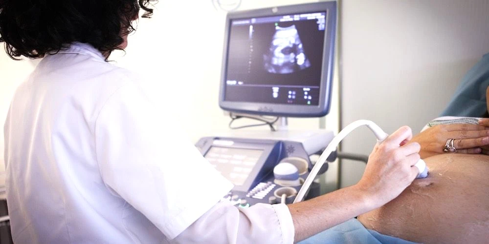
Ultrasound at Home Dubai: From AED 599 (2026)
Lire la suite
Neck MRI Dubai: What It Shows & Cost (2026)
Lire la suite
X-Ray Scan Dubai: Complete Guide (2026)
Lire la suite
MRI vs Ultrasound Dubai: Which Scan? (2026)
Lire la suite
MRI vs X-Ray Dubai: Which Scan Do You Need? (2026)
Lire la suite© 2026 Doctors Clinic Diagnostic Center (DCDC), Dubai Healthcare City. Originally published at https://doctorsclinicdubai.ae/blog/doppler-ultrasound-dvt-detection. All rights reserved. Unauthorized reproduction is prohibited.