Mga Pangunahing Punto
- MRI scans in Dubai cost AED 900 to 4,500 for individual regions and AED 5,000 to 15,000 for full body MRI, with most major insurance plans covering medically necessary scans
- Full body MRI screens your entire body from brain to pelvis without radiation, detecting tumors, aneurysms, organ abnormalities, and spinal conditions before symptoms appear
- MRI detects brain tumors with 95-99% sensitivity, liver cancer with 85-95% sensitivity, and kidney cancer with 90-95% sensitivity, though it has limitations for lung cancer and GI cancers
- MRI uses zero radiation unlike CT scans (10-30 mSv per scan), making it safe for repeated screening, children, and pregnant women after the first trimester
- A full body MRI takes 60 to 90 minutes with results reviewed by a consultant radiologist within 24 to 48 hours
- Open MRI is available for claustrophobic patients and works well for most diagnostic needs. For details, see our Open MRI guide
Whether your doctor ordered a single MRI scan or you are considering a full body MRI as a preventive health screening, this guide covers everything you need to know. From pricing and insurance to cancer detection rates and what happens inside the machine, we have compiled the most comprehensive MRI resource in Dubai based on our experience performing over 1,000 diagnostic scans per month at DCDC in Dubai Healthcare City since 2013.
This guide covers MRI scan costs in Dubai, types of MRI available, full body MRI screening, cancer detection effectiveness with sensitivity data, MRI vs CT and other imaging comparisons, who should consider full body MRI, the ROI of early detection, preparation steps, what to expect during your scan, managing claustrophobia, the DCDC protocol, and understanding your results.

What Is an MRI and How Does It Work?
MRI stands for Magnetic Resonance Imaging. Unlike X-rays or CT scans that use radiation, MRI uses powerful magnets and radio waves to create detailed images of your body's soft tissues. Your body is mostly water, and the hydrogen atoms in that water respond to the MRI's magnetic field. The machine detects those responses and converts them into incredibly detailed cross-sectional images of your organs, tissues, bones, and blood vessels.
This makes MRI particularly valuable for examining the brain and spinal cord (detecting tumors, strokes, multiple sclerosis, and other neurological conditions), joints (evaluating torn ligaments, cartilage damage, and arthritis), soft tissues (examining muscles, organs, and blood vessels), and the heart (assessing cardiac structure and function). MRI uses zero radiation, making it safe for repeated examinations and ideal for children and pregnant women after the first trimester.
"The real power of MRI lies in its ability to detect conditions that are completely silent clinically," explains Dr. Osama Elzamzami, Consultant Radiologist at DCDC. "I have seen cases where patients felt perfectly healthy, yet their scan revealed an early-stage renal mass or an unruptured brain aneurysm. These are findings that change lives when caught early."
Types of MRI Scans Available in Dubai
Different body regions require different MRI protocols optimized for the specific tissues and conditions being evaluated. Here are the most common types of MRI scans performed in Dubai:
- Brain MRI: The gold standard for brain tumor detection, stroke assessment, multiple sclerosis, aneurysms, and white matter changes. Can identify tumors as small as a few millimeters. Also evaluates the pituitary gland and vascular health.
- Spine MRI (Cervical, Thoracic, Lumbar): Evaluates disc herniations, spinal cord compression, spinal stenosis, nerve impingement, degenerative disc disease, and spinal tumors. The only imaging method that clearly visualizes the spinal cord itself.
- Knee and Joint MRI: Detects ACL and meniscal tears, cartilage damage, ligament injuries, bone marrow edema, and early signs of arthritis. Essential for sports injuries common in Dubai's active population.
- Abdominal and Pelvic MRI: Examines the liver, kidneys, pancreas, spleen, adrenal glands, bladder, and reproductive organs. Excellent for detecting cysts, tumors, fatty liver, and organ abnormalities.
- Cardiac MRI: Assesses heart chamber size, muscle thickness, valve function, and blood flow. Identifies cardiomyopathy, pericardial effusion, and structural abnormalities that echocardiograms may miss.
- Breast MRI: The most sensitive imaging method for breast cancer detection (90-95% sensitivity), particularly valuable for high-risk women and those with dense breast tissue.
- Full Body MRI: A comprehensive scan covering all major regions from brain to pelvis in a single session. Increasingly popular for preventive health screening. See our full body MRI cost guide for pricing details.
What Does a Full Body MRI Cover?
A full body MRI uses a structured, multi-sequence protocol to examine every major body region using imaging sequences optimized for each area. Here is what a comprehensive full body MRI typically covers:
- Brain and head: Detects tumors, aneurysms, white matter changes, stroke evidence, and pituitary abnormalities
- Cervical spine and neck: Evaluates disc herniations, spinal cord compression, and soft tissue masses
- Thoracic and lumbar spine: Identifies disc disease, vertebral fractures, nerve impingement, and degenerative conditions
- Chest: Assesses the heart, mediastinal structures, and major blood vessels (note: lung tissue is better evaluated by CT)
- Abdomen: Examines the liver, kidneys, pancreas, spleen, adrenal glands, and abdominal aorta for masses, cysts, or organ abnormalities
- Pelvis: Screens the bladder, reproductive organs, prostate (in men), and pelvic lymph nodes
- Major joints: Some protocols include shoulders, hips, and knees to detect cartilage damage, labral tears, or early arthritis
What Can a Full Body MRI Detect?
Full body MRI is exceptionally effective at detecting soft tissue abnormalities that other screening tools miss. Because MRI provides superior contrast resolution between different tissue types, it reveals conditions at very early stages.
- Tumors and masses: Both benign and malignant tumors in the brain, liver, kidneys, pancreas, breast, prostate, and pelvic organs
- Aneurysms: Bulging or weakened blood vessel walls in the brain (3-5% of the population have unruptured cerebral aneurysms) or aorta (80% mortality rate if ruptured)
- Organ abnormalities: Liver cysts, fatty liver (NAFLD affects 25-30% of adults), kidney stones, enlarged spleen, pancreatic lesions, and adrenal nodules
- Spinal conditions: Herniated discs, spinal stenosis, nerve compression, degenerative disc disease, and vertebral fractures
- Cardiovascular concerns: Heart muscle abnormalities, valve issues, aortic conditions, atherosclerosis, and vascular stenosis
- Neurological findings: White matter lesions, small vessel disease, early dementia markers, and demyelination patterns
- Inflammation and autoimmune markers: Sacroiliitis indicating ankylosing spondylitis, organ inflammation suggesting autoimmune conditions, and myocarditis
- Joint and musculoskeletal problems: Ligament tears, cartilage damage, bone marrow edema, meniscal tears, and early signs of arthritis
Full Body MRI for Cancer Screening
Cancer cells often have different water content and tissue characteristics compared to normal cells. MRI exploits these differences to create contrast between healthy and abnormal tissue. Advanced techniques such as diffusion-weighted imaging (DWI) further enhance the ability to identify areas of rapid cell division, a hallmark of malignant tumors. When contrast dye (gadolinium) is administered, MRI also reveals abnormal blood vessel patterns that tumors create to sustain their growth.
Cancers MRI Detects Well
- Brain tumors: MRI is the gold standard, with 95-99% sensitivity. It can identify tumors as small as a few millimeters.
- Liver cancer: MRI with contrast is highly sensitive (85-95%) for hepatocellular carcinoma and liver metastases, often outperforming CT.
- Kidney cancer: Renal cell carcinoma is well-visualized (90-95% sensitivity) with the ability to distinguish benign cysts from solid masses.
- Prostate cancer: Multiparametric MRI (mpMRI) achieves 80-90% sensitivity and has become a standard diagnostic tool.
- Breast cancer: MRI is the most sensitive imaging method (90-95%) for breast cancer detection, particularly for high-risk women with dense breast tissue.
- Bone and soft tissue sarcomas: MRI provides excellent visualization (90-95% sensitivity), clearly showing tumor extent and relationship to surrounding structures.
- Spinal cord tumors: MRI is the only reliable imaging method for detecting tumors within or adjacent to the spinal cord.
Cancers Where MRI Has Limitations
- Early-stage lung cancer: Small lung nodules (50-70% sensitivity) are better detected by low-dose CT due to air-filled lungs and respiratory motion.
- Colorectal cancer: Colonoscopy remains the standard. MRI is useful for rectal cancer staging but less effective for early polyp detection.
- Stomach and esophageal cancer: Endoscopy is far more effective for mucosal-based gastrointestinal cancers.
- Skin cancers: Melanoma and other skin cancers are diagnosed through visual examination and biopsy, not imaging.
| Cancer Type | MRI Sensitivity | MRI Specificity | Notes |
|---|---|---|---|
| Brain tumors | 95-99% | 90-95% | Gold standard imaging method |
| Liver cancer | 85-95% | 85-90% | With contrast enhancement |
| Kidney cancer | 90-95% | 85-90% | Excellent for mass characterization |
| Prostate cancer | 80-90% | 70-80% | Multiparametric MRI (PI-RADS) |
| Breast cancer | 90-95% | 70-80% | Highest sensitivity of any imaging |
| Bone metastases | 90-95% | 85-90% | Superior to bone scan |
| Lung cancer | 50-70% | 80-85% | CT preferred for lung screening |
Sensitivity and specificity values are approximate ranges based on published literature. Actual performance depends on equipment quality, radiologist experience, and tumor characteristics.
Whole Body Diffusion-Weighted MRI (WB-DWI)
WB-DWI is an advanced MRI technique that measures water molecule movement within tissues. Densely packed cancer cells restrict water movement, creating a bright signal that helps radiologists identify malignancies. Studies show WB-DWI performs comparably to PET-CT for staging certain cancers while avoiding radiation exposure entirely. This technique is available at DCDC's MRI department.
Who Should Consider a Full Body MRI?
Full body MRI is not a one-size-fits-all recommendation, but certain groups benefit significantly from comprehensive screening.
Age-Based Recommendations
- Ages 30-39: A baseline full body MRI is optional but valuable for establishing a reference point, especially for those with family history of cancer or cardiovascular disease.
- Ages 40-49: Strongly recommended as a baseline screening. This is when age-related conditions begin developing silently. Combine with comprehensive blood work for maximum insight.
- Ages 50 and above: Annual or biennial screening strongly recommended. This age group faces the highest risk for silent cancers, aneurysms, and organ disease.
Genetic and Family History Indicators
Individuals with hereditary cancer syndromes are at significantly elevated risk and benefit most from regular screening. These include BRCA1/BRCA2 mutations (breast, ovarian, prostate cancer risk), Lynch syndrome (colorectal and endometrial cancer risk), Li-Fraumeni syndrome (multiple cancer types), familial adenomatous polyposis, and Von Hippel-Lindau syndrome. Anyone with multiple first-degree relatives diagnosed with cancer, particularly at young ages, should discuss MRI screening with their physician.
Executive and High-Stress Professionals
A growing number of CEOs, business owners, and senior executives add full body MRI to their annual health routines. The executive lifestyle, which combines chronic stress, sedentary desk work for 10-14 hours daily, irregular high-calorie meals, frequent international travel, and limited time for medical appointments, creates a compounding health burden that standard checkups are not designed to catch. Full body MRI fills that gap by providing direct visualization of organs and tissues that blood tests cannot assess.
Dubai Expatriates
Expatriates new to the UAE healthcare system often lack continuity of medical records. A full body MRI establishes a comprehensive health baseline in a single visit, providing a reference point that makes future screening more informative.
Post-Treatment Cancer Surveillance
Cancer survivors use full body MRI to monitor for recurrence without repeated radiation exposure, allowing comparison scans that detect subtle changes over time.
What Standard Health Checkups Miss
Most annual health checkups consist of blood tests, blood pressure measurement, a physical examination, and perhaps an ECG. While these are useful baseline assessments, they have significant blind spots.
| Assessment Type | What It Detects | What It Misses |
|---|---|---|
| Blood Tests | Cholesterol, glucose, liver enzymes, inflammation markers | Tumors, aneurysms, structural organ changes |
| ECG | Heart rhythm abnormalities | Coronary artery narrowing, cardiac masses, early cardiomyopathy |
| Physical Exam | Blood pressure, BMI, visible conditions | Internal organ disease, early-stage cancer, spinal degeneration |
| Full Body MRI | Organ structure, tumors, vascular abnormalities, brain changes, spinal conditions | Requires clinical correlation for some findings |
Comparison of screening methods. Full body MRI complements standard checkups by adding structural imaging.
The ROI of Early Detection
The financial case for early detection through MRI is compelling. The cost of a full body MRI (AED 5,000-15,000) is a fraction of treating advanced-stage disease. More importantly, the personal consequences of a late diagnosis, including extended medical leave, reduced quality of life, and higher mortality, far exceed the investment in annual screening.
| Scenario | Estimated Cost (AED) | Recovery Time |
|---|---|---|
| Early-stage tumor (detected by MRI) | 50,000 – 100,000 | 2 – 4 weeks |
| Advanced-stage cancer (late detection) | 500,000+ | 6 – 12+ months |
| Aortic aneurysm (detected before rupture) | 80,000 – 150,000 | 4 – 6 weeks |
| Emergency aortic rupture repair | 300,000+ | 3 – 6 months |
| Early disc treatment (physiotherapy) | 20,000 – 50,000 | 4 – 8 weeks |
| Spinal surgery (late detection) | 200,000 – 400,000 | 3 – 6 months |
Estimated treatment costs illustrating the financial value of early detection through MRI screening.
Is a Full Body MRI Worth It?
This is one of the most debated questions in preventive medicine. The medical community holds varied opinions, and understanding both perspectives helps you make an informed decision.
The Case For Full Body MRI
- Detects conditions years before symptoms appear, when treatment is most effective and least invasive
- Zero radiation exposure makes it safe for annual repeat screening, unlike CT or PET scans
- Establishes a baseline for comparison in future scans, making subtle changes detectable over time
- Covers multiple organ systems in a single 60-90 minute session
The Case Against
- Incidental findings occur in approximately 40% of scans, some of which are benign but require follow-up, potentially causing anxiety
- Higher cost compared to standard screening methods (AED 5,000-15,000 vs AED 500-2,000 for blood work)
- Not all cancers are detectable (lung, GI, and skin cancers have lower MRI sensitivity)
- False positives may lead to unnecessary additional tests or biopsies
The bottom line: Full body MRI is most valuable for individuals with elevated risk factors, including those over 40, those with family history of cancer or cardiovascular disease, executives with high-stress lifestyles, and anyone seeking a comprehensive health baseline. For low-risk individuals under 40 with no family history, standard screening may be sufficient, though a baseline MRI still provides future comparison value.
Full Body MRI vs. CT Scan: Complete Comparison
MRI and CT are complementary technologies, not competitors. Each has distinct strengths. Understanding the differences helps you and your doctor choose the right tool for each clinical question.
| Factor | Full Body MRI | CT Scan |
|---|---|---|
| Radiation exposure | None (magnetic fields only) | Yes (10-30 mSv per scan) |
| Scan duration | 60-90 minutes | 5-15 minutes |
| Soft tissue detail | Excellent (superior) | Good |
| Bone detail | Good | Excellent (superior) |
| Brain imaging | Gold standard | Good for emergencies |
| Lung imaging | Limited (motion artifacts) | Excellent |
| Joint and ligament imaging | Excellent | Limited |
| Liver and kidney imaging | Excellent | Good |
| Spinal cord visualization | Excellent | Limited |
| Cost in Dubai | AED 5,000-15,000 (full body) | AED 1,500-4,000 (full body) |
| Contrast dye | Gadolinium (rare reactions) | Iodine-based (more common reactions) |
| Claustrophobia concern | Yes (enclosed tube or Open MRI) | Minimal (open ring design) |
| Metal implants | May be contraindicated | Generally safe |
| Emergency availability | Limited (long scan time) | Excellent (fast results) |
| Repeat screening safety | Very safe (no radiation) | Cumulative radiation risk |
MRI and CT are complementary. The right choice depends on the clinical question being asked.
When MRI Is the Better Choice
Choose MRI for cancer screening and surveillance (no radiation for repeated imaging), brain and neurological conditions (gold standard), spine and spinal cord evaluation (only method to visualize the cord), joint and soft tissue injuries (ligament tears, cartilage damage), liver and kidney characterization (superior to CT for lesion classification), pediatric and pregnancy imaging (no radiation), and proactive health screening.
When CT Is the Better Choice
Choose CT for emergencies and trauma (speed saves lives), lung conditions (far superior to MRI), bone fractures (clearest bone images), kidney stones (standard of care), acute stroke assessment (quick hemorrhage exclusion), and patients with MRI contraindications (pacemakers, certain implants).
Full Body MRI vs. PET-CT for Cancer
| Feature | Full Body MRI | PET-CT |
|---|---|---|
| Radiation exposure | None | Significant (CT + radiotracer) |
| Soft tissue detail | Excellent | Moderate |
| Metabolic information | Limited (DWI provides some) | Excellent |
| Brain tumor detection | Superior | Limited by normal brain uptake |
| Bone metastases | Excellent with DWI | Excellent |
| Lung nodule detection | Limited | Good (CT component) |
| Suitable for screening | Yes (no radiation) | Less ideal (radiation) |
| Scan duration | 60-90 minutes | 2-3 hours (including uptake) |
| Cost in Dubai | AED 5,000-15,000 | AED 8,000-15,000 |
For screening purposes, MRI is preferred due to no radiation. PET-CT is better for known cancer staging.
MRI vs. Ultrasound vs. X-Ray
Ultrasound is excellent for real-time imaging, pregnancy monitoring, and initial organ assessment, but provides less detail than MRI for deep tissues. X-Ray is the fastest and cheapest imaging option, ideal for bone fractures and chest assessment, but provides minimal soft tissue detail and uses radiation. MRI surpasses both in soft tissue resolution and diagnostic depth, which is why it remains the imaging method of choice when detailed characterization is needed.
MRI Scan Costs in Dubai: What You'll Actually Pay
MRI prices in Dubai vary based on the body part being scanned, whether contrast dye is needed, the machine type, and the facility. An MRI scan at DCDC Dubai Healthcare City offers competitive pricing with all-inclusive packages that cover the scan, consultant radiologist report, and digital images. Here are current price ranges:
| Body Part | Without Contrast | With Contrast |
|---|---|---|
| Brain MRI | AED 900 - 3,000 | AED 2,800 - 4,000 |
| Spine MRI (per region) | AED 900 - 3,000 | AED 2,800 - 4,000 |
| Knee / Shoulder MRI | AED 900 - 2,500 | AED 2,200 - 3,200 |
| Abdominal / Pelvic MRI | AED 2,500 - 3,500 | AED 3,200 - 4,500 |
| Cardiac MRI | AED 3,000 - 4,000 | AED 3,800 - 4,500 |
| Full Body MRI | AED 5,000 - 7,000 | AED 8,000 - 15,000 |
Typical MRI price ranges in Dubai (2026). Prices vary by facility. For detailed full body MRI pricing and package options, see our <a href="/blog/full-body-mri-cost-dubai" class="text-primary-600 hover:underline">complete MRI cost guide</a>.
What affects the price? Body part being scanned (brain and cardiac MRIs require specialized protocols), contrast dye (gadolinium contrast improves image clarity for certain conditions), machine type (Open MRI may have different pricing), and facility type (hospital-based MRI typically costs more than diagnostic centers).
How Long Does a Full Body MRI Take?
A full body MRI typically takes 60 to 90 minutes of active scanning time. The total appointment time, including check-in, preparation, and the scan itself, is usually 90 minutes to 2 hours. Here is a breakdown by body region:
| Body Region | Approximate Duration |
|---|---|
| Brain | 15-20 minutes |
| Cervical Spine | 10-15 minutes |
| Thoracic Spine | 10-15 minutes |
| Chest | 10-15 minutes |
| Abdomen and Pelvis | 15-20 minutes |
| Lumbar Spine | 10-15 minutes |
| Joints (each) | 10-15 minutes |
Individual region times are approximate. Full body scans proceed through regions sequentially.
The full timeline includes: check-in and safety screening (15-20 minutes before), the scan itself (60-90 minutes), and changing afterward (5-10 minutes). Results are typically available within 24-48 hours, with preliminary findings communicated within 24 hours for urgent cases. Ready to get started? Book your MRI scan at DCDC for same-week appointments with consultant radiologist reporting.
How to Prepare for Your MRI: Complete Checklist
The Day Before
- Check if fasting is required (usually for abdominal MRIs — 4-6 hours)
- Choose comfortable clothing without metal: yoga pants, sweatpants, cotton t-shirt
- Remove jewelry early: rings, watches, earrings, body piercings
- Skip caffeine — it increases anxiety and makes lying still harder
- Get a good night's sleep to help you stay calm and still during the scan
The Day Of
- Arrive 15-20 minutes early for paperwork and safety questionnaires
- Bring your referral letter and any previous imaging CDs
- Bring your Emirates ID and insurance card
- Leave valuables at home or use a locker — no metal in the scan room
- Use the bathroom before the scan (you'll be lying still for 20-90 minutes)
What to Tell the Technologist
Be completely honest on your safety questionnaire. The MRI's powerful magnets can interact dangerously with metal in your body. Tell them about pacemakers or cardiac devices, cochlear implants, any metal implants, plates, screws, or joint replacements, aneurysm clips or vascular stents, history of metal fragments (from welding, military service, or accidents), pregnancy or possibility of pregnancy, kidney problems (important for contrast decisions), and anxiety or claustrophobia.
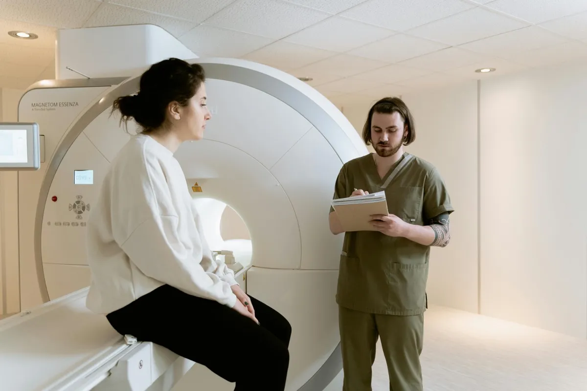
What Happens During the MRI: Step by Step
Knowing exactly what to expect dramatically reduces anxiety.
- 1. Changing and preparation: You may change into a hospital gown, though many patients wear their own metal-free clothing. All jewelry, watches, and pocket contents are removed.
- 2. Positioning: You lie on a padded table. Depending on the scan, a special coil may be placed around the body part being imaged. You receive earplugs or headphones (MRIs are loud) and a squeeze ball to signal the technologist.
- 3. The scan begins: The table slides into the machine (or in Open MRI, you are positioned within the open structure). The technologist monitors you through a window and intercom at all times.
- 4. What you experience: Loud sounds (banging, clicking, humming — completely normal), the need to stay very still, possible brief warmth, and scan sequences lasting 2-7 minutes each with short pauses between.
- 5. Contrast injection (if needed): Some scans require gadolinium contrast via IV. You may feel a cool sensation and brief metallic taste, both temporary and normal.
- 6. Completion: The table slides out, you get dressed, and can resume normal activities immediately unless sedation was used.
Managing Claustrophobia During MRI
Up to 37% of patients experience some MRI-related anxiety, but with proper support, the vast majority complete their scans successfully.
Before Your Appointment
- Request Open MRI — significantly more spacious and comfortable
- Discuss sedation options — a mild oral sedative taken before the scan can help significantly
- Practice relaxation techniques — breathing exercises and meditation apps in the days before
- Visit the facility beforehand — some patients find it helpful to see the machine and practice lying in it
During the Scan: 6 Comfort Tips
- Close your eyes before entering and keep them closed throughout
- Focus on slow, deep belly breathing (4 counts in, 6 counts out)
- Listen to music or a podcast through the provided headphones
- Wear comfortable, loose clothing without metal
- Communicate with the technologist — they can talk to you through the intercom at any time
- Remember it is temporary — most scans are 20-45 minutes for single regions, and you can pause anytime
The DCDC Full Body MRI Protocol
At Doctors Clinic Diagnostic Center in Dubai Healthcare City, full body MRI follows a structured, multi-sequence protocol developed over more than 13 years and refined through over 1,000 diagnostic scans performed monthly. International patients visit from around the world specifically for our diagnostic imaging expertise.
- Pre-scan consultation: A radiologist reviews your medical history, family history, and specific concerns to customize the imaging protocol.
- Multi-region sequencing: Dedicated imaging sequences are applied to each body region (brain, spine, chest, abdomen, pelvis) for optimal contrast and resolution.
- Advanced techniques: Diffusion-weighted imaging (DWI) for cancer screening, specific cardiac sequences when indicated, and specialized joint protocols as needed.
- Contrast enhancement (when indicated): Gadolinium-based contrast may be used for specific areas if initial sequences suggest further evaluation is needed.
- Consultant-led reporting: All images are reviewed and reported by a consultant radiologist with experience in whole-body imaging — not a general practitioner or junior radiologist.
- Results consultation: Findings are explained clearly during a follow-up consultation, with referral pathways arranged if any findings require specialist attention.
What Happens If Something Is Found?
If MRI reveals a suspicious finding, the radiologist classifies it based on its characteristics and provides a recommendation in the report. The pathway typically follows these steps:
- Radiologist review: Every image is analyzed by a consultant radiologist who classifies findings by urgency and clinical significance
- Multidisciplinary discussion: Complex findings may be discussed with relevant specialists (oncologists, cardiologists, neurologists) for optimal guidance
- Targeted follow-up imaging: Additional focused MRI with contrast, or complementary imaging (CT for lungs, ultrasound for superficial structures) as needed
- Biopsy referral: If a lesion requires tissue diagnosis, referral to the appropriate specialist is arranged
- Treatment planning: For confirmed diagnoses, the radiologist collaborates with the treating physician to establish a clear management plan
It is important to note that not all findings are concerning. Many incidental findings are benign and simply require monitoring. Your radiologist will help contextualize every result, clearly distinguishing between findings that need action and those that are normal variants.
Understanding Your MRI Results
After your scan, a specialized physician (radiologist) analyzes every image. Report generation is typically completed within 24-48 hours. For urgent cases, same-day or next-morning reporting may be available. The report goes to your referring physician, who explains the findings and discusses next steps during your follow-up.
Self-pay patients can often receive results directly. Insured patients typically get results through their referring physician. At DCDC, we offer a results consultation where the radiologist or referring physician explains your findings in plain language.
Special Considerations
MRI During Pregnancy
MRI is generally considered safe during pregnancy, especially after the first trimester, because it does not use radiation. However, gadolinium contrast is typically avoided in pregnant women unless absolutely necessary. Always inform the technologist if you are pregnant or might be.
MRI for Children
Children can have MRI scans safely. The main challenge is keeping them still. Options include scheduling during natural sleep time (for infants), using Open MRI where a parent can stay nearby, light sedation for very young or anxious children, and preparation with age-appropriate explanations.
MRI with Metal Implants
Many modern implants are MRI-safe or MRI-conditional, including most modern joint replacements, dental implants, and surgical hardware. However, you must disclose ALL metal in your body. Certain pacemakers, cochlear implants, and metallic foreign bodies may be absolute contraindications. The technologist will verify safety before proceeding.
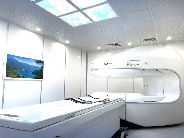
Open MRI vs. Traditional MRI
Open MRI has a wide, open design with space on the sides, providing excellent image quality for most diagnostic needs. It is ideal for claustrophobic patients, children, elderly patients, and larger individuals. Traditional closed MRI offers the highest image resolution and stronger magnetic fields (1.5T to 3T), which may be preferred for certain specialized examinations.
For most patients, Open MRI provides excellent diagnostic quality while dramatically improving comfort. Your doctor may recommend traditional MRI for highly specialized examinations, but for routine brain, spine, joint, and abdominal imaging, Open MRI works beautifully. Note that Open MRI scans may take slightly longer due to lower field strength. For a detailed comparison and pricing, read our complete Open MRI guide.
Insurance Coverage for MRI in Dubai
Most comprehensive health insurance plans in Dubai cover MRI scans when medically necessary. You will need a doctor's referral, pre-authorization from your insurer (which can take 24-72 hours), and must use an in-network facility for the best coverage.
Major insurers that commonly cover MRI include Daman, Saico, NAS, Aetna, BUPA, Cigna, and MetLife. Coverage levels vary: essential plans may cover 50-80% after deductible, enhanced plans often cover 80-100% at network providers, and premium plans typically provide 100% coverage with minimal co-pay. Note that full body MRI for screening purposes is typically not covered by insurance — it is considered elective. However, individual region MRI scans ordered for specific medical indications are usually covered.
Self-pay patients can often get same-day MRI appointments since they don't need to wait for insurance pre-authorization. Some companies now include executive MRI screening as a C-suite benefit alongside cardiac stress testing and comprehensive blood panels.
Limitations to Be Aware Of
- Lung tissue: MRI is less effective than CT for evaluating small lung nodules and lung parenchyma due to air-tissue interfaces and respiratory motion
- Bone cortex detail: While MRI detects bone marrow abnormalities well, fine cortical bone fractures may be better seen on CT or X-ray
- Incidental findings: Comprehensive scanning may reveal benign findings that require follow-up, potentially causing anxiety. An experienced radiologist contextualizes these results
- Scan duration: 60-90 minutes for full body MRI can be challenging for claustrophobic patients or those with difficulty lying still
- Metal implants: Certain metallic implants, pacemakers, or cochlear devices may be contraindications
- Not a replacement for organ-specific screening: Colonoscopy for colon cancer, mammography for breast cancer, and low-dose CT for lung cancer remain important complementary screening tools
Schedule Your MRI at DCDC
At Doctors Clinic Diagnostic Center in Dubai Healthcare City, we offer both standard and Open MRI options with experienced consultant radiologists, advanced imaging protocols including DWI for cancer screening, and fast turnaround on results. Same-day appointments often available for self-pay patients.
Kaugnay na Serbisyo sa DCDC
Dalubhasang pangangalaga at advanced diagnostics sa Dubai Healthcare City
Mga Madalas Itanong
Final Thoughts
An MRI scan is one of the most powerful diagnostic tools available, providing detailed images of your body's soft tissues without radiation exposure. Whether you need a single region scan for a specific condition or a full body MRI for comprehensive health screening, understanding costs, preparation, what MRI can and cannot detect, and what to expect helps you make informed decisions about your health.
Choosing the right facility matters. Look for experienced consultant radiologists (not junior doctors reviewing your images), modern equipment with advanced protocols like diffusion-weighted imaging, Open MRI options for comfort, and clear communication from booking to results. At Doctors Clinic Diagnostic Center in Dubai Healthcare City, we have been performing over 1,000 diagnostic scans monthly since 2013, providing both standard and Open MRI with consultant-led reporting and comprehensive health screening protocols that match the highest international standards.
Mga Sanggunian at Reperensya
Ang artikulong ito ay sinuri ng aming medikal na team at tumutukoy sa mga sumusunod na sanggunian:
- Dubai Health Authority - Diagnostic Imaging Regulations
- UAE Ministry of Health (MOHAP) - Diagnostic Imaging Standards
- American College of Radiology - MRI Safety & Screening Guidelines
- Radiological Society of North America - Whole Body MRI
- European Society of Radiology - Whole-Body MRI Position Statement
- RadiologyInfo.org - Patient MRI Safety Information
- American Cancer Society - Cancer Screening Guidelines
- National Comprehensive Cancer Network - Genetic Cancer Screening
- Journal of Magnetic Resonance Imaging - Whole-Body MRI Studies
Ang medikal na nilalaman sa site na ito ay sinusuri ng mga DHA-licensed na manggagamot. Tingnan ang aming patakarang editorial para sa higit pang impormasyon.

Isinulat ni
Dr. Osama Elzamzami
Consultant Radiologist
MD, Fellowship in Diagnostic Radiology
Dr. Osama Elzamzami is a Consultant Radiologist with extensive experience in MRI, CT, and ultrasound imaging. He specializes in musculoskeletal, neurological, and whole-body imaging at DCDC Dubai Healthcare City, with over a decade of experience interpreting complex diagnostic scans.
Related Articles
Full Body MRI Cost in Dubai: Complete Price Guide (2026)
Open MRI Dubai: Cost, Benefits & Who Should Choose It

Breast Cancer Screening: Complete Guide
blogPage.moreFromCategory
Knee MRI in Dubai: What It Shows, Cost & When You Need One
Basahin PaBrain MRI in Dubai: What It Detects, Cost & When You Need One
Basahin PaSpine MRI in Dubai: Cost, What It Shows & When You Need One
Basahin PaMRI with Contrast: What to Expect, Safety & Cost in Dubai
Basahin Pa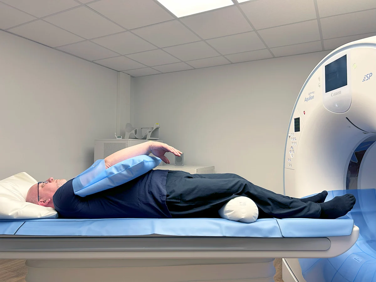
How to Prepare for a CT Scan: Fasting, Contrast & What to Expect
Basahin Pa