Mga Pangunahing Punto
- An OPG (orthopantomogram) is a panoramic X-ray that captures both jaws, all teeth, the TMJ joints, sinuses, and nerve canals in a single 15-second scan with no pain and no sensors inside the mouth
- The scan requires almost no preparation - just remove metal jewelry from the head and neck area; no fasting, injections, or contrast dye are needed
- OPG delivers only 10-20 microsieverts of radiation, equivalent to 1-2 days of natural background radiation and less than a single long-haul flight
- Dentists read OPG results systematically, evaluating teeth, bone levels, jaw joints, sinuses, and nerve canals for cavities, bone loss, impacted teeth, cysts, fractures, and TMJ disorders
- OPG is the essential first-line imaging study for dental implant planning, orthodontic assessment, wisdom tooth evaluation, jaw pain diagnosis, and routine dental screening
- When OPG identifies a finding requiring three-dimensional detail, a CBCT scan provides the supplementary precision needed for surgical planning
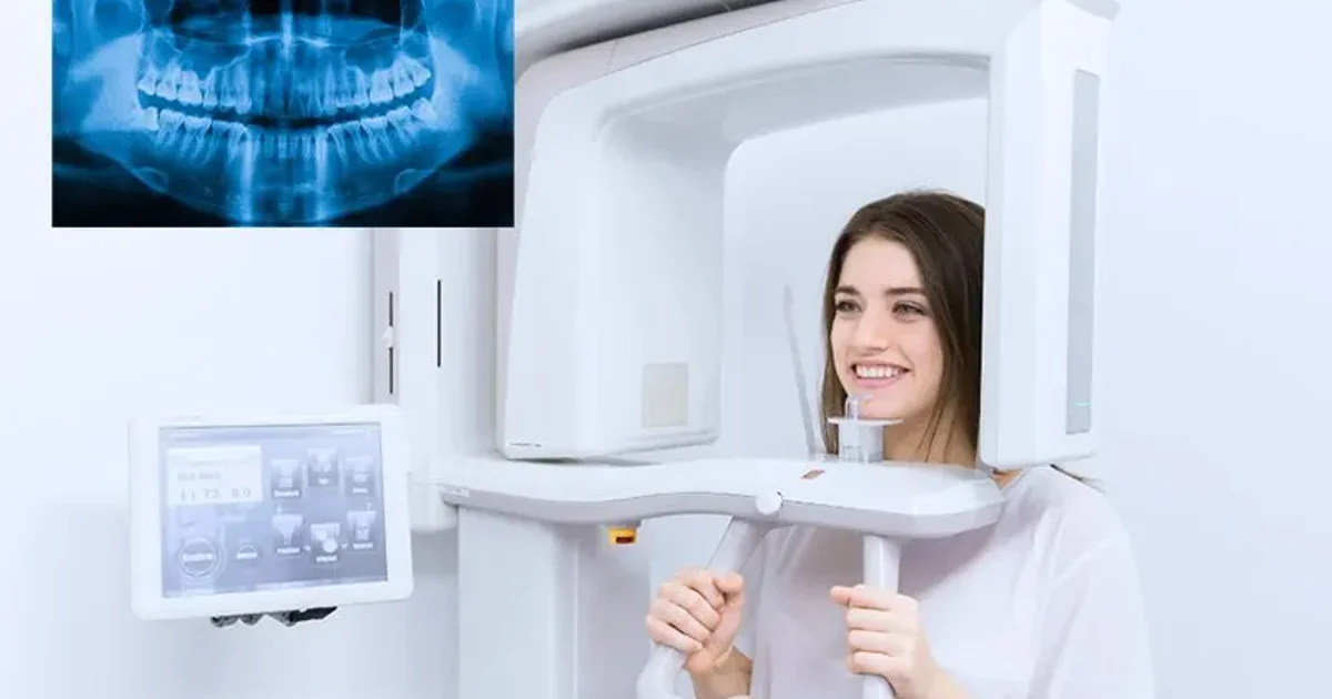
An OPG X-ray (orthopantomogram) is the most widely used extraoral dental imaging technique in the world. In a single painless scan lasting under 15 seconds, it captures a flat, two-dimensional panoramic image of both jaws, all teeth, the temporomandibular joints, maxillary sinuses, and surrounding bone from ear to ear. Whether you need an OPG for dental implants, orthodontic braces, wisdom tooth evaluation, or a routine dental checkup, this comprehensive guide covers everything: what the scan is, how it works, how to prepare, what happens during the procedure, how to understand your results, and how OPG serves each major dental specialty.
This 2026 mega-guide consolidates the complete body of OPG knowledge into a single resource reviewed by Dr. Osama Elzamzami, Consultant Radiologist (MD, FRCR) at Doctors Clinic Diagnostic Center (DCDC) in Dubai Healthcare City. Every section is clinically verified and includes comparison tables, report terminology explanations, patient stories, and specialist guidance to help you navigate your OPG experience from booking to results.
What Is an OPG X-Ray?
OPG stands for orthopantomogram, a compound term derived from the Greek words "ortho" (straight), "pan" (all), and "tomogram" (a sectional image) - collectively meaning a straight, all-encompassing sectional image of the jaws and teeth. In everyday clinical practice the scan is also called a panoramic radiograph, a panoramic X-ray, a pantomogram, or simply a "pano." Regardless of the name, the imaging principle is identical: a single X-ray beam sweeps around the patient's head to produce one continuous flat image that displays both the upper jaw (maxilla) and the lower jaw (mandible) from ear to ear. At DCDC, our digital OPG X-ray service uses modern equipment that produces high-resolution panoramic images at minimal radiation doses.
The concept was first developed by Finnish radiologist Yrjo Veli Paatero in the 1940s, and commercial OPG machines became widely available in dental clinics by the 1960s. Today, digital OPG systems have replaced film-based units in most modern clinics, producing higher-resolution images at lower radiation doses while allowing instant viewing on a computer screen.
How Does an OPG X-Ray Work?
An OPG X-ray works by using a technique called rotational tomography, in which an X-ray tube and a digital detector rotate simultaneously around the patient's head in a coordinated semicircular arc. As the tube and detector sweep from one side of the jaw to the other over approximately 15 to 20 seconds, the system captures a continuous series of narrow X-ray slices. Software then reconstructs these slices into a single panoramic image showing the full mandible, maxilla, teeth, TMJ joints, and adjacent structures.
The key principle is called narrow-beam tomography. Rather than exposing the entire head to radiation at once, the OPG machine uses a thin, focused beam that illuminates only a narrow vertical strip of anatomy at any given moment. This strip sweeps across the jaw as the machine rotates, building up the panoramic image incrementally. The result is a sharp image of the curved jaw structures projected onto a flat plane, with minimal superimposition of structures in front of or behind the focal zone.
Because the X-ray source remains outside the mouth throughout the procedure, the OPG is classified as an extraoral imaging technique. This is a significant advantage over intraoral X-rays, which require a sensor or film to be placed inside the mouth against the teeth. Patients who have a strong gag reflex, limited mouth opening, or dental anxiety consistently find the OPG far more comfortable than intraoral alternatives.
"The OPG is the dental world's most versatile screening tool," explains Dr. Osama Elzamzami, Consultant Radiologist at DCDC. "In a single 15-second scan, we capture both jaws, all teeth, the temporomandibular joints, and the surrounding bone - giving dentists a complete overview that no single intraoral X-ray can provide."
OPG vs Intraoral X-Ray vs CBCT
The simplest way to understand the difference between an OPG and an intraoral X-ray is to think of them as a wide-angle lens versus a macro lens. The OPG sweeps around the outside of your head and produces a single flat image showing both jaws from ear to ear. The intraoral X-ray places a small sensor inside your mouth and photographs just 2-4 teeth at extremely high resolution. A CBCT (cone-beam computed tomography) goes further, producing three-dimensional volumetric images that can be viewed in any plane. Neither type is universally "better" - they serve different diagnostic purposes and are frequently used together.
Intraoral X-rays come in three subtypes. A periapical X-ray captures the entire tooth from crown to root tip plus 2-3 mm of bone beyond the apex, and is the go-to image for root canal assessment and periapical infections. A bitewing X-ray captures the crowns of upper and lower teeth simultaneously and is the gold standard for detecting interproximal cavities. An occlusal X-ray uses a larger sensor placed on the biting surface to capture a broad view of one arch, primarily used in pediatric dentistry.
| Feature | OPG (Panoramic) | Periapical | Bitewing | CBCT |
|---|---|---|---|---|
| Coverage | Both jaws ear to ear, TMJ, sinuses | 2-3 teeth + root apices | Crowns of upper/lower premolars and molars | 3D volume of selected region or both jaws |
| Sensor location | Outside the mouth (extraoral) | Inside the mouth | Inside the mouth | Outside the mouth (extraoral) |
| Detail level | Moderate (broad overview) | Very high (tooth-specific) | Very high (crown and upper root) | Very high (3D cross-sections) |
| Radiation dose | 10-20 μSv | 1-8 μSv per image | 1-5 μSv per image | 30-200 μSv |
| Images for full mouth | 1 image | 14-20 images (full-mouth series) | 4 images (standard series) | 1 scan |
| Scan time | 15-20 seconds | < 1 second per exposure | < 1 second per exposure | 15-30 seconds |
| Patient comfort | Very comfortable; no sensor in mouth | Can be uncomfortable; gag reflex risk | Moderate; bite tab helps stability | Comfortable; no sensor in mouth |
| Best for | Screening, impacted teeth, fractures, orthodontics, implant overview, TMJ | Root canals, periapical infections, root fractures | Detecting early cavities, checking restoration fit | Implant planning, complex surgery, 3D anatomy, surgical guides |
| Approximate Dubai cost (AED) | 150-300 | 50-150 per image | 50-100 per image | 400-1,200 |
Master comparison of OPG panoramic X-ray, intraoral X-ray subtypes, and CBCT. Each serves a distinct diagnostic role. For detailed CBCT comparison, see our guide on <a href="/blog/cbct-vs-opg-xray" class="text-primary-600 hover:underline">CBCT vs OPG</a>.
From a cost-per-coverage perspective, the OPG is significantly more economical when a comprehensive view is needed. A single OPG at AED 150-300 covers both jaws and all teeth, while a full-mouth intraoral series achieving similar coverage costs AED 500-1,200. Most dental professionals use the OPG as a screening tool and then take periapical or bitewing intraoral X-rays of specific areas requiring closer inspection. For detailed pricing, see our guide on OPG X-ray cost in Dubai.
What Does an OPG Show?
An OPG X-ray shows the complete dental and maxillofacial anatomy in one image, enabling radiologists and dentists to identify a wide range of conditions that would otherwise require multiple separate intraoral films. The panoramic view is particularly powerful because it reveals the spatial relationship between structures across the entire jaw, not just one localized area.
Structures and conditions visible on a standard OPG include:
- All erupted teeth in both the upper and lower jaws, including their roots, crowns, and surrounding bone
- Unerupted and impacted teeth, especially wisdom teeth trapped beneath the gum or growing at an angle
- Developing teeth in children and adolescents, allowing dentists to assess eruption patterns and predict crowding
- Tooth decay (caries), particularly large interproximal cavities between teeth
- Periapical pathology such as dental abscesses, granulomas, and root-tip infections
- Bone loss from periodontal (gum) disease, visible as reduced bone height around tooth roots
- Jawbone abnormalities including cysts, tumors, and benign growths such as ameloblastomas or odontomas
- Jaw fractures from trauma affecting the mandible, condyles, or maxilla
- Temporomandibular joints (TMJ) showing signs of arthritis, erosion, condylar flattening, or dislocation
- Maxillary sinuses, revealing sinusitis, polyps, mucous retention cysts, or tooth roots projecting into the sinus floor
- Nasal cavity and nasal septum for gross deviation or obstruction
- Dental restorations such as fillings, crowns, bridges, and implants, and their relationship to adjacent anatomy
- The inferior alveolar nerve canal, critical for implant planning and wisdom tooth extraction
While the OPG provides an excellent overview, its two-dimensional nature means that some fine details - such as very early cavities or hairline root fractures - may require supplementary intraoral X-rays or CBCT imaging for confirmation. The OPG is best understood as a screening tool that identifies areas of concern for further detailed examination.
How to Prepare for an OPG
Preparing for an OPG X-ray is straightforward because the scan requires almost no special preparation. There is no fasting, no blood work, no injections, no contrast dye, and no recovery time. The single most important step is removing all metal objects from the head and neck area before the machine rotates around your jaw.
"The most common question patients ask us is whether they need to prepare anything before coming in for an OPG," says Dr. Osama Elzamzami. "The answer is refreshingly simple: remove your metal, stand still for 15 seconds, and the scan is done."
| Item to Remove | Why It Must Be Removed |
|---|---|
| Earrings (studs, hoops, ear cuffs) | Metal earrings sit directly in the scan path and produce bright white spots that can obscure the TMJ, condyle, and ramus of the mandible |
| Necklaces and chains | Necklaces drape across the cervical spine and lower jaw area, creating linear artifacts that can mimic or hide fractures |
| Facial and oral piercings (lip, tongue, nose, eyebrow) | Piercings inside or near the mouth create focal white artifacts that obscure teeth, bone, and soft tissue |
| Glasses and sunglasses | Metal frames and hinges project over the midface and sinuses, blocking the view of the upper jaw and nasal cavity |
| Hearing aids | Hearing aids contain metal components and batteries that produce dense artifacts overlapping the ear canal, TMJ, and posterior mandible |
| Removable dentures and retainers | Dentures contain metal clasps or dense acrylic that superimpose on natural teeth and bone, making evaluation impossible |
| Hairpins, clips, and hair ties with metal | Metal hair accessories create scattered artifacts across the image, particularly over the posterior skull and upper jaw |
| Head scarves with metal pins or embellishments | Metal pins generate pinpoint artifacts; the fabric itself can usually remain if all metal is removed |
Remove all metallic items from the head, neck, and upper chest before your OPG scan. Fixed dental work (crowns, bridges, braces, implants) does not need to be removed.
Eating, Drinking, and Medications
You can eat and drink completely normally before an OPG X-ray. There is no fasting requirement whatsoever. Take all regular medications as prescribed. No medication needs to be stopped or adjusted. If you take medication that causes dry mouth, bring a bottle of water to sip before positioning - this will make the bite block more comfortable.
Pregnancy and OPG Preparation
If you are pregnant or suspect you may be pregnant, inform the radiographer before the scan. Elective dental imaging is routinely postponed during pregnancy as a precautionary measure, especially during the first trimester. However, if there is a clinical emergency (such as a severe infection, abscess, or jaw fracture), an OPG can be performed safely with a lead apron shielding the abdomen. The radiation dose from an OPG (10-20 μSv) is directed at the jaw, far from the uterus, and is thousands of times below the threshold for fetal harm.
What to Tell the Radiographer
- Pregnancy or possible pregnancy - the team will decide whether to proceed, postpone, or apply additional shielding
- The reason for the scan - this helps the radiologist focus the report on what matters most (e.g., wisdom teeth, implant planning, orthodontic records)
- Previous jaw surgery or trauma - surgical hardware appears on the OPG and needs to be distinguished from pathology
- Difficulty standing still - the machine or patient support may be adjusted for movement disorders or chronic pain
- Previous dental imaging - bringing prior images allows the radiologist to compare and detect subtle changes over time
Practical tip: If you know you have an OPG appointment, consider leaving jewelry at home and wearing a simple top without a high metal zipper or collar clasp. This saves time at the clinic and eliminates the worry of misplacing valuables.
What Happens During an OPG
The complete OPG X-ray procedure takes under 5 minutes from the moment you enter the X-ray room to the moment you leave, with the actual scan occupying only 15 to 20 seconds. There is no preparation, no recovery, and no side effects afterward.
- Step 1 - Arrival and registration: Check in at reception with your Emirates ID or passport and referral letter (if applicable). Self-pay patients at DCDC can walk in without a referral. Registration takes 2-3 minutes.
- Step 2 - Remove metal objects: The radiographer asks you to remove all metallic items from the head and neck area. A small tray is provided for your belongings. This takes about 30 seconds.
- Step 3 - Wear protective shielding: A lead thyroid collar and/or lead apron is placed on you to shield the thyroid gland and body from scattered radiation, following the ALARA principle (As Low As Reasonably Achievable).
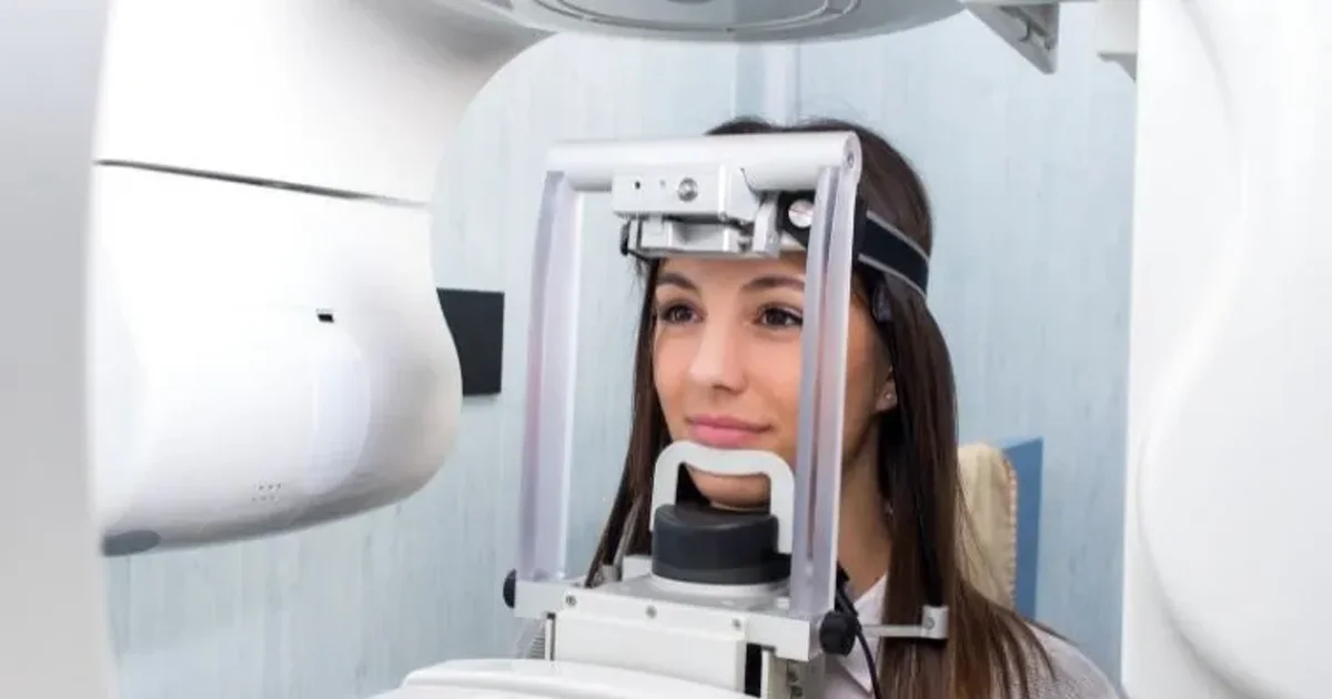
- Step 4 - Position in the machine: You step up to the OPG unit and rest your chin on a small chin rest. The radiographer adjusts the machine height and uses light-beam guides projected onto your face to align your head precisely. Two lateral head supports gently press against your temples to prevent movement. Correct positioning is the single most important factor for a high-quality image.
- Step 5 - Bite on the positioning guide: You gently bite down on a sterile plastic bite block that keeps your front teeth slightly apart and your jaws in the correct alignment. You close your lips around the block and press your tongue flat against the roof of your mouth - this prevents the tongue from creating a shadow that could obscure tooth roots.
- Step 6 - The scan: The radiographer steps behind a protective screen and activates the machine. The C-shaped arm begins rotating slowly around your head from one ear to the other. You hear a low humming sound. The scan takes 15 to 20 seconds. You must remain completely still and avoid swallowing, as any movement can blur the image.
- Step 7 - Scan complete: The machine stops. You step away. The digital image appears on the workstation within seconds. The radiographer performs a quick quality check. If satisfactory - which it is in the vast majority of cases - you are free to leave immediately. Total in-room time: 3 to 5 minutes.
There is no recovery time after an OPG. You can eat, drink, drive, and resume all normal activities immediately. The scan is completely painless - there are no needles, no injections, no contrast dye, and no sensors placed inside the mouth. You do not feel the X-rays passing through your body. Many patients report the OPG is significantly more comfortable than standard intraoral dental X-rays.
OPG for Children
Children as young as 5 years old can have an OPG, provided they can stand still for 15 to 20 seconds and follow simple instructions. Modern digital OPG machines include automatic pediatric dose-reduction protocols that lower the radiation output based on the child's size. A pediatric OPG delivers approximately 5 to 14 μSv - less than one day of natural background radiation. At DCDC, the radiographer takes extra time to explain the process in child-friendly language, and parents are welcome to stand in the room behind a protective screen.
Seven-year-old Youssef came to DCDC with his mother for his first-ever OPG scan, referred by his orthodontist. His mother was concerned about radiation and whether her son would be frightened. The radiographer spent a few minutes showing Youssef the machine, letting him touch the chin rest, and explaining he just needed to "stand like a statue." Youssef bit down on the positioning guide, stood perfectly still for 16 seconds, and the scan was done. "That was it?" he asked, looking surprised. His mother was relieved to learn the radiation dose was less than what Youssef would receive on a flight to visit his grandparents. The OPG revealed two permanent teeth developing at unusual angles, giving the orthodontist the information needed to plan early intervention.
Digital OPG X-Ray at DCDC Dubai Healthcare City
Get a high-resolution digital OPG scan with same-day radiologist reporting at Doctors Clinic Diagnostic Center. Walk-ins welcome. Located in Dubai Healthcare City, serving patients from Oud Metha, Umm Hurair 2, Karama, and across Dubai.
No referral required for self-pay patients
Reading Your OPG Results
Dentists and radiologists do not simply glance at an OPG and draw conclusions. They follow a structured, systematic approach that ensures every anatomical region is evaluated and no finding is overlooked. This disciplined reading method is what separates a thorough radiological assessment from a cursory look.
The Systematic Reading Approach
- Overall image quality check: The radiologist first confirms the OPG is properly exposed, correctly positioned, and free from motion blur or artifacts. A poor-quality image may need to be retaken.
- Symmetry assessment: Left and right sides should appear roughly symmetrical. Asymmetry in the ramus, condyles, or bone density may indicate pathology such as a tumor, fracture, or developmental abnormality.
- Teeth evaluation (quadrant by quadrant): Each tooth is examined individually across all four quadrants - upper right, upper left, lower left, lower right. Crown, root, and surrounding bone are assessed for cavities, fractures, root resorption, periapical lesions, and quality of existing restorations.
- Bone level assessment: Alveolar bone height around every tooth is evaluated. In health, bone reaches to within 1-2 mm of the cementoenamel junction. Reduced height indicates periodontal disease; the pattern (horizontal vs. vertical) guides treatment planning.
- TMJ evaluation: Both temporomandibular joints are examined for condylar shape, size, and symmetry. Flattening, erosion, osteophytes, or sclerosis suggest degenerative disease, arthritis, or trauma.
- Maxillary sinus review: The sinuses are checked for opacification, mucosal thickening, polyps, retention cysts, and proximity of upper tooth roots to the sinus floor.
- Nerve canal tracing: The inferior alveolar nerve canal is traced from the mandibular foramen to the mental foramen to assess its relationship to lower tooth roots, particularly wisdom teeth.
- Pathology identification: Any abnormal radiolucencies (dark areas) or radiopacities (bright white areas) are documented, measured, and correlated with symptoms.
This systematic reading typically takes a trained radiologist three to five minutes. At DCDC, every OPG is read by a consultant radiologist, not a technician, ensuring the highest standard of diagnostic accuracy.
"I follow the same checklist for every single OPG I read," says Dr. Osama Elzamzami. "Systematic reading is non-negotiable. If you jump straight to the area the patient is complaining about, you risk missing an incidental finding somewhere else that could be far more significant."
Common Findings on OPG X-Rays
| Finding | Appearance on OPG | What It Means |
|---|---|---|
| Dental cavities (caries) | Dark areas within the tooth crown or between adjacent teeth | Tooth decay; treatment ranges from a filling to root canal or extraction depending on extent |
| Bone loss (periodontal disease) | Reduced bone height around tooth roots | Gum disease causing bone recession; horizontal = generalized chronic periodontitis; vertical = localized defect possibly needing surgery |
| Impacted teeth | Tooth partially or fully trapped beneath gum or bone, often tilted | Most common with wisdom teeth; can cause pain, infection, damage to adjacent teeth, or cyst formation |
| Periapical pathology (abscess/granuloma) | Dark area at the tip of a tooth root | Infection spread from the tooth nerve into bone; requires root canal therapy or extraction |
| Cysts | Well-defined round/oval dark area within jawbone, often with thin white border | Fluid-filled sac (e.g., dentigerous cyst around impacted tooth); surgical removal usually required |
| TMJ abnormalities | Flattening, erosion, bone spurs, or asymmetry of condylar heads | Degenerative jaw joint changes causing clicking, locking, or pain; treatment from bite splints to surgery |
| Root resorption | Shortened or irregularly shaped root with blunted appearance | Root dissolving due to orthodontic pressure, trauma, impacted teeth, or unknown causes |
| Sinus abnormalities | Opacification or thickened lining within the maxillary sinus | Sinusitis, retention cysts, or tooth root projecting into sinus; dental causes must be ruled out |
Common OPG findings, their radiographic appearance, and clinical significance. Your radiologist documents all relevant findings in the formal written report.
Normal vs Abnormal OPG Results
A normal OPG displays: complete intact teeth with no dark spots, healthy fully formed tapered roots, even alveolar bone levels reaching to within 1-2 mm of the cementoenamel junction, symmetrical condyles with smooth rounded surfaces, clear air-filled sinuses with thin uniform lining, a visible inferior alveolar nerve canal with adequate distance from tooth roots, and no unexplained dark or bright white areas within the bone.
Abnormal findings include: dark areas (radiolucencies) within the jawbone suggesting cysts, tumors, or bone destruction; bright white masses (radiopacities) suggesting calcified tumors or foreign bodies; significant bone loss around multiple teeth; root shortening or irregularity; condylar asymmetry; widened periodontal ligament spaces; and opacified sinuses. It is important to note that "abnormal" does not always mean "dangerous" - many findings such as small mucous retention cysts in the sinus are incidental and require no treatment.
Understanding OPG Report Terminology
OPG reports use standardized medical terminology that can be confusing for patients. The table below translates the most common terms into plain language.
| Report Term | Plain Language Meaning |
|---|---|
| Radiolucent / Radiolucency | Dark area on the X-ray where the beam passed through easily - usually indicates bone destruction, cyst, abscess, or tumor |
| Radiopaque / Radiopacity | Bright white area that blocked the X-ray beam - metal fillings, implants, dense bone, or calcified tumors |
| Periapical pathology | Disease at the tip of a tooth root; dark area at root end typically means infection has spread from the pulp into bone |
| Resorption (root resorption) | Tooth root dissolving - appears as shortening, thinning, or irregular root contour |
| Horizontal bone loss | Uniform reduction in bone height across multiple teeth from chronic gum disease |
| Vertical (angular) bone loss | Localized V-shaped bone defect along one side of a tooth root; often requires targeted periodontal treatment |
| Impaction | Tooth that failed to erupt into its normal position - classified as mesioangular, distoangular, horizontal, or vertical |
| Well-defined vs ill-defined margins | Border of a lesion: well-defined (sharp) usually benign/slow-growing; ill-defined (fuzzy) raises concern for aggressive process |
| Osteosclerosis | Area of increased bone density appearing brighter white; usually benign and incidental, often near lower premolar or molar root tips |
| Pericoronitis | Infection of soft tissue around a partially erupted tooth, typically a wisdom tooth; recurrence often indicates extraction is needed |
| Dentigerous cyst | Fluid-filled sac around the crown of an unerupted tooth; appears as dark halo larger than 2.5 mm around the tooth on OPG |
Common OPG report terminology decoded. If your report contains a term not listed here, ask your dentist or radiologist to explain it during your consultation.
A 42-year-old woman visited DCDC for a routine dental checkup with no complaints. Her OPG revealed a well-defined radiolucent area measuring 2 cm surrounding the crown of an impacted lower premolar she never knew existed. The finding was consistent with a dentigerous cyst. A CBCT scan confirmed the diagnosis, and the patient was referred to an oral surgeon for elective removal before the cyst could expand. "This is exactly why we advocate for comprehensive panoramic imaging even in asymptomatic patients," says Dr. Elzamzami. "The most dangerous conditions are often the ones that cause no pain."
OPG for Dental Implants
The OPG is the standard first-line screening X-ray for dental implant candidates. Before a single implant can be placed, the surgeon needs answers to fundamental questions: Is there enough bone? Where is the nerve canal? How close is the sinus floor? Are neighboring teeth healthy? The OPG answers all of these in a single, rapid, low-cost scan - which is why it has been the universally accepted starting point for implant assessment for decades.
What OPG Reveals for Implant Assessment
- Vertical bone height: Standard dental implants range from 8-13 mm in length. The OPG displays the full alveolar ridge height from crest to the mandibular border or sinus floor, allowing a reliable initial measurement (with ~20-30% magnification correction).
- Inferior alveolar nerve canal position: The OPG shows the nerve canal as a radiolucent band through the lower jaw. Damaging this nerve during placement causes permanent numbness. If proximity is close on OPG, a CBCT is ordered for exact 3D measurement.
- Maxillary sinus floor position: The sinus can gradually expand downward after tooth loss (pneumatization), reducing available bone. The OPG shows whether a sinus lift procedure will be needed before upper posterior implants.
- Condition of adjacent teeth: The OPG reveals decay, periapical pathology, periodontal bone loss, and failed restorations near the implant site that must be addressed first.
- Existing pathology: Cysts, tumors, retained root fragments, and active infections at or near the planned site must be managed before surgery.
- Overall jaw architecture: For patients needing multiple implants or full-arch rehabilitation, the OPG provides a global view of ridge contour and bone resorption patterns.
OPG Limitations for Implants and When to Upgrade to CBCT
The OPG cannot show bone width (buccolingual dimension), bone density (Hounsfield units), or the exact 3D nerve position (buccal vs. lingual). These limitations mean the OPG functions as a triage tool: it separates straightforward cases from complex ones. Scenarios that almost always require a follow-up CBCT scan after the initial OPG include:
- Posterior mandibular implants near the nerve canal - when available bone height above the nerve appears less than 12-15 mm on OPG
- Upper posterior implants near the sinus - limited bone between ridge crest and sinus floor
- Narrow or atrophic ridges - significant ridge resorption visible on OPG suggesting bone grafting may be needed
- Multiple implants or full-arch cases - requiring precise 3D planning and possibly surgical guide fabrication
- Guided implant surgery - surgical guides are manufactured from CBCT data, not OPG
- Previous failed implants - CBCT evaluates the cause and guides revised planning
"I start every implant case with an OPG," says Dr. Elzamzami. "It is the fastest way to see the complete picture. Within seconds I can determine whether the patient is a clear candidate, whether they need bone grafting, or whether a CBCT is required for detailed surgical planning. Skipping the OPG and going straight to CBCT is like reading chapter five of a textbook without reading the introduction."
A 45-year-old patient visited DCDC for implant evaluation after losing two lower molars. The initial OPG immediately revealed adequate bone height above the nerve canal on the right side but borderline bone on the left. A targeted CBCT of the left mandible confirmed sufficient width for a standard implant on the right but identified a narrow ridge on the left requiring bone grafting first. "Without the OPG as the first step, we would not have known where to focus the CBCT," Dr. Elzamzami explains. "The OPG flagged the problem side, and the CBCT gave us millimeter-level detail to plan the graft and implant together."
OPG for Orthodontics
The OPG is the first imaging study every orthodontist requests before recommending braces, clear aligners, or any form of tooth movement. Orthodontic treatment is fundamentally about moving teeth through bone, and without the diagnostic information an OPG provides, no responsible orthodontist will begin treatment. The panoramic image answers critical questions: Are all permanent teeth present? Are any impacted or blocked? Are roots healthy enough to withstand orthodontic forces? Is there adequate bone support? Are the TMJ joints structurally normal?
What the Orthodontist Evaluates on Your OPG
| Structure / Condition | What the OPG Reveals | Why It Matters for Orthodontics |
|---|---|---|
| Erupted teeth | Position, alignment, and spacing in both arches | Confirms clinical findings; helps plan bracket placement and wire sequencing |
| Unerupted / impacted teeth | Location, angulation, and depth within bone (especially canines and wisdom teeth) | Impacted canines may need surgical exposure; impacted wisdom teeth may need extraction |
| Missing teeth (congenitally absent) | Absence of tooth buds for expected permanent teeth | Changes treatment goal - close the gap orthodontically or hold it open for a future implant |
| Supernumerary teeth | Extra teeth (mesiodens, paramolars) blocking normal eruption | Must be extracted before orthodontic alignment can proceed |
| Root length and morphology | Length, shape, and integrity of each tooth root | Short or dilacerated roots are at higher risk of resorption during tooth movement; forces must be reduced |
| Bone levels | Height of bone support around each tooth | Reduced bone limits safe tooth movement; periodontal treatment may be needed first |
| Jaw symmetry | Relative size and shape of left and right mandibular rami and condyles | Asymmetry may indicate skeletal problem requiring surgical correction |
| TMJ joints | Shape, surface, and position of condylar heads | Pre-existing degeneration or resorption can worsen during orthodontics if not identified early |
| Previous dental work | Crowns, bridges, root canals, posts, implants | Brackets cannot be bonded to implants; bridgework may need sectioning; root-treated teeth respond differently to force |
An OPG provides the orthodontist with a comprehensive diagnostic overview that no clinical examination alone can match.
OPG for Children's Orthodontic Assessment
The American Association of Orthodontists recommends an orthodontic evaluation by age 7. At this age, children are in the mixed dentition stage, and the OPG reveals developing tooth buds not yet erupted, allowing the orthodontist to predict crowding, identify ectopic canines, detect congenitally missing teeth, and spot supernumerary teeth years before the gap or obstruction becomes clinically visible. Early detection enables interceptive orthodontics that can reduce the complexity and duration of full braces in the teenage years.
OPG for Adult Orthodontics
Adult orthodontics presents unique challenges that make the pre-treatment OPG even more critical. Adults are more likely to have bone loss from periodontal disease, root-canal-treated teeth with compromised root structure, extensive dental restorations, and early TMJ degeneration. The OPG screens for all of these factors. For patients considering clear aligners such as Invisalign, the OPG is equally essential - while aligner companies use intraoral scans for tray fabrication, the orthodontist still needs the panoramic radiograph to evaluate root health, bone levels, and hidden pathology beneath the gum line.
The OPG is commonly combined with a lateral cephalometric X-ray for complete orthodontic records. The cephalometric image measures skeletal relationships (jaw sizes, angles, incisor inclination) that determine whether malocclusion is dental, skeletal, or both. At DCDC, both images can be taken in the same visit, often using the same machine.
A 13-year-old patient was referred to DCDC for an OPG before starting braces. The scan revealed both upper canines were impacted and migrating toward the roots of the lateral incisors - completely invisible on clinical examination because the baby canines were still in place. "Without the OPG, the orthodontist would have begun aligning visible teeth while the impacted canines continued resorbing adjacent roots," explains Dr. Elzamzami. "The OPG changed the entire treatment plan. The oral surgeon exposed both canines first, and the orthodontist guided them into the arch over 18 months."
OPG for Wisdom Teeth
The OPG is the standard imaging study for evaluating wisdom teeth (third molars) before deciding whether extraction is necessary. It is the only single-image dental X-ray that shows all four third molars, both jaws, the inferior alveolar nerve canals, and the maxillary sinuses simultaneously. International guidelines from the American Association of Oral and Maxillofacial Surgeons (AAOMS) and the Royal College of Surgeons of England both recommend the OPG as the first-line imaging modality for third molar assessment.
Wisdom Tooth Impaction Classification
The OPG clearly shows the angulation of each wisdom tooth relative to the adjacent second molar, which is the basis for classifying impaction type and predicting extraction difficulty.
| Impaction Type | OPG Appearance | Frequency | Extraction Difficulty | Key Finding |
|---|---|---|---|---|
| Mesioangular | Tilted forward toward second molar | 40-45% | Moderate | Crown pressing on distal surface of second molar; check for caries on adjacent tooth |
| Vertical | Upright but unerupted or partially erupted | 25-30% | Moderate to difficult | Tooth in correct axis but blocked; check follicle size for cyst development |
| Horizontal | Lying on its side, perpendicular to second molar | 10-15% | Difficult | Crown directed into second molar roots; assess for root resorption |
| Distoangular | Tilted backward toward ramus | 5-10% | Most difficult (lower jaw) | Crown pointing into ramus; limited access for extraction path |
| Buccal/Lingual (transverse) | Tooth displaced toward cheek or tongue | Rare | Variable | OPG may underestimate displacement; CBCT often needed |
Classification of wisdom tooth impaction based on OPG. Mesioangular is most common; distoangular is most surgically challenging in the mandible.
Nerve Proximity Signs on OPG
The inferior alveolar nerve canal appears as two parallel white lines on the OPG. When wisdom tooth roots appear to touch, overlap, or cross the canal, the risk of nerve injury during extraction increases. Specific high-risk signs that trigger a CBCT follow-up include:
- Darkening of the root at the canal crossing - suggests the canal may groove into or pass through the root
- Interruption or narrowing of the white canal lines - one of the most reliable OPG predictors of nerve proximity
- Deflection of the root where it meets the canal - may indicate the root has grown around the nerve
- Narrowing of the canal at the level of the wisdom tooth - suggests physical displacement of the nerve
- Diversion of the canal (course change) at the wisdom tooth level - strong indicator of intimate tooth-nerve relationship
- Superimposition of root apex on the canal - the 2D image cannot determine whether the nerve passes buccal, lingual, or through the root
OPG Signs That Wisdom Teeth Need Removal
- Recurrent pericoronitis: Partially erupted tooth with soft tissue pocket prone to infection; two or more episodes typically warrant extraction
- Caries on wisdom tooth or adjacent second molar: Food trapping between the two teeth causes decay; removing the wisdom tooth protects the more valuable second molar
- Root resorption of the second molar: Impacted wisdom tooth pressing against and dissolving the second molar root - a serious finding requiring prompt extraction
- Dentigerous cyst formation: Dark area surrounding the crown larger than 2.5 mm; can expand and weaken the jawbone if untreated
- Bone loss on distal surface of second molar: Periodontal defect caused by the impacted wisdom tooth compromising the second molar's long-term prognosis
- Orthodontic interference: Wisdom teeth likely to cause crowding or relapse after braces
A 22-year-old patient visited DCDC with recurring swelling behind the lower left molars. The clinical examination showed partially erupted soft tissue, but the full extent of the problem was not visible. An OPG immediately revealed a mesioangularly impacted wisdom tooth with a large radiolucent area around its crown consistent with a dentigerous cyst, and early root resorption of the adjacent second molar. "What looked like simple pericoronitis clinically turned out to be a cyst that had already started damaging the neighboring tooth," says Dr. Elzamzami. "The patient was referred for extraction within the week, and the second molar was saved."
Dental X-Ray Safety
If you have ever hesitated before a dental X-ray and wondered whether the radiation is truly safe, you are not alone. The short, definitive answer is: yes, dental X-rays are safe. The radiation dose from a standard OPG is approximately 10 to 20 microsieverts (μSv) - equivalent to roughly 1 to 2 days of natural background radiation that every person receives simply from living on Earth. For context, you absorb more radiation during a single long-haul flight from Dubai to London than you do from an OPG scan.
Radiation Dose Comparison
| Radiation Source | Approximate Dose (μSv) | Equivalent in OPG Scans |
|---|---|---|
| Eating 1 banana | 0.1 | 1/140th of an OPG |
| Single periapical dental X-ray | 1-8 | < 1 OPG |
| Single bitewing dental X-ray | 1-5 | < 1 OPG |
| One day of natural background radiation | 6.5 | About half an OPG |
| OPG (panoramic dental X-ray) | 10-20 | 1 OPG |
| Chest X-ray | 20 | ~1 OPG |
| Full-mouth intraoral series (14-20 films) | 35-170 | 2-12 OPGs |
| Dubai-to-London flight (cosmic radiation) | 40-80 | 3-6 OPGs |
| CBCT (dental cone beam) | 30-200 | 2-14 OPGs |
| Living in a concrete building for 1 year | 70 | 5 OPGs |
| Annual dose from food (potassium-40, carbon-14) | 300 | 21 OPGs |
| CT scan of the head | 2,000 | ~140 OPGs |
| Total average annual background radiation | 2,400 | ~170 OPGs |
Dental X-rays sit at the very bottom of the medical radiation spectrum. A single OPG delivers less radiation than a long-haul flight.
OPG Safety During Pregnancy
The American College of Obstetricians and Gynecologists (ACOG) and the American Dental Association (ADA) both state that dental X-rays can be performed safely during pregnancy when clinically indicated, with appropriate shielding. The radiation dose from an OPG (10-20 μSv) is far below the threshold for fetal harm - studies indicate at least 50,000 μSv to the uterus would be needed before any measurable risk. Scattered radiation reaching the uterus from a dental X-ray is estimated at less than 0.01 μSv. Elective imaging is typically postponed until after delivery as a precaution, but urgent diagnostic imaging should not be delayed - untreated dental infections can pose a greater risk to both mother and baby than the X-ray itself.
"The biggest risk during pregnancy is not the X-ray - it is the untreated dental infection that the X-ray would have diagnosed," explains Dr. Elzamzami. "I have seen cases where a pregnant patient delayed a necessary X-ray out of radiation fear, and the underlying abscess progressed to a systemic infection requiring emergency hospitalization."
OPG Safety for Children
Dental X-rays are safe for children when performed with appropriate technique and clinical justification. Modern digital machines include automatic pediatric dose-reduction protocols that lower radiation output by 30-50% based on the child's size. A pediatric OPG delivers approximately 5 to 14 μSv. All imaging follows the ALARA principle (As Low As Reasonably Achievable): X-rays are taken only when clinically necessary, the field of view is restricted, and every image uses proper shielding.
Lead Aprons and Thyroid Shields
In 2023, the American Dental Association updated its position statement to note that lead aprons and thyroid collars provide minimal additional protection during dental X-rays when modern digital equipment with rectangular collimation is used. The American Association of Physicists in Medicine similarly recommended discontinuing routine patient shielding for diagnostic X-rays. Modern machines are so precisely collimated that virtually no scattered radiation reaches the torso or thyroid. Many clinics, including DCDC, still offer lead shielding for patient comfort and reassurance, and it remains standard for pregnant and pediatric patients.
How Often Should You Get Dental X-Rays?
- New patients (all ages): A baseline set of X-rays (typically OPG and/or full-mouth series) at the initial visit
- Low caries risk adults: Bitewing X-rays every 24-36 months; OPG every 3-5 years or as needed
- High caries risk adults: Bitewing X-rays every 6-18 months; additional imaging as indicated
- Children with baby teeth: Bitewing X-rays every 12-24 months; OPG when permanent tooth development assessment is needed
- Adolescents: Bitewing X-rays every 18-36 months; OPG for orthodontic assessment or wisdom tooth evaluation, typically once between ages 16-20
- Periodontal disease patients: Periapical and/or bitewing X-rays every 12-24 months as determined by disease severity
Even for high-risk patients, the cumulative annual dose from dental X-rays remains extremely low. A patient receiving four bitewings and one OPG in a year accumulates approximately 30-40 μSv total - less than one week of natural background radiation.
Safe, Low-Dose Dental X-Rays at DCDC
DCDC uses the latest digital OPG and digital X-ray systems with automatic dose-reduction protocols for children and adults. Every scan is reported by a consultant radiologist. Walk-ins welcome at DCDC Dubai Healthcare City.
No referral required for self-pay patients
OPG X-Ray at DCDC Dubai Healthcare City
Doctors Clinic Diagnostic Center (DCDC) provides comprehensive digital OPG X-ray services at its facility in Dubai Healthcare City, combining advanced imaging technology with expert radiologist interpretation to deliver fast, accurate results for patients and referring clinicians.
- Complete imaging under one roof: OPG, CBCT, periapical X-rays, cephalometric imaging, CT, MRI, and ultrasound are all available on-site. Patients complete their full diagnostic workup in a single visit without traveling between facilities.
- Latest digital OPG technology: DCDC uses state-of-the-art digital panoramic systems that produce sharper images at lower radiation doses than older film-based machines. Images are available within seconds of the scan.
- Pediatric dose protocols: The OPG machine includes automatic dose-reduction settings for children, ensuring the lowest possible dose while maintaining diagnostic quality.
- Same-day consultant radiologist reporting: Every OPG at DCDC is reviewed and reported by a consultant radiologist specializing in dental and maxillofacial imaging - not a technician or general practitioner. Reports are typically completed the same day.
- Walk-in availability: While appointments are available, DCDC accepts walk-in patients for OPG scans throughout clinic hours. No referral is required for self-pay patients.
- Insurance accepted: DCDC works with major insurance providers in Dubai. Patients with insurance may need a referral from their dentist or GP to activate coverage.
- Convenient DHCC location: Located in Dubai Healthcare City, easily accessible from Oud Metha, Umm Hurair 2, Karama, Bur Dubai, and the wider Dubai metro area. Free parking and metro access available nearby.
Whether you need an OPG for wisdom tooth evaluation, orthodontic planning, implant assessment, jaw pain investigation, or a routine dental screening, DCDC provides a seamless experience from registration through to report delivery. For pricing details, see our guide on OPG X-ray cost in Dubai.
Book Your OPG Scan Today
Walk in or book ahead for a digital OPG X-ray at DCDC Dubai Healthcare City. Same-day results from a consultant radiologist. Call or WhatsApp to schedule.
Mga Madalas Itanong
Final Thoughts
The OPG X-ray remains one of the most valuable and versatile tools in dental and maxillofacial imaging. In less than 15 seconds, it provides a comprehensive panoramic view that helps dentists, orthodontists, implant surgeons, and oral surgeons diagnose conditions ranging from impacted wisdom teeth and bone loss to jaw fractures and TMJ disorders. With very low radiation (10-20 μSv), zero pain, and no special preparation required, it is also one of the most patient-friendly imaging studies available. For every major dental specialty - implant planning, orthodontic assessment, wisdom tooth evaluation, and routine screening - the OPG is the essential first step that determines the entire diagnostic and treatment pathway.
Whether your dentist has recommended an OPG, you are starting orthodontic treatment, planning dental implants, evaluating wisdom teeth, or simply need a comprehensive dental screening, Doctors Clinic Diagnostic Center in Dubai Healthcare City offers digital OPG scans with same-day consultant radiologist reporting and walk-in availability. All follow-up imaging including CBCT, CT, and MRI is available on-site. For detailed pricing, visit our guide on OPG X-ray cost in Dubai. Explore our full range of OPG X-ray services or contact us to book your scan today.
Mga Sanggunian at Reperensya
Ang artikulong ito ay sinuri ng aming medikal na team at tumutukoy sa mga sumusunod na sanggunian:
- American Dental Association - Dental Radiographic Examinations: Recommendations for Patient Selection and Limiting Radiation Exposure
- European Commission - Radiation Protection 136: European Guidelines on Radiation Protection in Dental Radiology
- National Council on Radiation Protection and Measurements (NCRP) - Report No. 177: Radiation Protection in Dentistry and Oral & Maxillofacial Imaging
- American Association of Oral and Maxillofacial Surgeons - White Paper on Third Molar Data
- American Association of Orthodontists - Clinical Practice Guidelines for Orthodontic Radiography
- European Association for Osseointegration (EAO) - Consensus Statements on Radiographic Examination for Implant Patients
- RadiologyInfo.org - Panoramic Dental X-ray (Orthopantomogram)
- American College of Obstetricians and Gynecologists - Guidelines for Diagnostic Imaging During Pregnancy and Lactation
Ang medikal na nilalaman sa site na ito ay sinusuri ng mga DHA-licensed na manggagamot. Tingnan ang aming patakarang editorial para sa higit pang impormasyon.

Isinulat ni
Dr. Osama Elzamzami
Diagnostic Radiology
MD, FRCR
Dr. Osama Elzamzami is Head of Radiology at DCDC Dubai Healthcare City, specializing in diagnostic imaging including X-ray, CT, MRI, OPG, CBCT, and ultrasound. He holds an MD and FRCR (Fellowship of the Royal College of Radiologists) and provides expert reporting for dental, maxillofacial, and general diagnostic imaging studies with a focus on patient safety and evidence-based practice.
Related Articles

OPG X-Ray Cost in Dubai: Updated Prices & Insurance Guide
CBCT vs OPG X-Ray: Which Dental Imaging Do You Need?
Kaugnay na Serbisyo sa DCDC
Dalubhasang pangangalaga at advanced diagnostics sa Dubai Healthcare City