Wichtigste Erkenntnisse
- An ultrasound scan uses high-frequency sound waves to create real-time images of organs, tissues, and blood vessels — it involves zero ionizing radiation, making it safe for all ages including pregnant women and children
- More than 10 types of ultrasound exist — abdominal, pelvic, thyroid, breast, pregnancy (2D/3D/4D), musculoskeletal, Doppler vascular, renal, scrotal, and echocardiography — each with specific preparation requirements
- Preparation varies by type: abdominal scans require 6-8 hours fasting, pelvic scans need a full bladder, while thyroid and musculoskeletal scans need no preparation at all
- Ultrasound has been used safely for 60+ years with no confirmed harmful effects — the WHO, ACOG, and AIUM all endorse it as the safest imaging modality
- The ultrasound cost in Dubai ranges from AED 300 to AED 800 depending on scan type, with most insurance plans covering the scan with a copay of AED 0-50
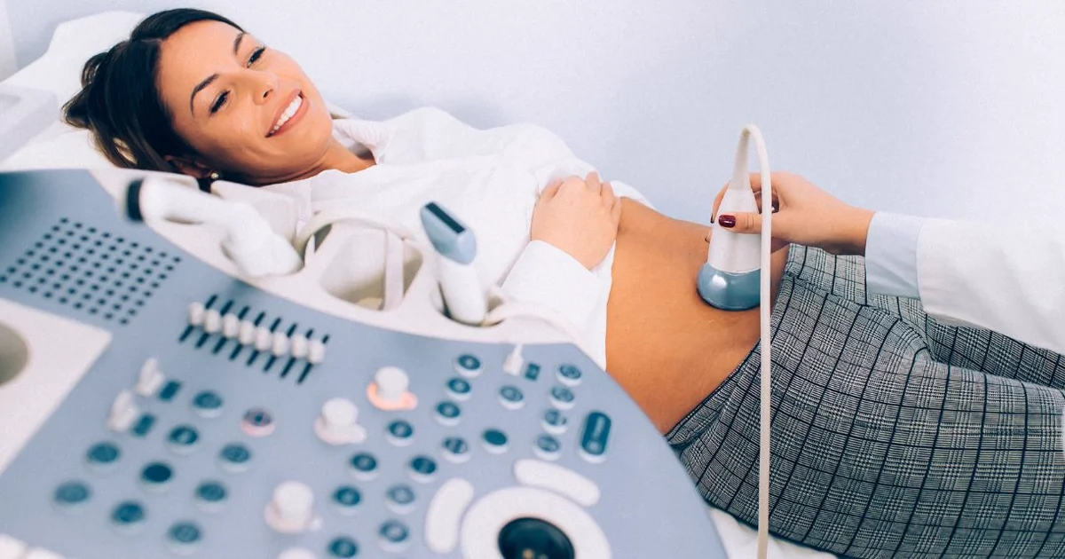
An ultrasound scan (sonography) is a diagnostic imaging procedure that uses high-frequency sound waves to create real-time images of organs, tissues, and blood vessels inside your body. Unlike X-rays or CT scans, ultrasound involves zero ionizing radiation, making it one of the safest medical imaging tools available for patients of all ages — including pregnant women, newborns, and children. The ultrasound cost in Dubai ranges from AED 300 to AED 800 depending on the type of scan, with most Dubai insurance plans covering the examination with a minimal copay.
This 2026 guide covers everything you need to know: how ultrasound technology works, the different types of scans and what each detects, how to prepare for every scan type, the evidence behind ultrasound safety, common myths debunked, how ultrasound compares to CT and MRI, a full pricing breakdown for Dubai, insurance coverage details, and what to expect at Doctors Clinic Diagnostic Center (DCDC) in Dubai Healthcare City.

What Is an Ultrasound and How Does It Work?
Ultrasound imaging, also called sonography, uses sound waves with frequencies above the range of human hearing (typically 2-18 megahertz) to create pictures of your internal structures. The technology was first adapted for medical use in the 1950s and has become one of the most widely used diagnostic tools in modern medicine, with UAE healthcare facilities performing over 2 million diagnostic imaging procedures annually according to the Dubai Health Authority. At DCDC, our ultrasound scan at DCDC uses state-of-the-art transducers to deliver high-resolution images across all scan types.
The process begins with a small handheld device called a transducer (probe), which the sonographer places against your skin after applying a water-based gel. The transducer emits brief pulses of high-frequency sound waves that travel into the body and bounce off internal structures — organs, tissues, fluid boundaries, and blood vessels. These returning echoes are captured by the same transducer and sent to a computer, which converts them into a detailed, real-time image displayed on a monitor.
Different tissues reflect sound waves differently, creating the characteristic greyscale ultrasound image. Dense structures like bone and kidney stones produce strong echoes and appear bright white. Fluid-filled structures such as cysts or the urinary bladder allow sound waves to pass through and appear dark or black. Soft tissue organs like the liver, kidneys, and thyroid fall in between, appearing in various shades of grey.
One of ultrasound's greatest advantages is real-time imaging. Unlike MRI or CT, which capture static snapshots, ultrasound shows movement as it happens — the beating of the heart, fetal movement, blood flowing through vessels, and tendon motion during physical examination. This real-time capability also makes ultrasound ideal for guiding procedures such as biopsies, fluid drainage, and needle placements.
"Ultrasound is the safest imaging tool in diagnostic radiology because it uses absolutely zero ionizing radiation," explains Dr. Osama Elzamzami, Consultant Radiologist at DCDC. "We can use it repeatedly on any patient, including pregnant women and newborns, without any cumulative risk. For many clinical questions, ultrasound gives us the answer faster and more safely than any other modality."
Types of Ultrasound Scans
Ultrasound technology is remarkably versatile. Different transducers and techniques allow radiologists to examine nearly every part of the body. The table below summarises the main types of diagnostic ultrasound scans, each designed for specific clinical purposes.
| Ultrasound Type | What It Examines | Common Indications | Duration |
|---|---|---|---|
| Abdominal | Liver, gallbladder, pancreas, spleen, kidneys, aorta | Abdominal pain, gallstones, fatty liver, kidney stones | 20-30 min |
| Pelvic (Female) | Uterus, ovaries, fallopian tubes, bladder | Abnormal bleeding, cysts, fibroids, fertility assessment | 15-25 min |
| Pelvic (Male) | Prostate, bladder, seminal vesicles | Urinary symptoms, prostate enlargement, post-void residual | 15-20 min |
| Thyroid | Thyroid gland, cervical lymph nodes | Thyroid nodules, goitre, thyroiditis, FNA guidance | 10-20 min |
| Breast | Breast tissue, axillary lymph nodes | Breast lumps, cysts, mammography follow-up, dense breasts | 15-25 min |
| Pregnancy (2D) | Fetus, placenta, amniotic fluid, uterus | Dating, anatomy survey, growth monitoring | 20-40 min |
| Pregnancy (3D/4D) | Fetal face, body surface, movements | Cleft lip detection, skeletal anomalies, bonding imaging | 20-30 min |
| Musculoskeletal | Joints, tendons, ligaments, muscles, soft tissue | Rotator cuff tears, tennis elbow, Achilles injuries, carpal tunnel | 15-30 min |
| <a href="/services/diagnostic-radiology/doppler-ultrasound" class="text-primary-600 hover:underline">Doppler Vascular</a> | Arteries, veins, blood flow | DVT, carotid stenosis, peripheral vascular disease, fetal blood flow | 20-40 min |
| Renal | Kidneys, ureters, bladder | Kidney stones, hydronephrosis, hematuria, kidney size | 15-20 min |
| Scrotal | Testes, epididymis, spermatic cord | Testicular pain, swelling, varicocele, torsion | 15-20 min |
| Echocardiography | Heart chambers, valves, blood flow | Heart murmurs, heart failure, valve disease, cardiomyopathy | 30-45 min |
Types of ultrasound scans available at DCDC Dubai Healthcare City. Duration is approximate and varies by clinical complexity.
Some examinations combine multiple techniques. A duplex ultrasound pairs standard B-mode (greyscale) imaging with Doppler blood flow assessment. Elastography measures tissue stiffness and is used in liver and thyroid assessment. Contrast-enhanced ultrasound (CEUS) uses microbubble contrast agents to characterise liver lesions without radiation or iodine-based contrast.
What Can Ultrasound Detect?
Ultrasound is the first-line investigation for a remarkably wide range of medical conditions. Its combination of safety, real-time imaging, affordability, and high diagnostic accuracy for soft tissue makes it the preferred starting point across primary care, internal medicine, obstetrics, surgery, and emergency medicine. Explore our full range of ultrasound scan services at DCDC.
| Body Area | Conditions Ultrasound Detects |
|---|---|
| Liver | Fatty liver, hepatitis, cirrhosis, liver cysts, haemangiomas, hepatocellular carcinoma screening, portal hypertension |
| Gallbladder / Bile Ducts | Gallstones (>95% sensitivity), cholecystitis, bile duct dilation, gallbladder polyps, sludge |
| Kidneys / Urinary Tract | Kidney stones, hydronephrosis, renal cysts, polycystic kidney disease, bladder masses, post-void residual |
| Thyroid | Thyroid nodules (detects as small as 2-3 mm), goitre, Hashimoto's thyroiditis, Graves' disease, suspicious lymph nodes |
| Uterus / Ovaries | Ovarian cysts, uterine fibroids, endometrial thickness, adenomyosis, ectopic pregnancy, IUD position |
| Breast | Breast cysts vs solid masses, fibroadenomas, ductal abnormalities, lymph node assessment, biopsy guidance |
| Blood Vessels | Deep vein thrombosis (DVT), carotid artery stenosis, peripheral arterial disease, aneurysms, varicose veins |
| Pregnancy | Fetal viability, dating, anatomy survey, growth monitoring, placenta position, amniotic fluid volume, fetal blood flow |
| Musculoskeletal | Rotator cuff tears, tendinitis, bursitis, Baker's cysts, muscle tears, joint effusions, hernias, foreign bodies |
| Scrotum / Testes | Testicular torsion, epididymitis, varicocele, hydrocele, testicular masses |
| Pancreas / Spleen | Pancreatitis, pancreatic cysts, splenomegaly, splenic cysts, pancreatic duct dilation |
| Aorta | Abdominal aortic aneurysm (AAA screening for men >65), aortic dissection evaluation |
Conditions detectable by diagnostic ultrasound, organised by body area.
For organ-specific detail, see our dedicated guides on abdominal ultrasound, thyroid ultrasound, pelvic ultrasound for women, and 3D/4D pregnancy ultrasound.
How to Prepare for an Ultrasound
Proper preparation is the single most important factor in obtaining clear, diagnostic-quality ultrasound images. Unlike X-rays or CT scans, ultrasound relies on sound waves that must pass through tissue without interference from gas, food residue, or an empty organ — so what you eat, drink, and wear before your appointment directly affects image quality. Incorrect preparation is the most common reason scans need to be repeated.
"The most common reason we need to reschedule or repeat an ultrasound is incorrect preparation," says Dr. Osama Elzamzami. "When patients follow the preparation instructions carefully, we get clear images on the first attempt, which means faster results and a better experience for everyone."
Preparation by Ultrasound Type
| Ultrasound Type | Fasting Required | Full Bladder Required | Special Instructions |
|---|---|---|---|
| Abdominal | Yes — 6-8 hours | No | No food, drink, chewing gum, or smoking. Sip water to swallow medications only |
| Pelvic (Transabdominal) | No | Yes — drink 1-1.5 L water 1 hr before | Do not urinate until after the scan is complete |
| Pelvic (Transvaginal) | No | No — empty bladder preferred | No special preparation required |
| Thyroid / Neck | No | No | Wear a top that does not cover the neck; avoid necklaces |
| Breast | No | No | No deodorant, lotion, or powder on chest area on day of scan |
| Pregnancy | No (but light meal recommended) | Varies — full bladder for early pregnancy | First trimester: drink 500 mL water 1 hr before. Second/third: no bladder prep |
| Musculoskeletal | No | No | Wear clothing that allows easy access to the joint/area being scanned |
| Renal / KUB | Mild — 4-6 hours | Yes — drink 500-750 mL water 1 hr before | Mild fasting improves renal visualisation; full bladder needed for bladder assessment |
| Doppler Vascular | Varies by type | No | Abdominal Doppler: fast 6-8 hrs. Leg/carotid Doppler: no fasting needed |
Ultrasound preparation requirements by scan type. Always confirm instructions with your imaging centre when booking.
Medications Before an Ultrasound
Most medications can be taken as normal before an ultrasound. If your scan requires fasting, swallow pills with a small sip of water only. Specific guidance by medication type:
- Blood pressure medications: Take as normal with a small sip of water
- Thyroid medications (e.g. levothyroxine): Take as normal
- Diabetes medications (oral): Take after the scan if fasting is required — carry snacks for after
- Insulin: Adjust dose as directed by your endocrinologist for fasting scans. Do not skip insulin entirely
- Pain medications: Take as normal; ibuprofen/paracetamol do not affect imaging
- Anticoagulants (blood thinners): Take as normal — ultrasound is non-invasive
- Liquid medications and syrups: Avoid if fasting; ask your doctor about timing
- Simethicone (anti-gas): Actually beneficial before abdominal ultrasound — reduces bowel gas for clearer images
What to Wear and Bring
- Clothing: Wear loose, comfortable, two-piece clothing so the sonographer can access the scan area without requiring you to change into a gown
- Avoid on skin: No lotions, creams, powders, or body oil over the scan area — these interfere with the ultrasound gel's ability to transmit sound waves
- Bring: Doctor's referral letter, insurance card, ID, and any previous imaging results for comparison
- Avoid: Chewing gum and smoking before fasting scans (both cause air swallowing). Avoid carbonated drinks for 6 hours before abdominal scans
- Arrive: 10-15 minutes early to complete registration and insurance verification
Patient Tip: Why Preparation Matters
A 45-year-old patient was referred to DCDC for an abdominal ultrasound after experiencing right upper quadrant pain. She had her scan at another facility the previous week, but the results were inconclusive because she had eaten breakfast two hours before the appointment. The gallbladder was contracted and could not be properly assessed. At DCDC, she followed the 8-hour fasting protocol, and the scan clearly revealed multiple gallstones ranging from 5 to 12 mm, along with mild gallbladder wall thickening. "The difference in image quality was dramatic," she shared. "I wish I had known how important fasting was the first time around."
What Happens During an Ultrasound Scan
Knowing what to expect during your ultrasound helps reduce anxiety and ensures you can cooperate effectively with the sonographer for the best possible images.
| Step | What Happens |
|---|---|
| 1. Check-in | You register at reception, present your referral and insurance card, and confirm your preparation (fasting status, bladder fullness) |
| 2. Positioning | You lie on a padded examination table, usually on your back. The sonographer may ask you to turn onto your side during the scan to visualise certain organs better |
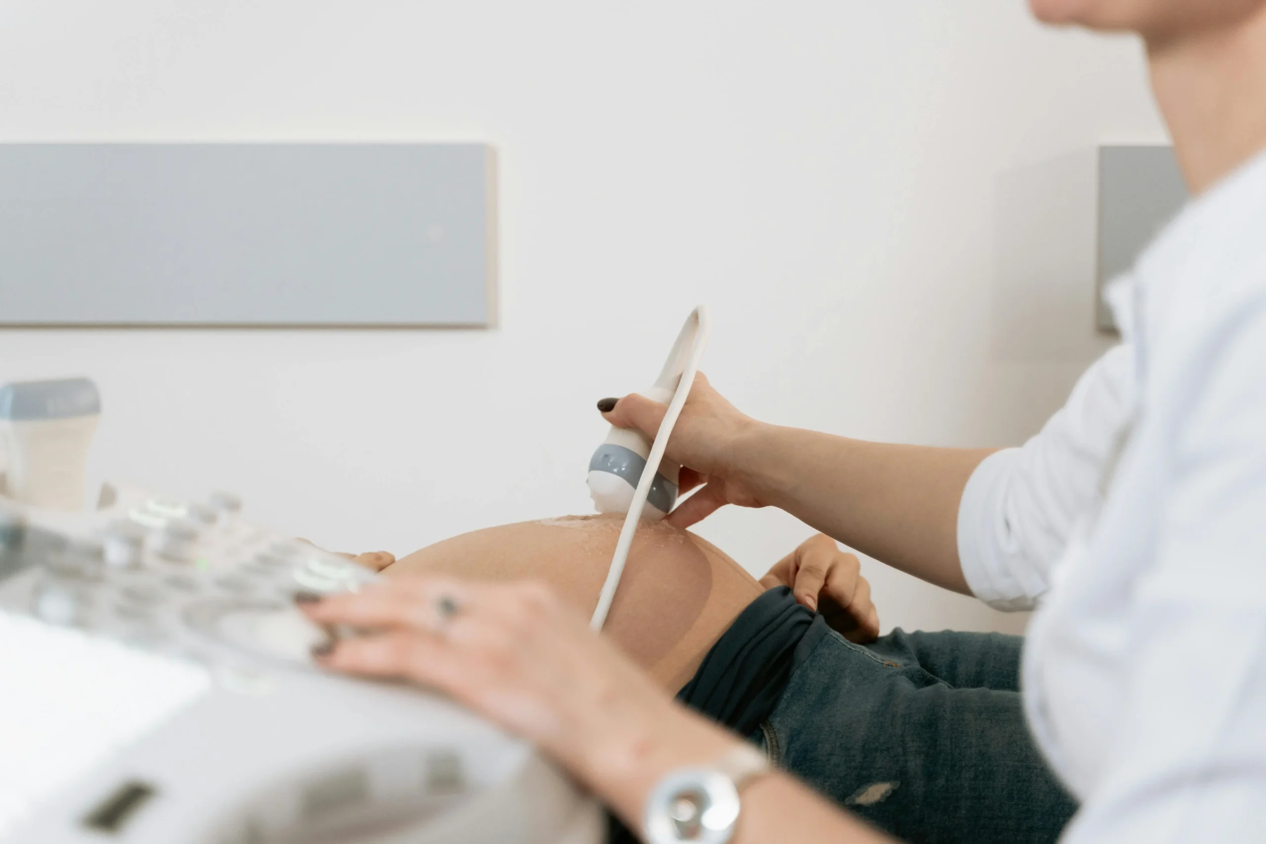
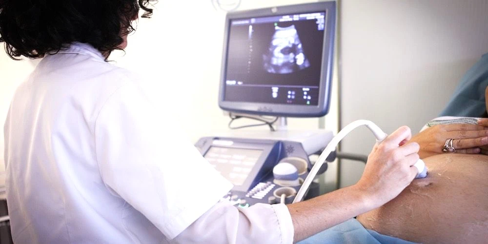
| 3. Gel application | A warm, water-based gel is applied to the skin over the scan area. The gel eliminates air between the transducer and your skin, ensuring clear sound wave transmission |
| 4. Scanning | The sonographer moves the transducer across the area, capturing images and measurements. You may be asked to hold your breath briefly for liver or kidney views |
| 5. Doppler assessment | If blood flow evaluation is needed, colour Doppler is activated — you may hear a whooshing sound representing blood flow through vessels |
| 6. Completion | The gel is wiped off, and you can return to normal activities immediately. There is no recovery time, no restrictions, and no side effects |
Step-by-step ultrasound procedure at DCDC.
Most ultrasound scans take 15-30 minutes. The examination is painless, though mild pressure from the transducer is normal. Transvaginal pelvic ultrasound uses a thin internal probe (2-3 cm diameter) for closer imaging of the uterus and ovaries — this is slightly uncomfortable but not painful. For all scan types, the sonographer or radiologist will explain the process and answer any questions before beginning.
Is Ultrasound Safe? Facts, Risks & Myths Debunked
Ultrasound is one of the safest medical imaging technologies available. It uses no ionizing radiation, requires no injections or contrast agents for most examinations, causes no pain, and has no confirmed harmful side effects when performed by qualified professionals. Ultrasound has been used in clinical medicine for over 60 years, during which extensive research involving millions of patients has consistently confirmed its safety.
Does Ultrasound Use Radiation?
No. Ultrasound does not use any form of ionizing radiation. This is the single most important safety fact. Unlike X-rays, CT scans, and fluoroscopy, which generate images using ionizing radiation, ultrasound creates images using mechanical sound waves. There are no X-ray photons, no gamma rays, and no electromagnetic radiation involved. Because there is no radiation, there is no cumulative dose to track — a patient who has ten ultrasound scans over a year has exactly the same radiation exposure as someone who has had none: zero.
Is Ultrasound Safe During Pregnancy?
Yes. Ultrasound is the primary imaging tool for prenatal care worldwide, endorsed by the WHO, ACOG, RCOG, and the AIUM. Obstetric ultrasound has been used since the 1960s, and in more than 60 years, no study has demonstrated a confirmed link between diagnostic ultrasound and any adverse outcome — no birth defects, no childhood cancer, no developmental delay, no hearing impairment, no neurological conditions. The one guideline is the ALARA principle (As Low As Reasonably Achievable): scans should be medically indicated, performed by trained professionals, and kept to the minimum time and power needed.
Is Ultrasound Safe for Children?
Yes. The European Society of Paediatric Radiology (ESPR) and the American College of Radiology (ACR) both recommend an "ultrasound first" approach for paediatric imaging whenever possible, precisely because there is no radiation. For conditions like appendicitis, intussusception, pyloric stenosis, and lymph node assessment in children, ultrasound is the first-choice imaging tool.
5 Common Ultrasound Myths Debunked
| Myth | Fact |
|---|---|
| Ultrasound uses radiation | Ultrasound uses sound waves, not radiation. It is fundamentally different from X-rays and CT scans. Zero ionizing radiation exposure, ever. |
| Too many ultrasounds are dangerous | There is no cumulative dose effect. Multiple scans have the same safety profile as a single scan. Even high-risk pregnancies receiving weekly ultrasounds show no adverse effects. |
| Ultrasound heats tissue and causes harm | Diagnostic ultrasound at standard settings produces thermal effects of less than 1°C — well below any harmful threshold. The Thermal Index (TI) is displayed on screen and kept below 1.0 by trained operators. |
| Ultrasound can cause cavitation (bubble damage) | Cavitation requires sustained, high-intensity ultrasound at levels far above diagnostic settings. The Mechanical Index (MI) is monitored to stay within safe limits. Diagnostic ultrasound has never produced clinically significant cavitation. |
| 3D/4D ultrasound is more dangerous than 2D | 3D/4D uses the same sound wave technology and power levels as 2D. The only concern is non-medical "keepsake" sessions that may use longer exposure times without clinical oversight — the FDA and AIUM discourage these. |
Common ultrasound safety myths corrected with evidence-based facts.
The Real Risk of Ultrasound: Operator Dependence
The most significant limitation of ultrasound is not a safety concern but a quality one: ultrasound is operator-dependent. The accuracy of the examination depends on the skill and experience of the person performing it. A poorly performed scan can miss findings that a skilled operator would detect. This is why having your ultrasound at a facility with experienced, fellowship-trained radiologists matters. At DCDC, every ultrasound is reviewed and reported by a consultant radiologist, not just a sonographer.
Ultrasound vs CT Scan vs MRI: Which Test Do You Need?
Ultrasound, CT, and MRI are the three most commonly used imaging modalities, and each excels at answering different clinical questions. None is universally superior — the best test is the one that answers your doctor's specific question. Here is how they compare:
| Feature | Ultrasound | CT Scan | MRI |
|---|---|---|---|
| Technology | High-frequency sound waves | Rotating X-ray beam | Magnetic field + radio waves |
| Ionizing Radiation | None | Yes (0.1-20 mSv depending on area) | None |
| Scan Duration | 10-40 minutes | 5-30 seconds per scan | 20-60 minutes |
| Cost in Dubai (AED) | 300-800 | 800-3,000 | 1,500-6,000 |
| Best For | Abdomen, thyroid, pregnancy, breast, real-time guidance | Lungs, bones, emergencies, cancer staging, vascular CTA | Brain, spine, joints, soft tissue, cardiac |
| Limitations | Bowel gas, deep structures, bone, lungs | Radiation, contrast allergy risk, less soft-tissue detail | Metal implants contraindicated, claustrophobia, slow |
| Contrast Agent | Microbubbles (rare, very safe) | Iodine-based (allergic reactions possible, kidney function required) | Gadolinium (generally safe, kidney function check needed) |
| Pregnancy Safe | Yes — standard of care | No — ionizing radiation | Generally avoided in 1st trimester as precaution |
| Real-Time Imaging | Yes — unique advantage | No | No (limited cine MRI) |
| Portability | Yes — can be done at bedside | No — fixed installation | No — fixed installation |
| Result Turnaround | Same day (hours) | Same day (hours) | Same day to 24 hours |
Head-to-head comparison of ultrasound, CT scan, and MRI for diagnostic imaging.
Which Test for Which Condition?
| Condition | Best First Test | Why |
|---|---|---|
| Abdominal pain | Ultrasound | No radiation, visualises gallstones/kidneys/liver with >95% accuracy for gallstones |
| Thyroid nodule | Ultrasound | Detects nodules as small as 2 mm, enables TI-RADS classification and FNA biopsy guidance |
| Pregnancy monitoring | Ultrasound | Only imaging modality considered safe standard of care throughout pregnancy |
| Breast lump | Ultrasound + Mammography | Differentiates solid vs cystic with high accuracy; first-line for women under 40 |
| Suspected DVT (blood clot) | Doppler Ultrasound | Non-invasive, real-time assessment of venous compressibility and blood flow |
| Knee/shoulder injury | MRI | Superior soft-tissue contrast for ligaments, cartilage, meniscus, and rotator cuff |
| Chest/lung symptoms | CT scan | Ultrasound cannot image lung tissue through air; CT provides detailed lung anatomy |
| Head injury / stroke | CT (acute) → MRI (follow-up) | CT detects acute bleeding within seconds; MRI gives superior brain detail for follow-up |
| Back pain / herniated disc | MRI | MRI provides detailed views of spinal discs, nerves, and soft tissues that ultrasound cannot reach |
| Bone fracture | X-ray | Fastest and cheapest for bone assessment; CT for complex fractures |
| Cancer staging | CT +/- MRI | CT for chest/abdomen staging; MRI for liver/brain/pelvic characterisation |
| Kidney stones | Ultrasound (initial) → CT (if needed) | Ultrasound first; non-contrast CT is gold standard if ultrasound is inconclusive |
Which imaging test is recommended for common conditions, with clinical rationale.
"Each imaging modality has a specific role in diagnosis," says Dr. Osama Elzamzami. "Choosing the right test is not about which machine is more advanced — it is about matching the technology to the clinical question. An ultrasound can answer in five minutes what an MRI would take forty-five minutes to show, and vice versa."
For more details on these modalities, see our guides on MRI scans in Dubai and CT angiography.
Book Your Ultrasound at DCDC
Doctors Clinic Diagnostic Center in Dubai Healthcare City offers same-day ultrasound scan appointments across all scan types with experienced consultant radiologists and competitive pricing. No long wait times. No hidden fees.
Or call us at +971 4 237 9939
Ultrasound Cost in Dubai: 2026 Prices by Scan Type
An ultrasound scan in Dubai costs between AED 300 and AED 800 for standard examinations, with specialised or multi-region scans reaching up to AED 1,000 at premium hospital facilities. The Dubai Health Authority regulates healthcare pricing, but ultrasound fees are not fixed — facilities set their own prices within competitive market ranges. Standalone diagnostic centres like DCDC consistently offer 20-40% lower pricing than hospital radiology departments while maintaining the same standard of equipment and radiologist expertise. When you book an ultrasound at DCDC, the quoted price includes the scan and a formal consultant radiologist report with no hidden charges.
| Ultrasound Type | Price Range (AED) | Common Indications | Scan Duration |
|---|---|---|---|
| Abdominal Ultrasound | 300-600 | Liver, gallbladder, kidneys, pancreas, spleen assessment | 20-30 min |
| Pelvic Ultrasound | 350-650 | Uterus, ovaries, bladder, prostate evaluation | 15-25 min |
| Thyroid Ultrasound | 300-500 | Thyroid nodules, goitre, thyroid gland assessment | 10-20 min |
| <a href="/services/gynecology-obstetrics/ultrasound-for-well-being-and-prenatal" class="text-primary-600 hover:underline">Pregnancy Ultrasound (2D)</a> | 400-650 | Fetal dating, anatomy survey, growth monitoring | 20-40 min |
| 3D/4D Pregnancy Ultrasound | 600-1,200 | 3D/4D baby imaging, facial features, anomaly detection | 20-30 min |
| Musculoskeletal Ultrasound | 350-600 | Joints, tendons, ligaments, soft tissue injuries | 15-30 min |
| Breast Ultrasound | 350-600 | Breast lumps, cysts, mammography follow-up | 15-25 min |
| Renal Ultrasound | 300-550 | Kidney stones, kidney size, hydronephrosis, bladder | 15-20 min |
| Doppler Vascular Ultrasound | 500-900 | DVT, carotid stenosis, peripheral vascular disease | 20-40 min |
| Scrotal Ultrasound | 350-550 | Testicular pain, swelling, varicocele, masses | 15-20 min |
Ultrasound scan prices in Dubai for 2026. Prices include scan and consultant radiologist report at accredited facilities.
What Affects Ultrasound Pricing?
- Scan complexity: A focused single-organ scan (e.g. thyroid, AED 300-500) costs less than a comprehensive multi-organ study (e.g. complete abdomen, AED 450-600) because it requires less time and generates a simpler report
- Facility type: Hospital radiology departments charge 20-50% more than standalone diagnostic centres like DCDC due to higher operational overhead
- Radiologist reporting: Some lower-priced services are performed by sonographers without consultant radiologist review. At DCDC, every scan includes a formal consultant radiologist report
- Doppler and advanced techniques: Adding Doppler blood flow assessment adds AED 100-200 to the base price. Elastography and contrast-enhanced ultrasound may add further
- Location: Facilities in Dubai Healthcare City (a medical free zone) may have different pricing structures than other areas
Does Insurance Cover Ultrasound in Dubai?
Most major health insurance plans in Dubai cover medically indicated ultrasound scans in full or with a copayment. The DHA Essential Benefits Plan (EBP) — the baseline for all insurance in Dubai — explicitly includes diagnostic radiology as a covered benefit. For insured patients, the effective out-of-pocket cost is typically just a copay of AED 0-50.
- Medical necessity: Insurance covers scans ordered by a physician for a documented medical indication. Purely elective scans (e.g. 3D/4D keepsake imaging) may not be covered
- Prior authorisation: Some plans require pre-approval. DCDC's insurance team handles this directly with your insurer
- Network status: DCDC is an approved provider with Daman, NAS, Oman Insurance, MetLife, AXA, and most other major UAE insurers
- Copayment: Most plans apply AED 0-50 for diagnostic imaging. Premium plans often have zero copay
- Self-pay: Transparent pricing with no hidden fees. Payment by cash, credit card, or Apple Pay. Total cost confirmed before scan begins
How to Save on Ultrasound Costs in Dubai
- Choose a standalone diagnostic centre: DCDC charges 20-40% less than hospital radiology departments for identical examinations with the same equipment and radiologist qualifications
- Verify insurance coverage before booking: Most plans cover ultrasound with a minimal copay. Confirming in advance prevents unexpected bills
- Use an in-network provider: Out-of-network facilities result in higher copayments or no coverage. DCDC is in-network with most major UAE insurers
- Ask about package pricing: If your doctor has ordered multiple scans (e.g. abdominal + pelvic), ask about bundled pricing for scans in the same visit
- Request only what is clinically necessary: Adding Doppler, 3D imaging, or additional regions increases cost. Your referral specifies what is needed
- Book directly: Some online booking platforms add service fees. Booking directly with DCDC via phone or WhatsApp ensures the actual listed price
Why Choose DCDC for Your Ultrasound in Dubai?
DCDC (Doctors Clinic Diagnostic Center) in Dubai Healthcare City performs over 1,000 ultrasound scans every month, making it one of the highest-volume ultrasound providers in the emirate. This volume translates directly into expertise across every ultrasound type.
- Experienced consultant radiologists: Every scan is reviewed and reported by a fellowship-trained consultant radiologist — not just a sonographer. Subtle findings are identified and documented
- Advanced equipment: High-resolution transducers, Doppler capability, elastography, 3D/4D imaging, and contrast-enhanced ultrasound (CEUS). Equipment regularly maintained and updated
- Same-day appointments and results: Walk-in and same-day appointments for most scan types. Reports typically completed within hours
- Competitive pricing: 20-40% lower than hospital radiology departments. Transparent self-pay pricing with no hidden fees
- Full insurance support: In-network with most major UAE insurers. Insurance team handles pre-authorisation and claims
- Safety standards: All equipment displays Thermal Index (TI) and Mechanical Index (MI) in real time. Regular maintenance protocols, calibrated to international AIUM and BMUS safety standards. Gel warmers for patient comfort
- Comprehensive imaging: If ultrasound results indicate further imaging is needed, DCDC offers CT, MRI, X-ray, mammography, DEXA bone density scans, and CBCT under the same roof — streamlining your diagnostic pathway
- Convenient location: Dubai Healthcare City, easily accessible from Oud Metha, Karama, Bur Dubai, Downtown Dubai, with ample parking
Patient Story: A Routine Scan That Caught a Problem Early
A 38-year-old marketing executive in Dubai had been experiencing intermittent upper abdominal discomfort for several weeks. Her GP ordered a routine abdominal ultrasound, and she chose DCDC for the same-day availability. The ultrasound, which took 25 minutes and cost AED 450, revealed multiple small gallstones along with mild wall thickening suggesting early cholecystitis. The radiologist's report was shared with her GP within two hours, and she was referred to a surgeon the following day. She underwent a straightforward laparoscopic cholecystectomy the following week and recovered fully within 10 days.
"What impressed me was how quickly everything moved," she later shared. "From the scan to the report to the surgeon consultation, I felt like the diagnostic centre genuinely prioritised getting me answers fast. The whole ultrasound experience was painless, affordable, and far more informative than I expected."
Patient Story: A Parent's Reassurance
The mother of a 3-year-old boy brought him to DCDC after his paediatrician recommended an abdominal ultrasound for recurrent tummy pain. She was concerned about radiation exposure for her young child. "When the radiologist explained that ultrasound uses absolutely no radiation — just sound waves like a sonar — I felt so much better," she said. The scan took 15 minutes, the child was calm throughout, and the results showed swollen lymph nodes in the abdomen (mesenteric lymphadenitis), a common and self-limiting condition in children. No further imaging was needed.
Get Your Ultrasound Scan at DCDC Today
DCDC offers competitive ultrasound pricing, same-day appointments, and fast results at our Dubai Healthcare City location. Our experienced radiology team performs over 1,000 ultrasound scans monthly across all body regions.
Same-day appointments available — Call +971 4 237 9939 or WhatsApp
Verwandte Leistungen im DCDC
Fachkundige Betreuung und moderne Diagnostik in Dubai Healthcare City
Häufig gestellte Fragen
Final Thoughts
Ultrasound is the most accessible, affordable, and versatile diagnostic imaging tool available in Dubai — providing real-time diagnostic information across virtually every medical specialty without any radiation exposure. Whether you need an abdominal ultrasound for AED 300-600, a pregnancy scan for AED 400-800, a thyroid ultrasound for AED 300-500, or any of the dozen other scan types available, the technology is safe, painless, and delivers results within hours. With most insurance plans covering the bulk of the expense, the effective out-of-pocket cost for insured patients is typically just AED 0-50.
At Doctors Clinic Diagnostic Center in Dubai Healthcare City, patients benefit from experienced consultant radiologists, advanced ultrasound equipment including Doppler and 3D/4D capability, same-day appointments, fast results, comprehensive insurance support, and pricing that is consistently 20-40% lower than hospital radiology departments. If you need an ultrasound in Dubai, contact DCDC via WhatsApp or call +971 4 237 9939 to schedule your appointment.
Quellen und Referenzen
Dieser Artikel wurde von unserem medizinischen Team überprüft und bezieht sich auf folgende Quellen:
- American Institute of Ultrasound in Medicine (AIUM) - Official Statement on Safety of Diagnostic Ultrasound
- World Health Organization (WHO) - Diagnostic Imaging: Ultrasound Safety
- American College of Obstetricians and Gynecologists (ACOG) - Ultrasound in Pregnancy
- RadiologyInfo.org - General Ultrasound
- American College of Radiology (ACR) - Appropriateness Criteria for Diagnostic Ultrasound
- British Medical Ultrasound Society (BMUS) - Guidelines for Professional Ultrasound Practice
- Dubai Health Authority (DHA) - Healthcare Facilities Regulations
- European Society of Paediatric Radiology (ESPR) - Ultrasound-First Recommendations
Medizinische Inhalte auf dieser Website werden von DHA-lizenzierten Ärzten überprüft. Siehe unsere redaktionelle Richtlinien für weitere Informationen.

Verfasst von
Dr. Osama Elzamzami
Diagnostic Radiology
MD, FRCR
Dr. Osama Elzamzami is a Consultant Radiologist specializing in diagnostic imaging including ultrasound, CT, MRI, and interventional radiology at DCDC Dubai Healthcare City.
Related Articles
Understanding Your Ultrasound Results: A Patient Guide
Abdominal Ultrasound: A Complete Guide
Thyroid Ultrasound in Dubai: When You Need It & What It Detects
blogPage.moreFromCategory
CT Scan Cost in Dubai: Prices, Types & What to Expect
Weiterlesen
DEXA Scan Dubai: Cost, T-Score Results & Who Needs It
WeiterlesenThyroid Ultrasound in Dubai: When You Need It & What It Detects
Weiterlesen
Open MRI Dubai | MRI Scan Cost, Uses & Benefits
Weiterlesen
MRI Cost in Dubai: AED 900-15,000 Price Comparison (2026)
Weiterlesen![Full Body MRI Cost Dubai: AED 5,000-15,000 [2026]](/wp-media/blog/full-body-mri-scan.webp)