Wichtigste Erkenntnisse
- Ein OPG (Orthopantomogramm) ist ein Panoramaroentgen, das beide Kiefer, alle Zaehne, TMJ-Gelenke, Nasennebenhoehlen und Nervkanaele in einem einzelnen 15-Sekunden-Scan ohne Schmerzen und ohne Sensoren im Mund erfasst
- Der Scan erfordert fast keine Vorbereitung - entfernen Sie nur Metallschmuck aus dem Kopf- und Halsbereich; kein Fasten, keine Injektionen oder Kontrastmittel erforderlich
- OPG liefert nur 10-20 Mikrosievert Strahlung, entsprechend 1-2 Tagen natuerlicher Hintergrundstrahlung und weniger als ein einzelner Langstreckenflug
- Zahnae lesen OPG-Ergebnisse systematisch und bewerten Zaehne, Knochenniveaus, Kiefergelenke, Nasennebenhoehlen und Nervkanaele auf Karies, Knochenverlust, impaktierte Zaehne, Zysten, Frakturen und TMJ-Stoerungen
- OPG ist die wesentliche Erstlinien-Bildgebung fuer Zahnimplantatplanung, kieferorthopaedische Beurteilung, Weisheitszahnbewertung, Kieferschmerzdiagnose und routinemaessiges zahnaerztliches Screening
- Wenn OPG einen Befund identifiziert, der dreidimensionale Details erfordert, liefert ein CBCT-Scan die ergaenzende Praezision fuer die chirurgische Planung
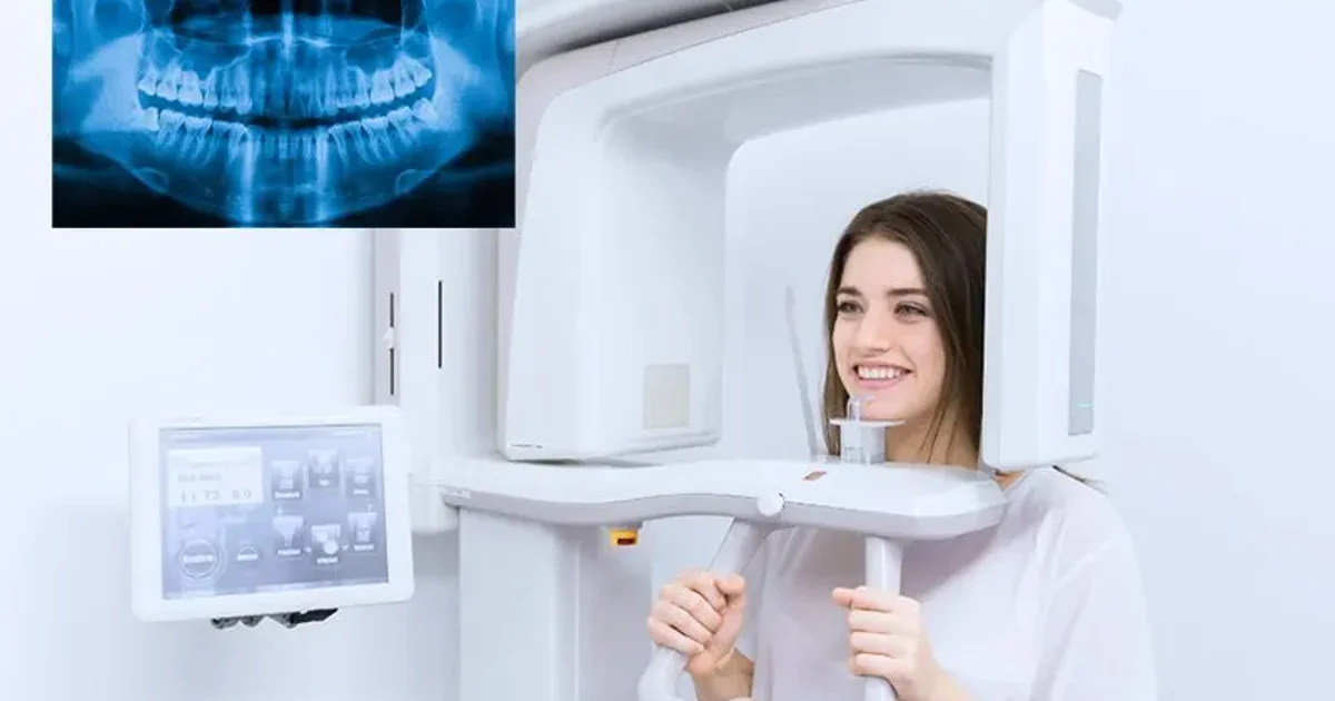
Ein OPG-Roentgen (Orthopantomogramm) ist die weltweit am haeufigsten verwendete extraorale zahnaerztliche Bildgebungstechnik. In einem einzigen schmerzlosen Scan von unter 15 Sekunden erfasst es ein flaches, zweidimensionales Panoramabild beider Kiefer, aller Zaehne, der Kiefergelenke, der Kieferhoehlen und des umgebenden Knochens von Ohr zu Ohr. Ob Sie ein OPG fuer Zahnimplantate, kieferorthopaedische Zahnspangen, Weisheitszahnbewertung oder eine routinemaessige zahnaerztliche Untersuchung benoetigen, dieser umfassende Leitfaden deckt alles ab.
Dieser 2026-Mega-Leitfaden konsolidiert das vollstaendige OPG-Wissen in einer einzigen Ressource, ueberarbeitet von Dr. Osama Elzamzami, Facharzt fuer Radiologie (MD, FRCR) bei Doctors Clinic Diagnostic Center (DCDC) in Dubai Healthcare City. Jeder Abschnitt ist klinisch verifiziert und enthaelt Vergleichstabellen, Erklaerungen zu Berichtsterminologie, Patientengeschichten und fachliche Anleitung.


Was ist ein OPG-Roentgen?
OPG steht fuer Orthopantomogramm, ein zusammengesetzter Begriff aus dem Griechischen "ortho" (gerade), "pan" (alles) und "tomogramm" (Schnittbild) - zusammen bedeutend ein gerades, umfassendes Schnittbild der Kiefer und Zaehne. In der klinischen Praxis wird der Scan auch als Panoramaradiographie, Panoramaroentgen oder einfach "Pano" bezeichnet.
Das Konzept wurde erstmals vom finnischen Radiologen Yrjo Veli Paatero in den 1940er Jahren entwickelt, und kommerzielle OPG-Geraete wurden in den 1960er Jahren weit verbreitet. Heute haben digitale OPG-Systeme filmbasierte Geraete in den meisten modernen Kliniken ersetzt.
Wie funktioniert ein OPG-Roentgen?
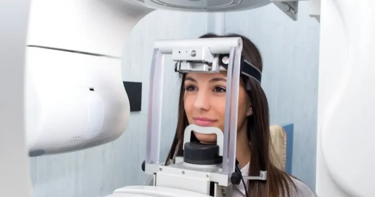
Ein OPG-Roentgen funktioniert durch eine Technik namens Rotationstomographie, bei der eine Roentgenroehre und ein digitaler Detektor gleichzeitig in einem koordinierten Halbbogen um den Kopf des Patienten rotieren. Die Software rekonstruiert dann diese Schnitte zu einem einzigen Panoramabild.
Das Schluesselprinzip wird Schmalstrahl-Tomographie genannt. Anstatt den gesamten Kopf auf einmal zu bestrahlen, verwendet das OPG-Geraet einen duennen, fokussierten Strahl, der nur einen schmalen vertikalen Streifen der Anatomie beleuchtet.
Da die Roentgenquelle waehrend des gesamten Verfahrens ausserhalb des Mundes bleibt, wird das OPG als extraorale Bildgebungstechnik klassifiziert. Patienten mit starkem Wuergereiz, eingeschraenkter Mundoeffnung oder Zahnarztangst finden das OPG deutlich komfortabler als intraorale Alternativen.
"Das OPG ist das vielseitigste Screening-Werkzeug der Zahnmedizin," erklaert Dr. Osama Elzamzami. "In einem einzigen 15-Sekunden-Scan erfassen wir beide Kiefer, alle Zaehne, die Kiefergelenke und den umgebenden Knochen."
OPG vs. Intraorale Roentgenaufnahme vs. CBCT
Der einfachste Weg, den Unterschied zu verstehen, ist, sie als Weitwinkelobjektiv versus Makroobjektiv zu betrachten. Das OPG umkreist die Aussenseite Ihres Kopfes und erzeugt ein einzelnes flaches Bild. Das intraorale Roentgen platziert einen kleinen Sensor in Ihrem Mund und fotografiert nur 2-4 Zaehne bei extrem hoher Aufloesung. Ein CBCT geht weiter und erzeugt dreidimensionale volumetrische Bilder.
Intraorale Roentgenaufnahmen gibt es in drei Untertypen. Eine periapikale Aufnahme erfasst den gesamten Zahn von Krone bis Wurzelspitze. Eine Bissfluegel-Aufnahme erfasst die Kronen von Ober- und Unterkieferzaehnen gleichzeitig. Eine Okklusalaufnahme verwendet einen groesseren Sensor auf der Kauflaeche.
| Merkmal | OPG (Panorama) | Periapikal | Bissfluegel | CBCT |
|---|---|---|---|---|
| Abdeckung | Beide Kiefer von Ohr zu Ohr, TMJ, Nasennebenhoehlen | 2-3 Zaehne + Wurzelspitzen | Kronen oberer/unterer Praemolaren und Molaren | 3D-Volumen ausgewaehlter Region oder beider Kiefer |
| Sensorposition | Ausserhalb des Mundes (extraoral) | Im Mund | Im Mund | Ausserhalb des Mundes (extraoral) |
| Detailstufe | Mittel (breiter Ueberblick) | Sehr hoch (zahnspezifisch) | Sehr hoch (Krone und obere Wurzel) | Sehr hoch (3D-Querschnitte) |
| Strahlendosis | 10-20 μSv | 1-8 μSv pro Bild | 1-5 μSv pro Bild | 30-200 μSv |
| Bilder fuer vollen Mund | 1 Bild | 14-20 Bilder (vollstaendige Serie) | 4 Bilder (Standardserie) | 1 Scan |
| Scanzeit | 15-20 Sekunden | < 1 Sekunde pro Belichtung | < 1 Sekunde pro Belichtung | 15-30 Sekunden |
| Patientenkomfort | Sehr komfortabel; kein Sensor im Mund | Kann unangenehm sein; Wuergereizrisiko | Mittel; Beisstab hilft bei Stabilitaet | Komfortabel; kein Sensor im Mund |
| Am besten fuer | Screening, impaktierte Zaehne, Frakturen, Kieferorthopaedie, Implantatuebersicht, TMJ | Wurzelkanaele, periapikale Infektionen, Wurzelfrakturen | Fruehe Karieserkennung, Kontrolle von Restaurationen | Implantatplanung, komplexe Chirurgie, 3D-Anatomie |
| Ungefaehre Dubai-Kosten (AED) | 150-300 | 50-150 pro Bild | 50-100 pro Bild | 400-1.200 |
Vergleich von OPG-Panoramaroentgen, intraorale Roentgen-Untertypen und CBCT. Fuer detaillierten CBCT-Vergleich siehe unseren Leitfaden zu <a href="/blog/cbct-vs-opg-xray" class="text-primary-600 hover:underline">CBCT vs. OPG</a>.
Aus der Kosten-pro-Abdeckung-Perspektive ist das OPG deutlich wirtschaftlicher. Fuer detaillierte Preise siehe unseren Leitfaden zu OPG-Roentgen Kosten in Dubai.
Was zeigt ein OPG?
Ein OPG-Roentgen zeigt die vollstaendige zahnaerztliche und kieferchirurgische Anatomie in einem Bild.
- Alle durchgebrochenen Zaehne in beiden Kiefern einschliesslich Wurzeln, Kronen und umgebendem Knochen
- Nicht durchgebrochene und impaktierte Zaehne, insbesondere Weisheitszaehne
- Sich entwickelnde Zaehne bei Kindern und Jugendlichen
- Zahnkaries, insbesondere grosse interproximale Kavitaeten
- Periapikale Pathologie wie Zahnabszesse und Granulome
- Knochenverlust durch parodontale Erkrankungen
- Kieferknochenanomalien einschliesslich Zysten, Tumoren und gutartige Wucherungen
- Kieferfrakturen durch Trauma
- Kiefergelenke (TMJ) mit Anzeichen von Arthritis, Erosion oder Dislokation
- Kieferhoehlen mit Sinusitis, Polypen oder Retentionszysten
- Nasenhoehle und Nasenscheidewand auf grobe Abweichung
- Zahnrestaurationen wie Fuellungen, Kronen, Bruecken und Implantate
- Der Nervus alveolaris inferior-Kanal, entscheidend fuer Implantatplanung und Weisheitszahnextraktion
Waehrend das OPG einen hervorragenden Ueberblick bietet, koennen aufgrund seiner zweidimensionalen Natur einige feine Details ergaenzende intraorale Roentgenaufnahmen oder CBCT-Bildgebung erfordern.
Vorbereitung auf ein OPG
Die Vorbereitung auf ein OPG-Roentgen ist unkompliziert, da der Scan fast keine spezielle Vorbereitung erfordert. Kein Fasten, keine Blutuntersuchung, keine Injektionen, kein Kontrastmittel und keine Erholungszeit. Der wichtigste Schritt ist das Entfernen aller Metallgegenstaende aus dem Kopf- und Halsbereich.
"Die haeufigste Frage, die Patienten stellen, ist, ob sie etwas vorbereiten muessen," sagt Dr. Elzamzami. "Die Antwort ist erfrischend einfach: Metall entfernen, 15 Sekunden stillstehen, und der Scan ist erledigt."
| Zu entfernender Gegenstand | Warum er entfernt werden muss |
|---|---|
| Ohrringe (Stecker, Creolen, Ohrmanschetten) | Metallohrringe sitzen direkt im Scanpfad und erzeugen helle weisse Flecken |
| Halsketten und Ketten | Halsketten erzeugen lineare Artefakte, die Frakturen imitieren koennen |
| Gesichts- und Mundpiercings | Piercings erzeugen fokale weisse Artefakte, die Zaehne und Knochen verdecken |
| Brillen und Sonnenbrillen | Metallrahmen verdecken den Oberkiefer und die Nasenhoehle |
| Hoergeraete | Erzeugen dichte Artefakte im Bereich des TMJ |
| Herausnehmbare Prothesen und Retainer | Metallklammern ueberlagern sich auf natuerliche Zaehne |
| Haarnadeln, Clips mit Metall | Erzeugen gestreute Artefakte im gesamten Bild |
| Kopftuecher mit Metallnadeln | Metallnadeln erzeugen punktuelle Artefakte; der Stoff kann bleiben, wenn alles Metall entfernt ist |
Entfernen Sie alle metallischen Gegenstaende aus Kopf, Hals und oberem Brustbereich. Fester Zahnersatz (Kronen, Bruecken, Zahnspangen, Implantate) muss nicht entfernt werden.
Essen, Trinken und Medikamente
Sie koennen vor einem OPG-Roentgen voellig normal essen und trinken. Kein Fasten erforderlich. Nehmen Sie alle regelmaessigen Medikamente ein.
Schwangerschaft und OPG-Vorbereitung
Wenn Sie schwanger sind oder vermuten, schwanger zu sein, informieren Sie die Radiographin vor dem Scan. Wahlaerztliche Zahnbildgebung wird waehrend der Schwangerschaft routinemaessig verschoben, insbesondere im ersten Trimester. Bei klinischem Notfall kann ein OPG jedoch sicher mit einer Bleischuerze durchgefuehrt werden. Die Strahlendosis (10-20 μSv) ist tausendmal unter der Schwelle fuer fetale Schaeden.
Was Sie dem Radiographen mitteilen sollten
- Schwangerschaft oder moegliche Schwangerschaft
- Den Grund fuer den Scan - hilft dem Radiologen, den Bericht zu fokussieren
- Fruehere Kieferoperationen oder Traumata
- Schwierigkeiten, still zu stehen
- Fruehere zahnaerztliche Bildgebung - fuer Vergleich
Praktischer Tipp: Lassen Sie Schmuck zu Hause und tragen Sie ein einfaches Oberteil ohne hohen Metallreissverschluss.
Was geschieht waehrend eines OPG
Das gesamte OPG-Verfahren dauert unter 5 Minuten, wobei der eigentliche Scan nur 15 bis 20 Sekunden dauert. Es gibt keine Vorbereitung, keine Erholung und keine Nebenwirkungen.
- Schritt 1 - Ankunft und Registrierung: Check-in mit Emirates ID oder Reisepass. 2-3 Minuten.
- Schritt 2 - Metallgegenstaende entfernen: Die Radiographin bittet Sie, alle metallischen Gegenstaende zu entfernen. Etwa 30 Sekunden.
- Schritt 3 - Schutzabschirmung tragen: Eine Bleischilddruese und/oder Bleischuerze wird angelegt.
- Schritt 4 - Im Geraet positionieren: Sie stellen sich ans OPG-Geraet und legen Ihr Kinn auf eine Kinnstütze. Die korrekte Positionierung ist der wichtigste Faktor fuer ein qualitativ hochwertiges Bild.
- Schritt 5 - Auf die Positionierungshilfe beissen: Sie beissen sanft auf einen sterilen Plastik-Beissblock. Sie schliessen die Lippen und druecken die Zunge flach gegen den Gaumen.
- Schritt 6 - Der Scan: Der C-foermige Arm dreht sich langsam um Ihren Kopf. Sie muessen vollstaendig still bleiben. 15 bis 20 Sekunden.
- Schritt 7 - Scan abgeschlossen: Das Geraet stoppt. Das digitale Bild erscheint innerhalb von Sekunden auf der Workstation. Gesamte Raumzeit: 3 bis 5 Minuten.
Es gibt keine Erholungszeit. Der Scan ist voellig schmerzfrei - keine Nadeln, keine Injektionen, kein Kontrastmittel und keine Sensoren im Mund.
OPG fuer Kinder
Kinder ab 5 Jahren koennen ein OPG erhalten. Moderne digitale OPG-Geraete umfassen automatische paediatrische Dosisreduktionsprotokolle. Ein paediatrisches OPG liefert etwa 5 bis 14 μSv - weniger als ein Tag natuerlicher Hintergrundstrahlung.
Der 7-jaehrige Youssef kam mit seiner Mutter zu DCDC. Die Radiographin nahm sich Zeit, ihm die Maschine zu zeigen. Youssef stand 16 Sekunden perfekt still, und der Scan war erledigt. Das OPG zeigte zwei bleibende Zaehne, die sich in ungewoehnlichen Winkeln entwickelten.
Digitales OPG-Roentgen bei DCDC Dubai Healthcare City
Erhalten Sie einen hochaufloesenden digitalen OPG-Scan mit Radiologen-Befund am selben Tag bei Doctors Clinic Diagnostic Center. Laufkundschaft willkommen.
Keine Ueberweisung fuer Selbstzahler erforderlich
Ihre OPG-Ergebnisse lesen
Zahnae und Radiologen folgen einem strukturierten, systematischen Ansatz, der sicherstellt, dass jede anatomische Region bewertet wird.
Der systematische Leseansatz
- Gesamte Bildqualitaetspruefung: Der Radiologe bestaetigt korrekte Belichtung und Positionierung.
- Symmetriebewertung: Links und rechts sollten ungefaehr symmetrisch erscheinen.
- Zaehne-Bewertung (Quadrant fuer Quadrant): Jeder Zahn wird individuell untersucht.
- Knochenniveau-Bewertung: Die Hoehe des Alveolarknochens um jeden Zahn wird bewertet.
- TMJ-Bewertung: Beide Kiefergelenke werden auf Form, Groesse und Symmetrie untersucht.
- Kieferhoehlen-Ueberpruefung: Die Nasennebenhoehlen werden auf Truebung und Polypen geprueft.
- Nervkanal-Verfolgung: Der Nervus alveolaris inferior-Kanal wird verfolgt.
- Pathologie-Identifizierung: Abnormale dunkle oder helle Bereiche werden dokumentiert.
Bei DCDC wird jedes OPG von einem Facharzt fuer Radiologie gelesen.
Haeufige Befunde auf OPG-Roentgenaufnahmen
| Befund | Erscheinung auf OPG | Was es bedeutet |
|---|---|---|
| Zahnkaries | Dunkle Bereiche innerhalb der Zahnkrone | Zahnverfall; Behandlung von Fuellung bis Wurzelbehandlung |
| Knochenverlust (Parodontitis) | Reduzierte Knochenhoehe um Zahnwurzeln | Zahnfleischerkrankung mit Knochenrueckgang |
| Impaktierte Zaehne | Zahn teilweise oder vollstaendig unter Zahnfleisch oder Knochen | Haeufig bei Weisheitszaehnen; kann Schmerzen oder Zystenbildung verursachen |
| Periapikale Pathologie | Dunkler Bereich an der Wurzelspitze | Infektion erfordert Wurzelbehandlung oder Extraktion |
| Zysten | Gut definierter runder dunkler Bereich im Kieferknochen | Fluessigkeitsgefuellter Sack; chirurgische Entfernung erforderlich |
| TMJ-Anomalien | Abflachung, Erosion oder Asymmetrie der Kondylen | Degenerative Kiefergelenksveraenderungen |
| Wurzelresorption | Verkuerzte oder unregelmaessig geformte Wurzel | Wurzelaufloesung durch verschiedene Ursachen |
| Nasennebenhoehlen-Anomalien | Truebung innerhalb der Kieferhoehle | Sinusitis oder Retentionszysten |
Haeufige OPG-Befunde und ihre klinische Bedeutung.
Normale vs. abnormale OPG-Ergebnisse
Ein normales OPG zeigt: intakte Zaehne ohne dunkle Flecken, gesunde Wurzeln, gleichmaessige Knochenniveaus, symmetrische Kondylen, klare luftgefuellte Nasennebenhoehlen und sichtbare Nervkanaele.
Abnormale Befunde umfassen: dunkle Bereiche im Kieferknochen, helle weisse Massen, signifikanten Knochenverlust, Wurzelverkuerzung und verdickte Nasennebenhoehlen.
OPG-Berichtsterminologie verstehen
| Berichtsbegriff | Bedeutung in einfacher Sprache |
|---|---|
| Radioluzent / Radioluzenz | Dunkler Bereich - zeigt Knochenzerstoerung, Zyste, Abszess oder Tumor an |
| Radioopak / Radioopazitaet | Heller weisser Bereich - Metallfuellungen, Implantate oder dichter Knochen |
| Periapikale Pathologie | Erkrankung an der Zahnwurzelspitze |
| Resorption | Wurzelaufloesung - erscheint als Verkuerzung oder unregelmaessige Kontur |
| Horizontaler Knochenverlust | Gleichmaessige Knochenhoehlenreduktion durch chronische Zahnfleischerkrankung |
| Vertikaler Knochenverlust | Lokalisierter V-foermiger Knochendefekt entlang einer Zahnwurzelseite |
| Impaktierung | Zahn, der nicht in seine normale Position durchgebrochen ist |
| Gut definierte vs. schlecht definierte Raender | Grenzen einer Laesion: scharf = meist gutartig; unscharf = Verdacht auf aggressiven Prozess |
| Osteosklerose | Bereich erhoehter Knochendichte; meist gutartig |
| Perikoronitis | Infektion um einen teilweise durchgebrochenen Zahn |
| Dentogene Zyste | Fluessigkeitsgefuellter Sack um die Krone eines nicht durchgebrochenen Zahns |
Haeufige OPG-Berichtsterminologie entschluesselt.
OPG fuer Zahnimplantate
Das OPG ist das Standard-Erstlinien-Screening-Roentgen fuer Zahnimplantatkandidaten.
Was OPG fuer die Implantatbeurteilung zeigt
- Vertikale Knochenhoehe: Das OPG zeigt die volle Alveolarkammhoehe.
- Position des Nervkanals: Das OPG zeigt den Nervkanal als strahlungsdurchlaessiges Band.
- Position des Kieferhoehlenbodens: Zeigt, ob ein Sinuslift-Verfahren noetig ist.
- Zustand benachbarter Zaehne: Karies, periapikale Pathologie und Knochenverlust.
- Bestehende Pathologie: Zysten, Tumoren, zurueckgebliebene Wurzelfragmente.
- Gesamte Kieferarchitektur: Fuer Patienten, die mehrere Implantate benoetigen.
OPG-Einschraenkungen und wann ein CBCT-Upgrade noetig ist
Das OPG kann Knochenbreite, Knochendichte oder die genaue 3D-Nervposition nicht zeigen. Szenarien, die fast immer ein Follow-up-CBCT erfordern, umfassen: hintere Unterkiefer-Implantate nahe dem Nervkanal, obere hintere Implantate nahe dem Sinus und mehrere Implantate.
OPG fuer Kieferorthopaedie
Das OPG ist die erste Bildgebung, die jeder Kieferorthopäde vor der Empfehlung von Zahnspangen anfordert.
| Struktur / Zustand | Was das OPG zeigt | Warum es fuer die Kieferorthopaedie wichtig ist |
|---|---|---|
| Durchgebrochene Zaehne | Position, Ausrichtung und Abstand | Bestaetigt klinische Befunde; hilft bei der Bracket-Platzierung |
| Nicht durchgebrochene / impaktierte Zaehne | Lage, Winkelung und Tiefe | Impaktierte Eckzaehne benoetigen moeglicherweise chirurgische Freilegung |
| Fehlende Zaehne (angeboren) | Fehlen von Zahnkeimen | Aendert das Behandlungsziel - Luecke schliessen oder offenhalten |
| Ueberzaehlige Zaehne | Zusaetzliche Zaehne, die den normalen Durchbruch blockieren | Muessen vor der Ausrichtung entfernt werden |
| Wurzellaenge und -morphologie | Laenge, Form und Integritaet jeder Wurzel | Kurze Wurzeln haben hoeheres Resorptionsrisiko |
| Knochenniveaus | Hoehe der Knochenunterstuetzung | Reduzierter Knochen begrenzt sichere Zahnbewegung |
| Kiefersymmetrie | Relative Groesse von linkem und rechtem Unterkieferast | Asymmetrie kann skelettales Problem anzeigen |
| TMJ-Gelenke | Form und Position der Kondylen | Bestehende Degeneration kann sich waehrend der Kieferorthopaedie verschlechtern |
| Fruehere Zahnbehandlungen | Kronen, Bruecken, Wurzelbehandlungen, Implantate | Brackets koennen nicht auf Implantate geklebt werden |
OPG bietet dem Kieferorthopaeden einen umfassenden diagnostischen Ueberblick.
OPG fuer Weisheitszaehne
Das OPG ist die Standard-Bildgebung fuer die Bewertung von Weisheitszaehnen (dritten Molaren) vor der Entscheidung, ob eine Extraktion noetig ist.
| Impaktierungstyp | OPG-Erscheinung | Haeufigkeit | Extraktionsschwierigkeit | Hauptbefund |
|---|---|---|---|---|
| Mesioangular | Nach vorne zum zweiten Molaren geneigt | 40-45% | Mittel | Krone drueckt auf zweiten Molaren |
| Vertikal | Aufrecht aber nicht durchgebrochen | 25-30% | Mittel bis schwierig | Korrekte Achse aber blockiert |
| Horizontal | Auf der Seite liegend | 10-15% | Schwierig | Krone auf Wurzeln des zweiten Molaren gerichtet |
| Distoangular | Nach hinten zum Ramus geneigt | 5-10% | Am schwierigsten (Unterkiefer) | Begrenzter Zugang fuer Extraktionsweg |
| Bukkal/Lingual (transversal) | Zur Wange oder Zunge verschoben | Selten | Variabel | OPG kann Verschiebung unterschaetzen; CBCT oft noetig |
Klassifizierung der Weisheitszahnimpaktierung basierend auf OPG.
Zahnroentgen-Sicherheit
Ja, Zahnroentgenaufnahmen sind sicher. Die Strahlendosis eines Standard-OPG betraegt etwa 10 bis 20 Mikrosievert (μSv) - entsprechend 1 bis 2 Tagen natuerlicher Hintergrundstrahlung.
| Strahlungsquelle | Ungefaehre Dosis (μSv) | Äquivalent in OPG-Scans |
|---|---|---|
| Eine Banane essen | 0,1 | 1/140 eines OPG |
| Einzelne periapikale Aufnahme | 1-8 | < 1 OPG |
| Ein Tag natuerliche Hintergrundstrahlung | 6,5 | Etwa ein halbes OPG |
| OPG (Panoramazahnroentgen) | 10-20 | 1 OPG |
| Thorax-Roentgen | 20 | ~1 OPG |
| Dubai-nach-London-Flug | 40-80 | 3-6 OPGs |
| CBCT | 30-200 | 2-14 OPGs |
| CT-Scan des Kopfes | 2.000 | ~140 OPGs |
| Gesamte jaehrliche Hintergrundstrahlung | 2.400 | ~170 OPGs |
Zahnroentgenaufnahmen stehen am unteren Ende des medizinischen Strahlungsspektrums.
OPG-Sicherheit waehrend der Schwangerschaft
Die ACOG und ADA bestaetigen beide, dass Zahnroentgenaufnahmen waehrend der Schwangerschaft bei klinischer Indikation sicher durchgefuehrt werden koennen. Dringende diagnostische Bildgebung sollte nicht verzoegert werden.
OPG-Sicherheit fuer Kinder
Zahnroentgenaufnahmen sind fuer Kinder sicher. Moderne Geraete haben automatische paediatrische Dosisreduktionsprotokolle.
Sichere, niedrig dosierte Zahnroentgenaufnahmen bei DCDC
DCDC verwendet die neuesten digitalen OPG- und digitalen Roentgensysteme. Laufkundschaft willkommen bei DCDC Dubai Healthcare City.
Keine Ueberweisung fuer Selbstzahler erforderlich
OPG-Roentgen bei DCDC Dubai Healthcare City
Doctors Clinic Diagnostic Center (DCDC) bietet umfassende digitale OPG-Roentgen-Dienste in Dubai Healthcare City.
- Vollstaendige Bildgebung unter einem Dach: OPG, CBCT, periapikale Roentgenaufnahmen, kephalometrische Bildgebung, CT, MRT und Ultraschall vor Ort.
- Neueste digitale OPG-Technologie: Schaerfere Bilder bei niedrigeren Strahlendosen.
- Paediatrische Dosisprotokolle: Automatische Dosisreduktion fuer Kinder.
- Radiologen-Befund am selben Tag: Jedes OPG wird von einem Facharzt fuer Radiologie bewertet.
- Walk-in-Verfuegbarkeit: Keine Ueberweisung fuer Selbstzahler erforderlich.
- Versicherung akzeptiert: DCDC arbeitet mit den grossen Versicherungsanbietern in Dubai.
- Praktische DHCC-Lage: Leicht erreichbar von Oud Metha, Umm Hurair 2, Karama.
Fuer Preisdetails siehe unseren Leitfaden zu OPG-Roentgen Kosten in Dubai.
Buchen Sie Ihren OPG-Scan heute
Walk-in oder vorbestellen fuer ein digitales OPG-Roentgen bei DCDC Dubai Healthcare City. Ergebnisse am selben Tag vom Facharzt fuer Radiologie.
Verwandte Leistungen im DCDC
Fachkundige Betreuung und moderne Diagnostik in Dubai Healthcare City
Frequently Asked Questions
Abschliessende Gedanken
Das OPG-Roentgen bleibt eines der wertvollsten und vielseitigsten Werkzeuge in der zahnaerztlichen und kieferchirurgischen Bildgebung. In weniger als 15 Sekunden liefert es einen umfassenden Panoramaueberblick. Mit sehr niedriger Strahlung (10-20 μSv), null Schmerzen und ohne besondere Vorbereitung ist es eine der patientenfreundlichsten Bildgebungsstudien.
Ob Ihr Zahnarzt ein OPG empfohlen hat oder Sie eine umfassende zahnaerztliche Untersuchung benoetigen, Doctors Clinic Diagnostic Center in Dubai Healthcare City bietet digitale OPG-Scans mit Radiologen-Befund am selben Tag und Walk-in-Verfuegbarkeit. Alle Folgebildgebungen einschliesslich CBCT, CT und MRT sind vor Ort verfuegbar. Fuer detaillierte Preise besuchen Sie unseren Leitfaden zu OPG-Roentgen Kosten in Dubai.
Quellen und Referenzen
Dieser Artikel wurde von unserem medizinischen Team überprüft und bezieht sich auf folgende Quellen:
- American Dental Association - Dental Radiographic Examinations: Recommendations for Patient Selection and Limiting Radiation Exposure
- European Commission - Radiation Protection 136: European Guidelines on Radiation Protection in Dental Radiology
- NCRP Report No. 177: Radiation Protection in Dentistry and Oral & Maxillofacial Imaging
- AAOMS - White Paper on Third Molar Data
- AAO - Clinical Practice Guidelines for Orthodontic Radiography
- EAO - Consensus Statements on Radiographic Examination for Implant Patients
- RadiologyInfo.org - Panoramic Dental X-ray
- ACOG - Guidelines for Diagnostic Imaging During Pregnancy and Lactation
Medizinische Inhalte auf dieser Website werden von DHA-lizenzierten Ärzten überprüft. Siehe unsere redaktionelle Richtlinien für weitere Informationen.

Verfasst von
Dr. Osama Elzamzami
Diagnostische Radiologie
MD, FRCR
Dr. Osama Elzamzami ist Leiter der Radiologie bei DCDC Dubai Healthcare City, spezialisiert auf diagnostische Bildgebung einschliesslich Roentgen, CT, MRT, OPG, CBCT und Ultraschall. Er besitzt einen MD und FRCR (Fellowship of the Royal College of Radiologists) und bietet Expertenbefundung fuer zahnaerztliche, kieferchirurgische und allgemeine diagnostische Bildgebungsstudien.
Verwandte Artikel

OPG-Roentgen Kosten in Dubai: Aktuelle Preise und Versicherungsleitfaden
CBCT vs. OPG-Roentgen: Welche zahnaerztliche Bildgebung brauchen Sie?
© 2026 Doctors Clinic Diagnostic Center (DCDC), Dubai Healthcare City. Originally published at https://doctorsclinicdubai.ae/blog/opg-xray-results-explained. All rights reserved. Unauthorized reproduction is prohibited.