Key Takeaways
- A CBCT (Cone Beam Computed Tomography) scan is a specialized 3D X-ray that produces detailed three-dimensional images of teeth, jawbone, nerve pathways, sinuses, airways, and surrounding facial structures in a single 20-to-40-second rotation
- CBCT delivers 50 to 100 times less radiation than a conventional medical CT scan, with typical doses of 30 to 200 microsieverts compared to approximately 2,000 microsieverts for a standard head CT
- CBCT is essential for dental implant planning (changes the surgical plan in approximately 30% of cases), wisdom tooth nerve mapping (reduces nerve injury by 25-30%), root canal diagnosis (detects up to 34% more canals), and orthodontic treatment planning
- The scan requires no special preparation, no fasting, no injection, and no sedation — the entire appointment takes 10 to 15 minutes with no recovery time
- DCDC in Dubai Healthcare City offers advanced CBCT imaging with same-day results, dose-optimized protocols, pediatric settings, and experienced consultant radiologist reporting
A CBCT scan (Cone Beam Computed Tomography) is a specialized type of X-ray technology that produces detailed, three-dimensional images of the teeth, jawbone, soft tissues, nerve pathways, and surrounding facial structures in a single rotation. Unlike conventional dental X-rays that produce flat, two-dimensional images, CBCT dental imaging captures a complete volumetric dataset that allows dentists, oral surgeons, orthodontists, endodontists, and radiologists to view the anatomy from any angle and in any cross-sectional plane. This technology has transformed modern dentistry by providing the precision needed for complex procedures such as dental implant placement, wisdom tooth extraction planning, root canal treatment, and orthodontic assessment.
This comprehensive guide covers everything patients and referring clinicians need to know about CBCT scanning in 2026: what CBCT stands for and how the technology works, how it compares to regular dental X-rays and medical CT, the different types of CBCT scans by field of view, specific uses in implant planning, wisdom teeth, root canal treatment, and orthodontics, other clinical applications, how to prepare for a scan, what happens during the procedure step by step, radiation safety and dose comparisons, and where to get a CBCT scan at Doctors Clinic Diagnostic Center (DCDC) in Dubai Healthcare City.

What Is a CBCT Scan?
CBCT stands for Cone Beam Computed Tomography, a diagnostic imaging technique that uses a cone-shaped X-ray beam to capture a cylindrical volume of data in a single 360-degree rotation around the patient's head. The name describes the geometry of the X-ray beam: rather than the narrow, fan-shaped beam used in traditional medical CT scanners, a CBCT machine emits a wide, cone-shaped beam that captures an entire region of interest in one pass. This fundamental difference in beam geometry is what makes CBCT faster, more compact, and lower in radiation dose than conventional CT.
CBCT technology was first introduced for dental and maxillofacial imaging in the late 1990s and has since become a standard diagnostic tool in dental clinics, oral surgery practices, and diagnostic imaging centers worldwide. The technology bridges the gap between limited two-dimensional dental X-rays (such as periapical and panoramic radiographs) and full medical CT scans, providing three-dimensional detail at a fraction of the radiation dose and cost of a hospital CT scanner.
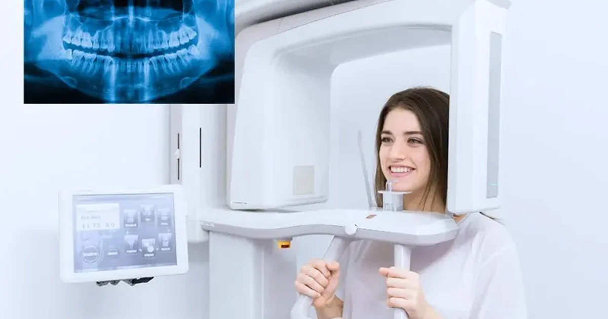
A CBCT scan works by rotating a compact X-ray source and a flat-panel digital detector around the patient's head in a single arc, capturing between 200 and 600 individual projection images over the course of 20 to 40 seconds. These raw projection images are then processed by sophisticated reconstruction algorithms that convert them into a three-dimensional volumetric dataset composed of hundreds of cross-sectional slices, each as thin as 0.1 to 0.3 millimeters.
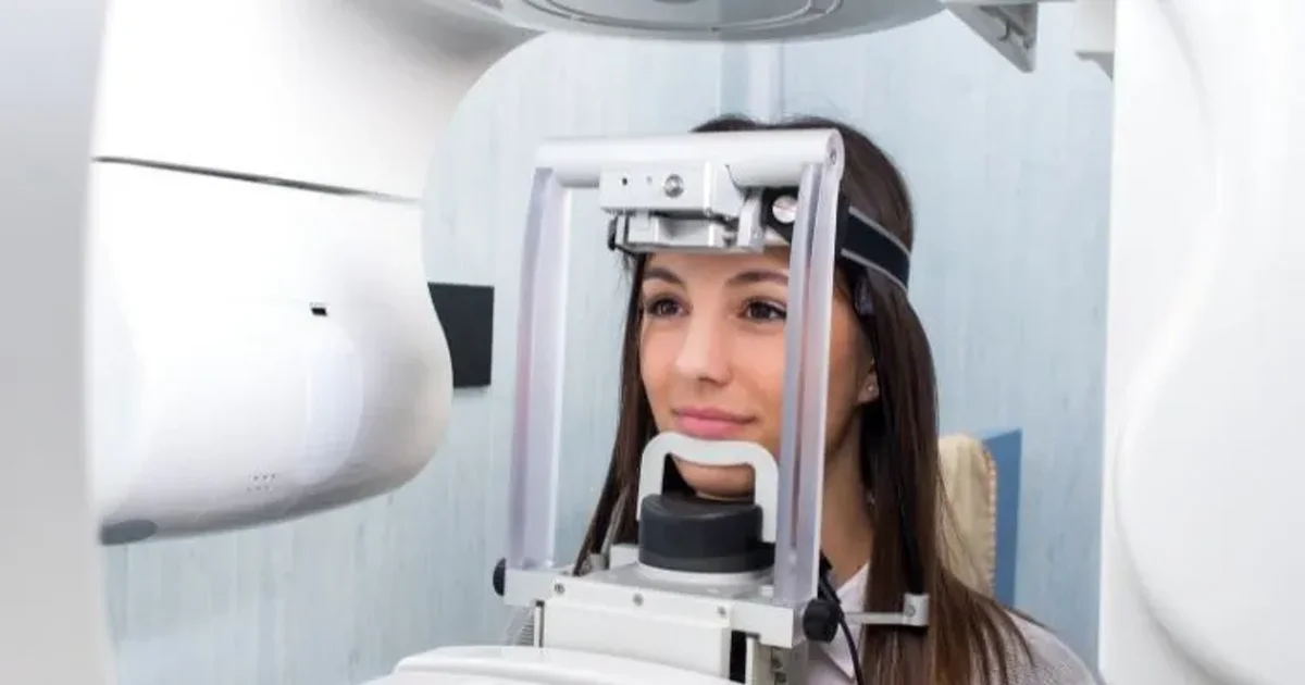
During the scan, the patient stands upright or sits in a chair with their head stabilized by a chin rest and head support. The C-arm of the CBCT machine rotates smoothly around the head, and the patient simply needs to remain still for the duration of the rotation. There is no tunnel or enclosed space, making CBCT significantly more comfortable than a traditional medical CT scanner, especially for patients who experience claustrophobia.
Once the raw data is captured, the computer reconstructs the images into three standard viewing planes: axial (horizontal slices from top to bottom), coronal (front-to-back slices), and sagittal (side-to-side slices). Clinicians can also generate curved panoramic reconstructions, cross-sectional views along the arch of the jaw, and full 3D volume renderings. This versatility is what makes CBCT indispensable for treatment planning across multiple dental specialties.
"At DCDC, we use CBCT technology that captures a complete 3D image of the jaw in under 40 seconds with radiation levels significantly lower than a standard medical CT scan," explains Dr. Osama Elzamzami, Consultant Radiologist at DCDC. "This means patients get the precise imaging their dentist needs without unnecessary exposure."
What Does a CBCT Scan Show?
A CBCT scan reveals a comprehensive range of dental and maxillofacial structures with exceptional clarity, providing diagnostic information that is simply not available from conventional two-dimensional X-rays:
- Teeth and tooth roots: Position, shape, number, length, curvature, root fractures, and supernumerary or unerupted teeth
- Jawbone (alveolar bone): Bone height, width, density, and volume available for dental implant placement or bone grafting
- Nerve canals: The exact course of the inferior alveolar nerve, mental foramen, and other critical nerve structures that must be avoided during surgery
- Maxillary sinuses: Sinus floor position, membrane thickness, mucous retention cysts, and sinusitis
- Airways: Upper airway dimensions relevant to sleep apnea assessment and orthodontic treatment planning
- Temporomandibular joints (TMJ): Condylar morphology, joint space, degenerative changes, and signs of temporomandibular disorders
- Pathology: Cysts, tumors, periapical lesions, bone infections (osteomyelitis), root resorption, and other jaw abnormalities
- Impacted teeth: The precise three-dimensional position of impacted wisdom teeth and canines relative to adjacent teeth and nerves
CBCT vs Regular Dental X-Ray vs Medical CT
Understanding the differences between a CBCT scan, a regular dental X-ray, and a medical CT scan helps patients appreciate why their dentist may recommend one type of imaging over the other. Each technology has legitimate roles, and the choice depends on the clinical question being asked. The fundamental difference between CBCT and medical CT lies in the shape of the X-ray beam, the size of the machine, the body region it images, and the radiation dose it delivers.
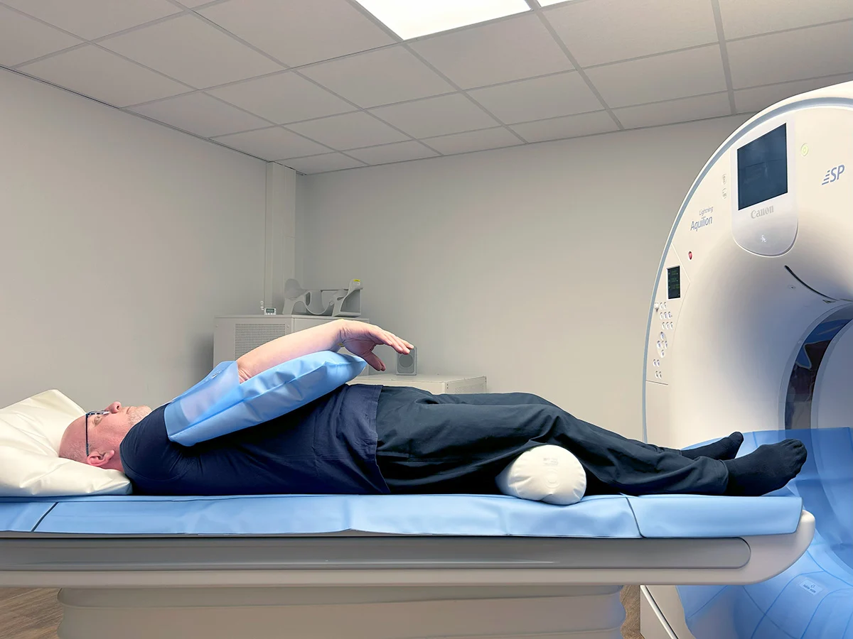
A CBCT (Cone Beam CT) machine uses a wide, cone-shaped X-ray beam paired with a flat-panel detector. In a single rotation, it captures a complete three-dimensional volume in 20 to 40 seconds. CBCT machines are compact, with patients standing or sitting upright in an open design. A medical CT scanner uses a narrow, fan-shaped beam that rotates continuously while the patient lies on a table moving through a large tunnel-shaped gantry, acquiring thin slices through hundreds of rotations. This approach delivers significantly more radiation but captures exceptional soft tissue contrast needed for brain, organ, and vascular imaging.
Another key distinction is spatial resolution. CBCT scanners achieve 0.1 to 0.3 millimeter voxel sizes, ideal for visualizing fine bone structures, individual tooth roots, thin cortical plates, and narrow nerve canals. Medical CT scanners typically have 0.5 to 1.0 millimeter resolution for bone imaging. This means CBCT actually produces sharper images of bony anatomy than medical CT, despite using far less radiation.
| Feature | Regular Dental X-Ray (2D) | CBCT Scan (3D) | Medical CT Scan |
|---|---|---|---|
| Dimensions | Two-dimensional flat image | Three-dimensional volumetric data | Three-dimensional volumetric data |
| Radiation dose | 5-20 uSv (periapical/OPG) | 30-200 uSv | 2,000-20,000 uSv |
| Bone resolution | Limited, overlapping structures | 0.1-0.3 mm voxels (superior) | 0.5-1.0 mm voxels |
| Soft tissue contrast | None | Limited (not diagnostic) | Excellent (diagnostic quality) |
| Scan time | < 1 sec to 15 sec | 20-40 seconds | 5-30 seconds per region |
| Patient position | Standing or seated | Standing or seated, open design | Lying down, enclosed tunnel |
| Contrast dye | Not used | Not used | Frequently used (IV iodinated) |
| Nerve canal visibility | Approximation only | Exact 3D course and dimensions | Visible but lower bone resolution |
| Bone measurement | Estimated (magnification error) | Precise to 0.1 mm, true 1:1 | 0.5-1.0 mm accuracy |
| Best for | Routine check-ups, cavities | Implants, surgery, complex dental | Soft tissue, trauma, cancer staging |
| Cost range (Dubai) | AED 50-200 | AED 500-1,500 | AED 2,000-5,000+ |
Comparison of dental X-ray, CBCT, and medical CT scan. CBCT provides the best balance of bone detail, low radiation, and cost for dental and maxillofacial imaging.
"The easiest way to understand the difference is this: CBCT is a specialist tool built specifically for teeth and jaws, while medical CT is a generalist tool designed to image everything from the brain to the pelvis," says Dr. Osama Elzamzami. "For dental and maxillofacial diagnosis, CBCT gives us the bone detail we need at a fraction of the radiation. Medical CT becomes necessary only when we need to see soft tissues or areas outside the head and neck."
For a detailed side-by-side comparison of CBCT and panoramic X-rays specifically, see our dedicated article on CBCT vs OPG X-ray: which do you need?
Types of CBCT Scans: Small, Medium & Large Field of View
Not all CBCT scans are the same. The field of view (FOV) determines how much anatomy is captured and directly affects the radiation dose, image resolution, and cost. Your dentist or radiographer selects the smallest FOV that covers the clinical area of interest, following the ALARA principle to minimize radiation while maintaining diagnostic quality.
| FOV Type | Coverage Area | Typical Size | Radiation Dose | Approximate Cost (Dubai) | Common Uses |
|---|---|---|---|---|---|
| Small FOV | A few teeth and surrounding bone | 4x4 to 5x5 cm | 30-80 uSv | AED 500-800 | Single implant site, endodontic assessment, localized pathology, root fracture |
| Medium FOV | One complete jaw or quadrant | 8x8 to 10x10 cm | 50-150 uSv | AED 700-1,000 | Multiple implants in one jaw, wisdom teeth, orthodontic assessment of one arch |
| Large FOV | Both jaws and full facial skeleton | 15x15 to 20x17 cm | 100-200 uSv | AED 900-1,500 | Full-arch implants (All-on-4/6), surgical planning, TMJ bilateral, full orthodontic records |
CBCT field of view options with corresponding radiation doses, costs, and clinical indications. Smaller FOV means lower radiation and higher resolution for the target area.
A patient needing assessment of a single implant site receives a small-field scan, not a full-jaw scan, reducing radiation by up to 60 percent compared to a large FOV. Modern CBCT machines also offer dedicated pediatric dose reduction protocols that lower the tube current and voltage to match smaller body sizes, reducing the effective dose by 30 to 40 percent compared to adult settings.
For detailed pricing information, see our guide on CBCT scan cost in Dubai.
CBCT for Dental Implants
Dental implant placement is the single most common indication for a CBCT scan dental examination. Implant surgery involves drilling into living bone and inserting a titanium post that must integrate with surrounding tissue over several months. The margin for error is extremely small. Placing an implant even 1 to 2 millimeters off course can damage a nerve, perforate the sinus floor, or result in insufficient bone support leading to implant failure. A CBCT scan eliminates much of this risk by providing the surgeon with a complete anatomical map before surgery begins.
Traditional 2D dental X-rays, including panoramic radiographs (OPG), compress three-dimensional anatomy into a flat image. A panoramic X-ray shows height but not the buccolingual (cheek-to-tongue) dimension of the jaw. It also magnifies structures by 15 to 30%, making measurements unreliable. Since an implant occupies space in all three dimensions, planning with only two dimensions is fundamentally incomplete.
What CBCT Reveals for Implant Planning
- Available bone volume: Precise height, width, and depth measurements at each proposed implant site, accurate to 0.1 mm
- Bone density and quality: Differentiation between dense cortical bone and spongy trabecular bone, which affects implant stability and healing time
- Inferior alveolar nerve canal position: Exact location and course of the nerve that provides sensation to the lower lip and chin, critical for mandibular implants
- Mental foramen location: The point where the nerve exits the jawbone, which must be avoided during premolar-region implants
- Maxillary sinus floor height: The distance between the ridge crest and the sinus floor, determining whether a sinus lift is needed
- Neighboring tooth roots: Exact root positions and angulations to ensure adequate spacing between implants and natural teeth
- Existing pathology: Cysts, granulomas, residual root tips, and bony lesions that need treatment before implant placement
Published research in the International Journal of Oral & Maxillofacial Implants reports that implants placed with CBCT-guided planning achieve success rates exceeding 97%, compared to approximately 90 to 95% when placed using only 2D imaging. CBCT imaging changes the surgical plan in approximately 30% of dental implant cases, revealing bone that is thinner than expected, nerves positioned higher than suggested by the panoramic film, or sinus anatomy that requires modification of the treatment approach.
Guided Implant Surgery
Guided implant surgery represents the most advanced application of CBCT data. The CBCT scan is imported into implant planning software such as Nobel Clinician, Blue Sky Plan, or coDiagnostiX. The surgeon virtually places each implant in the ideal position, then a surgical guide is 3D-printed that snaps onto the patient's teeth or gums. During surgery, the guide directs the drill at the exact angle and depth planned digitally, achieving placement accuracy within 0.5 mm of the planned position.
When Is CBCT Mandatory for Implants?
- Implants near the inferior alveolar nerve: Any implant in the posterior mandible where the nerve canal's position is uncertain on 2D imaging
- Sinus lift procedures: When the maxillary sinus floor is low and elevation is required to create sufficient bone height
- Bone grafting cases: When the ridge is too narrow or too short and augmentation is needed
- Full-arch rehabilitation (All-on-4, All-on-6): Complex procedures requiring precise angulation of 4 to 6 implants
- Immediate implant placement: Inserting an implant at the same appointment as tooth extraction requires detailed socket dimension knowledge
- History of failed implants: Understanding why a previous implant failed requires 3D assessment of residual bone
The European Association for Osseointegration (EAO) and the International Team for Implantology (ITI) both recommend CBCT imaging whenever conventional radiography does not provide sufficient information for safe implant placement, which in practice means the majority of implant cases.
Planning Dental Implants? Get a CBCT Scan First
At DCDC in Dubai Healthcare City, our CBCT scan gives your implant surgeon the precise 3D data needed for safe, accurate implant placement. Same-day appointments and digital report delivery to your dentist.
CBCT for Wisdom Teeth
Wisdom tooth extraction is one of the most commonly performed oral surgery procedures worldwide, yet it carries a measurable risk of injury to the inferior alveolar nerve (IAN), the nerve that provides sensation to the lower lip, chin, and gums. A standard panoramic X-ray can suggest that a wisdom tooth root may be close to the nerve canal, but it cannot confirm the three-dimensional relationship. The roots may appear to overlap with the canal on the flat image, but in reality they may pass buccally or lingually without actually touching the nerve. Conversely, a 2D X-ray can make a tooth appear safe when the roots are in fact compressing the nerve canal.
A CBCT scan for wisdom teeth eliminates this guesswork. Research published in the British Journal of Oral and Maxillofacial Surgery demonstrates that CBCT assessment before wisdom tooth extraction reduces the incidence of inferior alveolar nerve injury by 25-30% compared to surgery planned on panoramic radiographs alone.
What CBCT Shows About Wisdom Teeth
- Root number and morphology: Wisdom teeth can have one to five roots, which may be straight, curved, hooked, dilacerated, or fused. CBCT reveals the exact shape and curvature of each root.
- Root-nerve relationship: Whether the roots are separated from the nerve canal by intact bone, in direct contact, or whether the canal passes between divergent roots
- Impaction depth and angulation: Precise measurements of how deeply the tooth is embedded and at what angle it tilts relative to the adjacent second molar
- Relationship to the second molar: Detection of even early-stage resorption (erosion) of the adjacent second molar root, invisible on panoramic X-rays
- Associated pathology: Dentigerous cysts, tumors, or infections associated with the impacted tooth, with precise extent measurements
- Lingual bone plate thickness: A thin or absent lingual plate increases lingual nerve injury risk, and CBCT identifies this before surgery
Wisdom Tooth Impaction Types on CBCT
| Impaction Type | Angulation | Frequency | Clinical Significance |
|---|---|---|---|
| Mesioangular | Tilted forward toward the second molar | 43% of impactions | Most common; moderate difficulty; risk of second molar damage |
| Distoangular | Tilted backward into the ramus | Less common | Most difficult extraction; higher risk of mandibular angle fracture |
| Horizontal | Lying sideways parallel to the jaw | Variable | Highest risk of nerve injury and second molar resorption; coronectomy often considered |
| Vertical | Upright but unable to erupt | Variable | Least complicated; nerve risk depends on root depth relative to canal |
Classification of wisdom tooth impaction types. CBCT classifies impaction with far greater accuracy than panoramic X-rays by eliminating superimposition and distortion.
When to Upgrade from OPG to CBCT for Wisdom Teeth
A panoramic X-ray remains the standard first-line imaging for wisdom tooth assessment. CBCT is recommended when the OPG shows radiographic signs of nerve proximity (root darkening at the canal crossing, canal deflection or narrowing, diversion of the canal, or narrowing of the root tip), deep horizontal or distoangular impaction, unusual root morphology, suspected pathology, second molar root resorption, or when the patient has high anxiety about nerve risk. International guidelines from NICE and the European Association for Cranio-Maxillo-Facial Surgery recommend CBCT in these scenarios.
"In approximately 30-40% of cases where the panoramic X-ray suggests nerve proximity, the CBCT reveals that the nerve is actually separated from the roots by intact bone and the extraction can proceed normally," notes Dr. Osama Elzamzami. "In the remaining cases, the CBCT confirms true intimate contact, and the surgeon can decide to perform a coronectomy or modify the surgical approach to protect the nerve. Either way, the patient benefits from a decision based on evidence rather than guesswork."
CBCT for Root Canal Treatment
Root canal treatment relies on the endodontist's ability to locate, clean, and seal every canal inside a tooth. When a canal is missed or an infection goes undetected, the treatment fails. CBCT for root canal treatment has transformed endodontics by giving clinicians a complete three-dimensional view of root anatomy, periapical pathology, and surrounding structures that conventional two-dimensional X-rays cannot provide.
A landmark study published in the Journal of Endodontics found that CBCT imaging changed the diagnosis in 62% of endodontic cases compared to periapical radiography alone. Research in the International Endodontic Journal demonstrates that CBCT identifies up to 34% more root canals than conventional radiography, particularly the second mesiobuccal canal (MB2) of upper first molars, which is present in approximately 90% of teeth but detected on periapical X-rays in fewer than 55% of cases.
What CBCT Reveals for Endodontics
- Extra and hidden root canals: The MB2 canal and other accessory canals that are the single most common reason for root canal treatment failure when missed
- Periapical lesions and infections: CBCT detects periapical pathology at a significantly earlier stage than 2D X-rays, which can only show lesions after cortical bone erosion. CBCT finds periapical lesions in up to 34% more roots.
- Vertical root fractures: Periapical X-rays have only 20-30% sensitivity for vertical root fractures; CBCT achieves 70-80% sensitivity by providing cross-sectional views that eliminate superimposition
- Root resorption: Definitive visualization of internal vs. external resorption, its location and extent, determining whether the tooth is restorable
- Canal anatomy and curvatures: Complete tracing of each canal from pulp chamber to apical foramen, identifying sharp curvatures, S-shaped bends, and canal bifurcations
CBCT for Root Canal Retreatment
Root canal retreatment is one of the strongest clinical indications for CBCT imaging. When initial root canal therapy fails, the endodontist must determine why. A conventional periapical X-ray can show existing filling material and a periapical radiolucency but cannot reliably answer this question. CBCT reveals missed canals that were never treated, separated instruments lodged in canals, voids and gaps in the root canal filling, perforation sites, and the true three-dimensional extent of persistent infection. The American Association of Endodontists specifically recommends CBCT for retreatment cases.
"A two-dimensional X-ray shows you what it can, but a CBCT scan shows you what you need to see," explains Dr. Osama Elzamzami. "In endodontics, the difference between a visible and a hidden canal can be the difference between a successful treatment and a tooth that eventually needs extraction."
When CBCT Is Recommended for Root Canal
- Complex root anatomy: Upper molars (MB2 canal), mandibular premolars with multiple canals, mandibular molars with extra roots (radix entomolaris)
- Retreatment cases: When initial treatment has failed and the cause is unclear on conventional radiographs
- Suspected root fracture: Patients with symptoms suggestive of a vertical root fracture (isolated deep pocket, sinus tract, pain on biting)
- Persistent symptoms after treatment: Pain, swelling, or sinus tract despite apparently adequate root canal treatment
- Pre-surgical planning (apicoectomy): Precise root tip position, proximity to nerves, and cortical bone thickness
- Dental trauma: Luxation injuries, horizontal root fractures, and root resorption not visible on periapical X-rays
CBCT for Orthodontics
Orthodontic treatment involves deliberately moving teeth through bone over months or years. Every millimeter matters. If the orthodontist underestimates bone thickness surrounding a tooth root, the root can be pushed through the outer bone plate, causing a dehiscence or fenestration. A CBCT scan for orthodontics gives the orthodontist a complete three-dimensional view of teeth, roots, jawbone, TMJ, and upper airway, providing diagnostic detail that traditional panoramic and cephalometric X-rays cannot deliver.
Research published in the American Journal of Orthodontics and Dentofacial Orthopedics confirms that CBCT imaging changes the orthodontic diagnosis or treatment plan in up to 36% of cases compared to conventional 2D imaging alone.
What CBCT Reveals for Orthodontic Planning
- Root positions and morphology: Exact length, curvature, and 3D position of every tooth root, critical for determining safe movement directions
- Alveolar bone thickness: Buccal and lingual bone thickness around each tooth with sub-millimeter precision, identifying thin areas before treatment begins
- Impacted canines: Exact 3D position of impacted canines relative to adjacent roots. CBCT detected root resorption of adjacent teeth caused by impacted canines in 48% of cases where resorption was invisible on panoramic X-rays
- Airway dimensions: Volumetric airway measurement in cubic millimeters for patients with suspected obstructive sleep apnea or narrow airways
- Skeletal asymmetry: Precise 3D cephalometric analysis that captures true skeletal asymmetry, which 2D cephalograms mask by superimposing left and right sides
- TMJ assessment: Condylar shape, joint space, and degenerative changes visible before orthodontic treatment begins
- Pre-existing root resorption: Baseline detection of root resorption from previous treatment, trauma, or idiopathic causes, influencing force levels selected
CBCT for Braces vs Clear Aligners
For fixed braces, CBCT identifies teeth with short or dilacerated roots vulnerable to resorption under heavy forces, maps bone topography for expansion vs. extraction decisions, and shows ideal insertion sites for temporary anchorage devices (TADs/mini-screws). For surgical orthodontics, CBCT is indispensable for virtual surgical planning, osteotomy simulation, and surgical splint fabrication.
For clear aligner treatment (Invisalign, Spark), CBCT adds a critical layer. Most aligner software works from intraoral scans that capture only crown surfaces, not roots or bone. When CBCT data is merged with the intraoral scan, the orthodontist sees the complete picture: crown position, root position, and bone boundaries together. This enables verification that planned movements will not push roots outside the bone envelope, reducing iatrogenic side effects.
Is CBCT Necessary for Every Orthodontic Case?
No. CBCT is not needed for every orthodontic patient. For straightforward cases such as mild crowding, simple spacing, or Class I alignment in an adolescent with no impacted teeth, a panoramic X-ray and lateral cephalometric film typically provide sufficient diagnostic information. CBCT is specifically recommended for impacted teeth, skeletal asymmetry, surgical orthodontics, TAD placement planning, airway assessment, cleft lip and palate cases, and complex root anatomy.
Other CBCT Applications
TMJ Disorders
Patients experiencing jaw pain, clicking, locking, or limited mouth opening benefit from CBCT evaluation of the temporomandibular joint. The scan reveals bony changes in the condyle such as flattening, erosion, osteophyte formation, and fractures that indicate degenerative joint disease. While MRI remains the gold standard for soft tissue evaluation of the TMJ disc, CBCT provides superior visualization of hard tissue components and is often the first imaging step in a TMJ workup. A focused CBCT of both TMJs gives high-resolution images at a fraction of the radiation dose and cost of a medical CT.
Jaw Surgery (Orthognathic Surgery)
Orthognathic surgery, jaw reconstruction after trauma, and the removal of tumors or cysts all require detailed three-dimensional imaging of the facial skeleton. CBCT data can be imported into surgical planning software to create virtual surgical simulations, custom cutting guides, and 3D-printed surgical models. This level of planning precision reduces operating time, improves surgical outcomes, and minimizes the risk of complications.
Trauma and Fractures
Dental and maxillofacial trauma, including root fractures, alveolar bone fractures, and dentoalveolar injuries, benefit from CBCT assessment. The 3D images reveal the full extent of fracture lines, the displacement of fragments, and the involvement of adjacent teeth and nerve structures that may not be apparent on conventional radiographs.
Pathology Detection
CBCT can detect dental infections (periapical abscesses, osteomyelitis), cysts (dentigerous cysts, odontogenic keratocysts), and tumors (ameloblastoma, odontoma) in the jawbone. The 3D images reveal the size, location, and extent of pathology more accurately than 2D X-rays. However, for soft tissue tumors or suspected malignancies, an MRI or contrast-enhanced medical CT may be recommended for further evaluation.
Periodontal Bone Loss
CBCT reveals the true three-dimensional pattern of bone loss around teeth, including buccal and lingual defects that are invisible on standard 2D X-rays. This information helps periodontists plan regenerative procedures and assess the true extent of periodontal destruction.
How to Prepare for a CBCT Scan
The good news is that CBCT scan preparation is remarkably straightforward. Unlike many medical imaging procedures that require fasting, contrast dye injections, or lengthy pre-scan protocols, a CBCT scan requires almost no advance preparation at all.
What You Can Do Normally
- Eat and drink normally: There are no fasting requirements. In fact, eating a light meal before your appointment is encouraged to help you feel steady during the scan.
- Take your regular medications: Continue all prescribed medications as normal. There are no drug interactions because no contrast agents or sedatives are used.
- Drive to and from the appointment: No sedation is involved, so you can drive yourself.
- Return to normal activities immediately: You can go straight back to work, school, or exercise after the scan. There is no recovery period.
What to Remove Before the Scan
The most important preparation step is removing all metal objects from the head and neck area. Metal creates bright white streaks and dark shadows on the 3D images called artifacts that can obscure the structures your dentist needs to see.
| Item to Remove | Why It Must Be Removed |
|---|---|
| Earrings (studs, hoops, dangles) | Scatter artifacts distort images of the ear region, TMJ, and upper jaw |
| Necklaces and chains | Streak artifacts obscure the lower jaw, chin, and cervical spine area |
| Eyeglasses and sunglasses | Metal frames create artifacts near the upper jaw, sinuses, and orbital region |
| Hearing aids | Electronic and metal components produce significant artifacts near the ear canal and TMJ |
| Hairpins, clips, and barrettes | Even small metal clips cause localized streak artifacts across the scan volume |
| Removable dentures (partial or full) | Metal clasps and frameworks create extensive artifacts obscuring tooth and bone detail |
| Orthodontic retainers (removable) | Metal wires generate artifact lines reducing image clarity |
| Facial piercings (nose, lip, tongue) | Any metal piercing within the scan field produces localized artifacts |
| Scarves/hijabs with metal fasteners | Metal pins or brooches must be removed; fabric without metal is fine |
Items to remove before a CBCT scan. Fixed dental work (crowns, bridges, implants, fixed braces) does not need to be removed.
What to Bring
- Referral letter: If your dentist provided a referral, bring it. This helps the radiographer set the correct scan parameters and field of view.
- Previous imaging: If you have previous dental X-rays or CBCT scans from another facility, bring them so the radiologist can compare findings.
- Insurance card and identification: Emirates ID or passport for patient registration.
Special Considerations
Pregnancy: CBCT scans are generally avoided during pregnancy as a precaution. If you are pregnant or suspect you might be, inform the radiographer before the scan. In most non-emergency situations, the scan can be postponed until after delivery. Children: CBCT can be safely performed on children when clinically justified. Modern machines offer pediatric protocols with reduced radiation settings. Children may need a parent present for reassurance.
What Happens During a CBCT Scan: Step-by-Step Procedure
The CBCT scan procedure is straightforward, non-invasive, and remarkably quick. From arrival to departure, the entire appointment typically takes 10 to 15 minutes, with the actual scan lasting only 20 to 40 seconds. The procedure is completely painless -- there is no injection, no contrast dye, no sedation, and no physical sensation during the scan itself.
| Step | What Happens | Duration |
|---|---|---|
| 1. Arrival & Registration | Check in at reception, provide referral and identification. No forms for allergies or medical history specific to the scan because no contrast or sedation is used. | 2-3 minutes |
| 2. Preparation | Remove all metal objects from head and neck area. A lead thyroid collar may be placed around your neck to shield the thyroid gland from scatter radiation. | 1-2 minutes |
| 3. Positioning | Stand or sit at the CBCT machine. Chin rests on a padded support, forehead stabilizer keeps head in position. Some machines use a bite block for additional stability. | 1-2 minutes |
| 4. The Scan | C-arm rotates smoothly around your head. Quiet humming sound. Keep tongue pressed against roof of mouth, lips closed, breathe through nose. Completely painless. | 20-40 seconds |
| 5. Image Reconstruction | Computer reconstructs 3D images. Radiographer checks scan quality for adequate coverage and sharpness. If motion artifact is detected, a repeat takes only 20-40 seconds more. | 3-5 minutes |
| 6. Reporting & Results | Consultant radiologist reviews all slices and reconstructions. Detailed written report prepared and shared digitally with your referring dentist. | Same day at DCDC |
Complete CBCT scan procedure timeline from arrival to results. Total appointment time is approximately 10-15 minutes.
Unlike MRI (lying in a narrow tube for 20-60 minutes) or contrast-enhanced CT (requiring an IV injection), a CBCT scan requires nothing more than standing or sitting still for half a minute. There is no enclosed space, no tunnel, and no feeling of confinement. Patients who are anxious about dental procedures or claustrophobic often find the CBCT scan is the most comfortable part of their entire dental treatment journey.
After Your CBCT Scan
There is no recovery time, no side effects, and no restrictions after the scan. You can immediately put your jewelry back on, reinsert dental appliances, and go about your day. Results are delivered in two formats: a written radiologist report summarizing all findings, and the complete 3D image dataset in DICOM format that your dentist or surgeon can load into their planning software. At DCDC, both are shared digitally with your referring clinician via secure electronic transfer.
Book Your CBCT Scan at DCDC
At Doctors Clinic Diagnostic Center in Dubai Healthcare City, the entire CBCT scan appointment takes just 10-15 minutes with same-day results. Our experienced radiology team ensures a comfortable, efficient experience from start to finish.
CBCT Radiation Safety
Patients who are told they need a CBCT scan often ask: is CBCT safe? The short answer is that a CBCT scan is one of the safest advanced imaging technologies in modern dentistry. The CBCT radiation dose ranges from 30 to 200 microsieverts, which is roughly equivalent to 1 to 3 days of the natural background radiation every person absorbs from the environment. By comparison, a standard medical CT scan of the head delivers approximately 2,000 microsieverts, meaning CBCT exposes the patient to 50 to 100 times less radiation.
Radiation Dose Comparison Table
| Imaging Type | Effective Dose (uSv) | Equivalent Background Radiation | Relative to CBCT |
|---|---|---|---|
| Single periapical X-ray | 5 | ~1 day | ~10x lower |
| Bitewing X-rays (4 films) | 20 | ~3 days | ~5x lower |
| Panoramic X-ray (OPG) | 10-20 | ~2-3 days | ~5-10x lower |
| Cephalometric X-ray | 5-10 | ~1-2 days | ~10-20x lower |
| CBCT scan (small FOV) | 30-80 | ~5-12 days | Baseline (small) |
| CBCT scan (medium FOV) | 50-150 | ~8-23 days | Baseline (medium) |
| CBCT scan (large FOV) | 100-200 | ~15-30 days | Baseline (large) |
| Chest X-ray | 20 | ~3 days | ~5x lower |
| Medical CT scan (head) | 2,000 | ~10 months | ~10-60x higher |
| Medical CT scan (abdomen) | 8,000-10,000 | ~3-4 years | ~50-300x higher |
| Annual background radiation (UAE) | 2,400/year | 365 days | Reference |
Radiation dose comparison across dental and medical imaging modalities. CBCT sits between conventional dental X-rays and medical CT, but far closer to dental X-rays.
A full-mouth series of periapical X-rays (18-20 films) delivers approximately 90-100 microsieverts, which overlaps with the dose range of a medium-field CBCT scan. In cases where both a full-mouth series and a panoramic X-ray would be needed, a single CBCT scan can replace both while providing superior diagnostic information at a comparable total dose.
The ALARA Principle
Every CBCT scan follows the ALARA principle: As Low As Reasonably Achievable. This means a scan is recommended only when the clinical benefit of 3D imaging clearly outweighs the minimal radiation exposure. The radiographer selects the smallest field of view and lowest dose settings that produce a diagnostically adequate image. At DCDC, every scan request is reviewed to confirm clinical justification before proceeding.
CBCT Safety for Children
Children are more radiosensitive than adults because their tissues are still growing and cells are dividing more rapidly. However, CBCT is safe for children when clinically justified. Modern CBCT machines offer dedicated pediatric dose reduction protocols that can reduce the effective dose by 30 to 40 percent compared to adult settings. A small FOV CBCT in a child using pediatric settings may deliver as little as 15 to 40 microsieverts, comparable to two or three standard dental X-rays. The European Academy of DentoMaxilloFacial Radiology (EADMFR) and the American Academy of Oral and Maxillofacial Radiology (AAOMR) both recommend that CBCT in children should only be performed when conventional radiography does not provide sufficient information.
Pregnancy Considerations
CBCT scans are generally avoided during pregnancy as a precaution. The radiation dose from a dental CBCT is directed at the head and jaw, far from the abdomen. Research published in Radiation Protection Dosimetry confirmed that the fetal dose from a dental CBCT scan is less than 0.01 microsieverts, thousands of times below the threshold of 100,000 microsieverts at which any measurable fetal risk has been observed. Despite this extremely low risk, standard practice is to postpone elective CBCT scans until after delivery. In genuine dental emergencies during pregnancy, the scan may be performed with additional protective measures including a lead apron.
How Often Can You Have a CBCT Scan?
There is no fixed limit on how many CBCT scans a patient can have, but every scan must be clinically justified. Most patients need only one or two CBCT scans per treatment episode. Even three medium-field scans in a year (approximately 300-450 microsieverts total) would increase annual radiation exposure by only 12 to 19 percent above the natural background level. For comparison, a single round-trip flight between Dubai and London adds approximately 60 to 80 microsieverts of cosmic radiation.
"The radiation from a typical dental CBCT scan is so low that it is biologically comparable to the natural radiation you absorb during a short domestic flight or a few days of simply living on Earth," explains Dr. Osama Elzamzami. "When a CBCT is clinically indicated, the diagnostic benefit overwhelmingly outweighs this minimal exposure."
CBCT Scan at DCDC Dubai Healthcare City
DCDC (Doctors Clinic Diagnostic Center) in Dubai Healthcare City is a leading diagnostic imaging facility offering advanced CBCT scanning alongside a comprehensive range of dental and medical imaging services. The center combines state-of-the-art cone beam CT technology with the expertise of experienced consultant radiologists who specialize in dental and maxillofacial imaging interpretation.
Technology and Safety Protocols
DCDC uses a modern CBCT system capable of capturing high-resolution 3D images with adjustable fields of view, from small focused scans of a few teeth to full maxillofacial volumes covering both jaws and the entire facial skeleton. The machine operates with optimized dose protocols aligned with international guidelines from the ICRP, EADMFR, and the Dubai Health Authority (DHA).
- Clinical justification review: Every scan request is assessed to confirm that 3D imaging is necessary and that the clinical question cannot be answered by lower-dose conventional X-rays
- Optimized dose settings: Radiographers use the lowest kV and mA settings that produce diagnostically acceptable images, with dedicated pediatric protocols for younger patients
- Smallest field of view: The scan is limited to the smallest area covering the clinical region of interest, reducing radiation by up to 60 percent compared to full-field scans
- Lead thyroid collar: Placed over the thyroid gland during every scan to shield this radiation-sensitive organ from scatter radiation
- Pregnancy screening: All female patients of childbearing age are asked about pregnancy before any X-ray examination
- Consultant radiologist oversight: Every scan is reviewed and reported by a consultant radiologist with expertise in dental and maxillofacial imaging
Same-Day Results and Digital Sharing
In most cases, the radiologist's report and the 3D image dataset are available on the same day as the scan. Digital images can be shared directly with your referring dentist, oral surgeon, orthodontist, or endodontist through secure electronic transfer in DICOM format, compatible with all major treatment planning software including Nobel Clinician, Blue Sky Plan, Dolphin, InVivoDental, and Anatomage. This eliminates the need to carry physical films between appointments.
Location and Booking
DCDC is located in Dubai Healthcare City, one of the region's premier medical free zones, easily accessible from Oud Metha, Karama, Bur Dubai, Downtown, and the wider UAE. The center is open six days a week, and CBCT appointments can be booked by phone, WhatsApp, or through the online booking form. Walk-in patients are also accommodated subject to availability. For pricing details, see our guide on CBCT scan cost in Dubai.
With over 13 years of operation and more than 1,000 diagnostic scans performed every month, DCDC has established a reputation for accurate, timely, and patient-centered diagnostic imaging. Whether you need a CBCT scan for implant planning, wisdom teeth, orthodontics, root canal treatment, TMJ evaluation, or any other dental indication, DCDC's radiology team ensures you receive the precise imaging your clinician needs to deliver the best possible care.
Patient Stories: How CBCT Changed Treatment Plans
Sinus Discovery That Changed an Implant Plan
A 45-year-old Dubai resident needed dental implants in his upper jaw but had been told by a previous clinic, based on a standard panoramic X-ray, that he had sufficient bone for the procedure. When he sought a second opinion at DCDC, a CBCT scan revealed a critical finding that the two-dimensional X-ray had completely missed: the floor of the maxillary sinus was significantly lower than expected, leaving far less bone height than the panoramic X-ray had suggested. "The panoramic X-ray showed what appeared to be 10 millimeters of bone below the sinus. The CBCT revealed only 3 millimeters of actual bone height," says Dr. Osama Elzamzami. The surgeon planned a sinus lift procedure first, built the necessary bone height, and then placed the implants safely several months later with no complications.
CBCT Reveals Nerve Wrapped Around Wisdom Tooth Root
A 28-year-old patient was referred to DCDC after her panoramic X-ray showed wisdom tooth roots overlapping directly with the inferior alveolar nerve canal. The CBCT revealed that the nerve canal did not simply pass below the roots -- it tracked directly between the two divergent roots with the cortical bone boundary completely absent on both sides. Based on this finding, the oral surgeon recommended a coronectomy, removing only the crown while leaving the roots undisturbed to protect the nerve. The procedure was successful with no nerve symptoms. A follow-up CBCT six months later confirmed the retained roots had migrated away from the nerve. Without the CBCT, a complete extraction would have carried a high probability of permanent lip numbness.
Hidden Canal Saves a Tooth From Extraction
A 38-year-old patient experienced persistent pain in a lower molar that had undergone root canal treatment two years earlier. Multiple periapical X-rays showed an apparently well-treated tooth with no visible periapical radiolucency. The CBCT scan revealed two findings the X-rays had completely missed: a missed distal canal that was never treated during the original procedure, and a 4 mm periapical lesion situated entirely within the buccal cancellous bone, invisible on the standard X-ray projection. The patient underwent retreatment targeting the untreated canal, and her symptoms resolved completely within weeks. Without the CBCT, the tooth would likely have been extracted.
Get Your 3D Dental Scan at DCDC
DCDC in Dubai Healthcare City offers advanced CBCT scan services with same-day results, experienced consultant radiologists, dose-optimized protocols, and transparent pricing. Walk-in or book ahead for your appointment.
Related Services at DCDC
Expert care and advanced diagnostics at Dubai Healthcare City
Frequently Asked Questions
Final Thoughts
A CBCT scan represents one of the most significant advancements in dental diagnostic imaging, providing three-dimensional detail that fundamentally changes how dentists, oral surgeons, endodontists, and orthodontists plan and execute treatment. From the precision required for safe dental implant placement and the nerve mapping essential for wisdom tooth extraction, to the hidden canal detection that saves teeth during root canal treatment and the skeletal analysis that guides orthodontic decisions, CBCT delivers diagnostic information that two-dimensional X-rays simply cannot match. With radiation doses 50 to 100 times lower than medical CT, scan times under 40 seconds, and no preparation or recovery required, CBCT has become an essential tool in modern evidence-based dentistry.
At Doctors Clinic Diagnostic Center in Dubai Healthcare City, patients benefit from advanced CBCT technology, strict ALARA safety protocols, pediatric dose settings, experienced radiology specialists, and same-day results that keep treatment plans on track. If your dentist has recommended a CBCT scan, or if you are planning dental implants, orthodontic treatment, wisdom tooth extraction, or root canal therapy and want to ensure the most accurate diagnostic foundation, booking a CBCT scan in Dubai at DCDC is a practical and straightforward step. For information on pricing, visit our guide on CBCT scan cost in Dubai, compare CBCT with panoramic X-rays in our CBCT vs OPG X-ray guide, or explore our dedicated CBCT imaging service page for more details on what DCDC offers.
Sources & References
This article was reviewed by our medical team and references the following sources:
- American Dental Association (ADA) - Dental Radiographic Examinations: Recommendations for Patient Selection and Limiting Radiation Exposure
- European Academy of DentoMaxilloFacial Radiology (EADMFR) - Basic Principles for Use of Dental Cone Beam CT
- International Commission on Radiological Protection (ICRP) - Publication 103: The 2007 Recommendations
- Ludlow JB, Timothy R, Walker C, et al. - Effective dose of dental CBCT: a meta-analysis. Dentomaxillofacial Radiology, 2015; 44(1): 20140197
- International Journal of Oral & Maxillofacial Implants - CBCT in Implant Dentistry: Systematic Review
- British Journal of Oral and Maxillofacial Surgery - CBCT Assessment of Mandibular Third Molars and the Inferior Alveolar Nerve
- American Association of Endodontists (AAE) - Position Statement on Use of CBCT in Endodontics
- American Journal of Orthodontics and Dentofacial Orthopedics - Impact of CBCT on Orthodontic Diagnosis and Treatment Planning
- RadiologyInfo.org - Cone Beam CT (CBCT)
Medical content on this site is reviewed by DHA-licensed physicians. See our editorial policy for more information.

Written by
Dr. Osama Elzamzami
Consultant Radiologist
MD, FRCR
Dr. Osama Elzamzami is a Consultant Radiologist specializing in diagnostic imaging including CBCT, CT, MRI, and ultrasound at DCDC Dubai Healthcare City. He holds an MD and FRCR (Fellowship of the Royal College of Radiologists) and has extensive experience in dental and maxillofacial imaging.
Related Articles

CBCT Scan Cost in Dubai: Pricing Guide

CBCT vs OPG X-Ray: Which Do You Need?
OPG X-Ray Results Explained: Complete Guide
More in Diagnostic Imaging
CT Scan Cost in Dubai: Prices, Types & What to Expect
Read More
DEXA Scan Dubai: Cost, T-Score Results & Who Needs It
Read More
Thyroid Ultrasound in Dubai: When You Need It & What It Detects
Read More
Open MRI Dubai | MRI Scan Cost, Uses & Benefits
Read More
MRI Cost in Dubai: AED 900-15,000 Price Comparison (2026)
Read More![Full Body MRI Cost Dubai: AED 5,000-15,000 [2026]](/wp-media/blog/full-body-mri-scan.webp)