Points cles
- A mammogram is the standard first-line breast cancer screening tool for women aged 40 and older, using low-dose X-rays to detect calcifications and masses at an early stage.
- A breast MRI uses magnetic fields and radio waves with no ionizing radiation and has a higher sensitivity (approximately 94-99%) than mammography (approximately 85-90%), making it better at detecting small cancers especially in dense breast tissue.
- Breast MRI is specifically recommended for women at high lifetime risk of breast cancer (20% or greater), including those with BRCA1/BRCA2 mutations, a strong family history, or prior chest radiation therapy.
- Mammograms are more widely available, cost significantly less (AED 400-800 vs AED 3,000-6,000 for breast MRI in Dubai), and have higher specificity, meaning fewer false-positive results that lead to unnecessary biopsies.
- Many high-risk women benefit from getting both tests annually, with mammography and breast MRI performed on an alternating six-month schedule to maximize early detection.
Choosing between a mammogram vs breast MRI is one of the most common questions women face when planning breast cancer screening. Both imaging methods detect breast cancer, but they work in fundamentally different ways, carry different costs, and are recommended for different patient populations. A mammogram remains the gold standard screening tool for women at average risk, while a breast MRI offers superior sensitivity for high-risk women. Understanding each test's strengths and limitations helps women and their physicians make informed screening decisions.
This article provides a detailed comparison of mammograms and breast MRI, covering how each technology works, when breast MRI is specifically recommended, accuracy differences in sensitivity and specificity, cost considerations in Dubai, whether you can get both tests, and how to determine which imaging approach your doctor is likely to recommend based on your individual risk profile.
Mammogram vs Breast MRI: Overview
A mammogram is a specialized X-ray examination of the breast that has been the cornerstone of breast cancer screening for over four decades. It uses low-dose ionizing radiation to produce detailed images of breast tissue and is the only screening modality proven in large randomized trials to reduce breast cancer mortality. Mammograms are highly effective at detecting microcalcifications, tiny calcium deposits that can be an early sign of ductal carcinoma in situ (DCIS) or invasive cancer. Modern digital mammography and 3D tomosynthesis have further improved detection in dense breast tissue.
A breast MRI (Magnetic Resonance Imaging) uses a powerful magnetic field, radio frequency pulses, and an intravenous gadolinium-based contrast agent to produce highly detailed cross-sectional images of the breast without ionizing radiation. It is the most sensitive imaging tool available for detecting breast cancer, with a sensitivity of approximately 94 to 99 percent. However, breast MRI has a lower specificity than mammography, meaning it identifies more findings that turn out to be benign, which can lead to additional imaging and biopsies.
The fundamental difference between a mammogram vs MRI lies in what each test does best. Mammography excels at detecting calcifications, is widely available, and costs relatively little. Breast MRI excels at detecting soft tissue abnormalities, small invasive cancers, and cancers hidden within dense breast tissue, but it requires intravenous contrast, takes longer, and costs substantially more. Neither test is universally superior; the right choice depends on a woman's age, breast density, genetic risk, personal medical history, and the clinical question being asked.
"The decision between mammogram and breast MRI is not an either-or question for many patients," says Dr. Osama Elzamzami, Head of Radiology at DCDC. "For average-risk women, mammography is the evidence-based standard. For high-risk women, breast MRI adds a critical layer of detection that mammography alone cannot provide. At DCDC, we help each patient understand which imaging pathway is most appropriate for her specific situation."
How Each Imaging Method Works
How a Mammogram Works
During a mammogram, each breast is positioned on a flat support plate and gently compressed by a paddle to spread the tissue evenly. Compression is essential because it reduces breast thickness, separates overlapping tissue structures, reduces the radiation dose needed, and improves image clarity. Two standard views are taken of each breast: the craniocaudal (CC) view from above and the mediolateral oblique (MLO) view from the side. The entire examination takes approximately 15 to 20 minutes, with each compression lasting only a few seconds.
Modern mammography systems use either full-field digital mammography (FFDM) or digital breast tomosynthesis (DBT), commonly known as 3D mammography. In tomosynthesis, the X-ray tube moves in an arc over the breast, capturing multiple low-dose images from different angles that are reconstructed into thin slices. This significantly reduces the masking effect of overlapping tissue and improves cancer detection in women with dense breasts. The radiation dose for a standard two-view mammogram is approximately 0.4 millisieverts (mSv), equivalent to about seven weeks of natural background radiation.
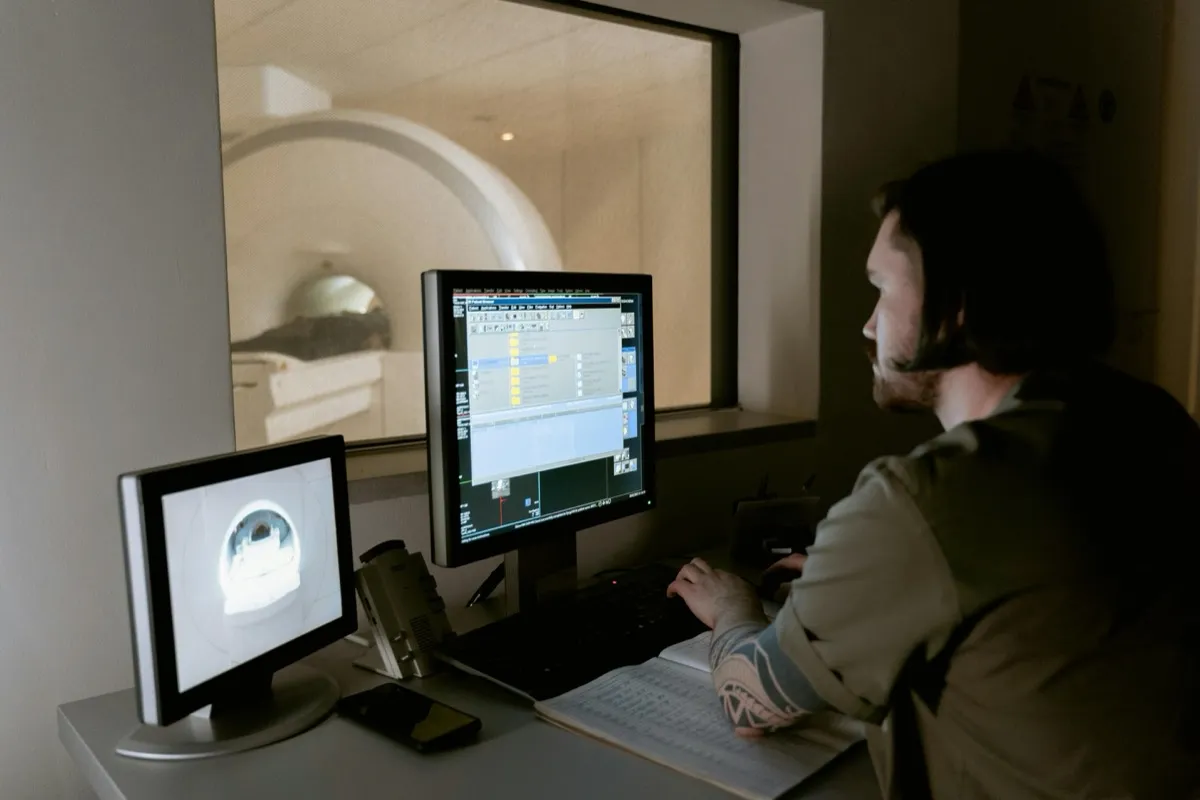
How a Breast MRI Works
A breast MRI requires the patient to lie face down on a padded table with the breasts positioned in a dedicated breast coil, which contains openings that allow the breasts to hang freely without compression. The table slides into the MRI scanner, a cylindrical magnet that generates a powerful magnetic field. Before the scan begins, an intravenous line is placed so that a gadolinium-based contrast agent can be injected during the examination.
The MRI scanner captures images before and after the contrast injection. Cancerous tissue takes up the contrast agent more rapidly than normal tissue due to angiogenesis (new blood vessel formation). By comparing pre-contrast and post-contrast images, the radiologist identifies areas of abnormal enhancement that may indicate cancer. The examination takes approximately 30 to 45 minutes, during which the patient must remain still. The machine produces loud knocking sounds, for which earplugs or headphones are provided.
Breast MRI does not use ionizing radiation, and the magnetic field and radio waves are not known to cause biological harm. However, the gadolinium contrast agent carries a small risk of allergic reaction (approximately 1 in 1,000 patients) and requires normal kidney function to be cleared from the body. Patients with severe kidney disease, certain metallic implants, or pacemakers may not be eligible for breast MRI.
When Is Breast MRI Recommended?
Breast MRI is not recommended as a screening tool for all women. Its high sensitivity comes at the cost of lower specificity, higher expense, and the need for intravenous contrast. Major medical organizations including the American Cancer Society (ACS), the NCCN, and the European Society of Breast Imaging (EUSOBI) recommend breast MRI screening specifically for women at elevated lifetime risk.
Breast MRI is recommended for the following groups of women:
- BRCA1 or BRCA2 gene mutation carriers: Women who carry these inherited genetic mutations have a lifetime breast cancer risk of 45 to 72 percent. Annual breast MRI beginning at age 25 to 30, in addition to mammography, is the standard of care for this population.
- Women with a lifetime risk of 20% or higher: Risk assessment tools such as the Tyrer-Cuzick model can estimate lifetime breast cancer risk based on family history, reproductive history, and other factors. Women who meet the 20% or higher threshold qualify for supplemental MRI screening.
- Women with a first-degree relative with a BRCA mutation: Even if a woman has not been tested herself, having a mother, sister, or daughter with a confirmed BRCA mutation places her at significantly elevated risk.
- Women who received chest radiation between ages 10 and 30: Treatment for conditions such as Hodgkin lymphoma with mantle field radiation substantially increases the lifetime risk of breast cancer, and annual MRI screening is recommended beginning eight years after radiation or at age 25, whichever comes later.
- Women with Li-Fraumeni syndrome, Cowden syndrome, or Bannayan-Riley-Ruvalcaba syndrome: These rare hereditary conditions carry a significantly elevated risk of breast cancer, and MRI screening is part of the recommended surveillance protocol.
- Women with extremely dense breast tissue and additional risk factors: While breast density alone does not automatically warrant MRI screening, women with very dense tissue who also have other risk factors may benefit from supplemental MRI, particularly when mammography has limited sensitivity in their breast tissue.
In addition to screening, breast MRI is used diagnostically to evaluate the extent of a newly diagnosed cancer before surgery, assess tumor response to neoadjuvant chemotherapy, investigate discrepancies between clinical findings and other imaging, evaluate breast implant integrity, and search for an occult primary tumor when cancer is found in a lymph node but not visible on mammography or ultrasound.
Accuracy Comparison: Sensitivity and Specificity
Understanding the accuracy of breast MRI vs mammogram requires examining two key metrics: sensitivity (how well the test detects cancer when present) and specificity (how well the test correctly identifies normal tissue when no cancer is present). An ideal test would score high on both, but in practice there is always a trade-off.
| Feature | Mammogram | Breast MRI |
|---|---|---|
| Radiation | Low-dose ionizing radiation (0.4 mSv) | None (uses magnetic field and radio waves) |
| Contrast agent | Not required | Intravenous gadolinium required |
| Cost in Dubai | AED 400-800 | AED 3,000-6,000 |
| Duration | 15-20 minutes | 30-45 minutes |
| Sensitivity (overall) | 85-90% | 94-99% |
| Sensitivity (dense breasts) | 48-64% | 77-94% |
| Specificity | 88-95% | 72-85% |
| Best for detecting | Calcifications, architectural distortions | Soft tissue masses, invasive cancers |
| Best for | Average-risk screening, calcification detection | High-risk screening, dense breast tissue, cancer staging |
| Compression required | Yes | No |
| Availability | Widely available in most clinics | Specialized centers with breast coil |
Head-to-head comparison of mammogram and breast MRI across key clinical and practical factors. The optimal test depends on the individual patient's risk profile.
Mammography has an overall sensitivity of approximately 85 to 90 percent. However, this drops significantly in women with dense breast tissue, falling to 48 to 64 percent. Dense tissue appears white on a mammogram, and since cancers also appear white, tumors can be masked by surrounding tissue, sometimes described as looking for a snowball in a snowstorm.
Breast MRI has a sensitivity of approximately 94 to 99 percent across all breast tissue types, including dense tissue. This means breast MRI misses very few cancers. The difference is particularly dramatic in women with dense breast tissue, where MRI's sensitivity remains high (77 to 94 percent) while mammography's sensitivity drops substantially. For high-risk women, this additional detection capability can be lifesaving.
The trade-off lies in specificity. Mammography has a specificity of approximately 88 to 95 percent, while breast MRI's specificity is lower at approximately 72 to 85 percent. This means breast MRI produces more false-positive results, where the test flags an abnormality that turns out to be benign. Each false positive can lead to additional imaging, anxiety, and potentially an unnecessary biopsy. This is the primary reason breast MRI is not recommended for routine screening in average-risk women.
"Sensitivity tells us how good a test is at finding cancer, and in that regard breast MRI is unmatched," says Dr. Osama Elzamzami, Head of Radiology at DCDC. "But specificity tells us how good a test is at avoiding false alarms, and mammography performs better on that measure. For high-risk women, the higher sensitivity of MRI outweighs the cost of more false positives. For average-risk women, mammography strikes the right balance."
Cost Comparison in Dubai
The cost difference between a mammogram and a breast MRI in Dubai is substantial, and this is an important practical factor in screening decisions. A standard digital mammogram in Dubai typically costs between AED 400 and AED 800, depending on the facility, whether 2D or 3D tomosynthesis is performed, and whether a radiologist report is included. A breast MRI in Dubai typically costs between AED 3,000 and AED 6,000, reflecting the more expensive equipment, longer scan time, cost of the gadolinium contrast agent, and the specialized radiologist interpretation required.
Insurance coverage varies. Most health insurance plans in the UAE cover annual screening mammography for women aged 40 and older as a standard preventive benefit. Coverage for breast MRI is more variable and often requires pre-authorization with documented evidence of high-risk status, such as a confirmed genetic mutation or a risk assessment showing 20% or greater lifetime risk. At DCDC, the patient services team assists with insurance pre-authorization and can advise on out-of-pocket costs before booking.
For women who require both tests annually, performing both at the same center can reduce overall costs. The financial cost of screening must always be weighed against the potential cost of a delayed diagnosis, which carries far greater medical, emotional, and financial consequences.
Book Breast Imaging at DCDC
At Doctors Clinic Diagnostic Center in Dubai Healthcare City, our radiology team provides both digital mammography and breast MRI with expert radiologist interpretation. Learn more about our mammogram screening services. Get accurate breast imaging with personalized care and timely results.
Can You Get Both Tests?
Yes, many women benefit from getting both a mammogram and a breast MRI, and for high-risk women this dual approach is the recommended standard of care. The two tests are complementary because they detect different abnormalities: mammography is superior at detecting microcalcifications, while breast MRI is superior at detecting soft tissue masses, particularly small invasive cancers that do not produce calcifications. Studies confirm that combining both tests detects more cancers than either alone.
The recommended approach for high-risk women is to alternate the two tests on a six-month schedule: for example, a mammogram in January and a breast MRI in July. This means breasts are screened every six months rather than annually, effectively doubling surveillance frequency. Research published in the Journal of Clinical Oncology confirms that this alternating strategy detects more interval cancers than either test performed annually alone.
Patient Scenario: When Both Tests Are Needed
Consider a 42-year-old woman in Dubai whose mother was diagnosed with breast cancer at age 48 and whose maternal aunt had ovarian cancer at 55. Genetic testing revealed she carries a BRCA2 mutation, placing her lifetime risk at approximately 50 percent. Her mammograms had been normal for two years, but her breast tissue was classified as heterogeneously dense (BI-RADS category C), reducing mammographic sensitivity.
Her physician at DCDC recommended alternating mammography and breast MRI every six months. During her first breast MRI, a small 8-millimeter enhancing lesion was identified in the upper outer quadrant of her left breast. This lesion had not been visible on her mammogram six months earlier, nor was it palpable. An MRI-guided biopsy confirmed early-stage invasive ductal carcinoma, Grade 1, with no lymph node involvement.
"This case illustrates precisely why breast MRI is essential for high-risk women," says Dr. Osama Elzamzami, Head of Radiology at DCDC. "The cancer was small, early stage, and completely invisible on mammography due to the masking effect of dense breast tissue. Without the MRI, it would likely have grown undetected for another year or more. Because it was caught early, the patient had a lumpectomy with an excellent prognosis. Early detection directly translates into better outcomes and more treatment options."
The patient continues with her alternating screening schedule and remains cancer-free three years after treatment. Her case is not unusual; studies consistently demonstrate that breast MRI detects cancers in high-risk women that mammography misses, and that these MRI-detected cancers are typically smaller and earlier stage, leading to better survival outcomes.
Which Test Does Your Doctor Recommend?
The choice between mammogram and breast MRI depends on individual factors your doctor evaluates during a breast health consultation. The recommendation is tailored to each patient's risk profile, medical history, and breast characteristics.
Your doctor will likely recommend mammography alone if:
- You are a woman aged 40 or older at average risk of breast cancer with no significant family history
- You have no known genetic mutations associated with breast cancer
- Your lifetime breast cancer risk is estimated to be below 20 percent
- You have fatty or scattered fibroglandular breast tissue where mammography performs well
- You are seeking standard annual screening as recommended by major medical guidelines
Your doctor will likely recommend mammography plus breast MRI if:
- You carry a BRCA1, BRCA2, or other high-penetrance genetic mutation
- Your lifetime breast cancer risk is 20 percent or higher based on a validated risk model
- You have a first-degree relative with a known BRCA mutation
- You received radiation therapy to the chest between ages 10 and 30
- You have a hereditary syndrome associated with elevated breast cancer risk (Li-Fraumeni, Cowden, or Bannayan-Riley-Ruvalcaba)
- You have extremely dense breast tissue combined with other risk factors that reduce mammographic sensitivity
Your doctor may recommend breast MRI diagnostically (not for screening) if:
- You have been newly diagnosed with breast cancer and the extent of the disease needs to be assessed before surgery
- You are undergoing neoadjuvant chemotherapy and your oncologist needs to evaluate treatment response
- There is a discrepancy between clinical findings and mammography or ultrasound results
- You have breast implants and implant integrity needs to be evaluated
- Cancer has been found in an axillary lymph node but no primary breast tumor is visible on other imaging
If you are unsure whether you qualify for breast MRI screening, the first step is a breast health risk assessment with your physician. This involves reviewing your personal and family history and potentially undergoing genetic counseling. At DCDC, the radiology team works closely with referring physicians to ensure each patient receives the most appropriate imaging.
Regardless of which imaging modality is recommended, the most important step is to screen consistently and on schedule. Breast cancer detected at an early stage has a five-year survival rate exceeding 99 percent. Both mammography and breast MRI serve the same goal: finding cancer as early as possible, when treatment is most effective.
Consult With Our Radiology Team
Not sure which breast imaging test is right for you? The radiology specialists at DCDC in Dubai Healthcare City can review your risk profile and recommend the most appropriate screening strategy. Book a consultation today.
Call or WhatsApp us for guidance on mammogram and breast MRI options.
Questions frequentes
Final Thoughts
The choice between a mammogram vs breast MRI is not about which test is inherently better, but about which test or combination is most appropriate for your risk profile. Mammography remains the proven, cost-effective standard for average-risk screening. Breast MRI provides unmatched sensitivity for high-risk women and plays an essential diagnostic role in cancer staging and treatment monitoring. For high-risk women, combining both tests on an alternating schedule offers the most comprehensive surveillance available.
At Doctors Clinic Diagnostic Center in Dubai Healthcare City, our radiology team provides both digital mammography and breast MRI with experienced radiologist interpretation and personalized patient care. Whether you need routine screening, high-risk surveillance, or diagnostic imaging, DCDC ensures that you receive the right test at the right time, with results that help your treating physician make the best possible decisions for your breast health. To learn more about breast imaging, explore our guides on what a mammogram is and breast cancer screening.
Sources et references
Cet article a ete revise par notre equipe medicale et fait reference aux sources suivantes :
- American Cancer Society - Breast MRI Screening Recommendations
- Saslow D, et al. American Cancer Society Guidelines for Breast Screening with MRI as an Adjunct to Mammography. CA: A Cancer Journal for Clinicians. 2007;57(2):75-89
- National Comprehensive Cancer Network (NCCN) - Breast Cancer Screening and Diagnosis Guidelines
- European Society of Breast Imaging (EUSOBI) - Recommendations for Breast MRI Screening
- RadiologyInfo.org - Breast MRI
Le contenu medical de ce site est revise par des medecins agrees DHA. Voir notre politique editoriale pour plus d'informations.

Redige par
Dr. Osama Elzamzami
Diagnostic Radiology
MD, FRCR
Dr. Osama Elzamzami is a Consultant Radiologist and Head of Radiology at DCDC Dubai Healthcare City, specializing in diagnostic imaging including mammography, breast MRI, CT, and ultrasound.